
Environmental Fate of Metal Nanoparticles in Estuarine Environments


1
Department of Earth Sciences, Environment and Resources, University of Naples Federico II, Via Cintia 21, 80126 Naples, Italy
2
Department of Chemical Sciences, University of Naples Federico II, Via Cintia 21, 80126 Naples, Italy
*
Author to whom correspondence should be addressed.
Water 2022, 14(8), 1297; https://doi.org/10.3390/w14081297
Submission received: 21 March 2022
/
Revised: 10 April 2022
/
Accepted: 13 April 2022
/
Published: 15 April 2022
(This article belongs to the Special Issue New Insights on Pollution and Remediation of Trace Elements in Coastal and Estuarine Sediments)
Abstract
:In the last decade, metal engineered nanomaterials (ENMs) have seen an exponential use in many critical technologies and products, as well an increasing release into the environment. Coastal ecosystems worldwide may receive ENM-polluted waters and wastes, with a consequent alteration of habitats and contamination of aquatic biota. There is a scarcity of data regarding the fate of these emerging contaminants in such environments. Open issues include the determination of the sources, the quantification of the interactions with marine sediments, the bioaccumulation pathways, the ecotoxicology on marine fauna and the identification of the principal biotic and abiotic factors that may alter metal ENMs toxicity. Little is known about their potential transference into the food web, as well toxicity features and co-stressors of single or multiple ENMs under laboratory and real environmental conditions for various taxonomic phyla. This review reports current knowledge on the ecological impact of ENMs under the complex environmental conditions of estuary systems, identifies gaps in current knowledge and provides directions for future research.
1. Introduction
One of the fastest expanding industries in the last few years is represented by the nanotechnology industry [1]. The synthesis and manipulation of nanomaterials (NMs) is very intriguing, as they present critical physical phenomena based on their size [2]. The European Union defines nanomaterials as those having particles with one or more dimensions in the size range of 1–100 nm [3]. They present unique electronic, magnetic and optical features and are involved in biological, surface and interface processes that otherwise would not be possible if they were in the bulk state [2]. Their reactivity can be quite singular when they are below <5 nm [2]. An important group of NMs with large commercial use is given by metal engineered nanoparticles (ENMs). Based on the production levels, ENMs are ordered in the following descending order: titanium dioxide (nTiO2), silicon dioxide (nSiO2), iron (nFe) and iron oxide (nFexOy), zinc oxide (nZnO), silver (nAg), carbon nanotubes (CNTs), cerium oxide (nCeO2), copper (nCu) and copper oxide (nCuO), metal sulphides, selenides and tellurides [4,5,6,7]. Metal and metal oxide nanoparticles of Ti, Zn and Ag are engineered nanoparticles that are used in many consumer and industrial products [8]. Their use extends over ever more industrial sectors due to their extraordinary properties, i.e., in new marine nanotechnologies as pollution remediation products [9]. These nanoscale products and by-products inevitably end up in rivers, lakes, estuaries and coastal waters during their production, use and end of life via wastewater, atmospheric deposition and other routes [7,8,10,11].
The very tiny dimension of these products affects their stability, dispersion, aggregate dimension, sedimentation and, ultimately, final bioaccumulation and bioavailability [12]. In the aqueous phase, ENMs are transported along with the waters and the suspended particulate matter (SPM), causing unusual toxicity [11,13]. The tiny size of these materials allows them to easily penetrate tissues through fluid-phase endocytosis and caveolae, diffusing through pores and ion transport systems, with the possibility to transport other toxic contaminants [11,13]. They can cross bio-barriers, such as skin, blood, brain, intestine and maternal foetus [14]. Their highly reactive surfaces can generate reactive oxygen radicals (ROS), with damage at the cellular levels of DNA, proteins and membranes, and interfere with electron transport processes [15].
The research on the ecotoxicological effects of ENMs is extensive for freshwater systems, from reduced swimming, feeding, growth and alteration of reproduction behaviour and digestive stress [16,17,18,19]. However, the increasing release of ENMs in estuary and coastal waters requires more research on their fate, behaviour and ecotoxicological consequences [15,20]. Some authors [21] maintain that ENM levels are far from being lethal, and the knowledge of the sub-lethal implications for marine population stability is poor. There is a lack of knowledge on median lethal or effective concentration (LC/EC50), exposure limits and effects ranges for various saltwater organisms [12]. The lack of information on the chronic and acute toxicity patterns of ENMs limits the definition of standardised experimental conditions, as well as specific analytical quantitation conditions, making the exposure scenario in the marine environment rather obscure [12]. The definition of toxicity is hard to set up, even for simplified biological models, due to the complex conditions of seawater, with its very high salinity and ionic strength [22].
This review reports current knowledge on the ecological impact of ENMs under the complex environmental conditions of estuary systems, identifies gaps in current knowledge and provides directions for future research.
1.1. Synthesis, Properties and Nature of NPs
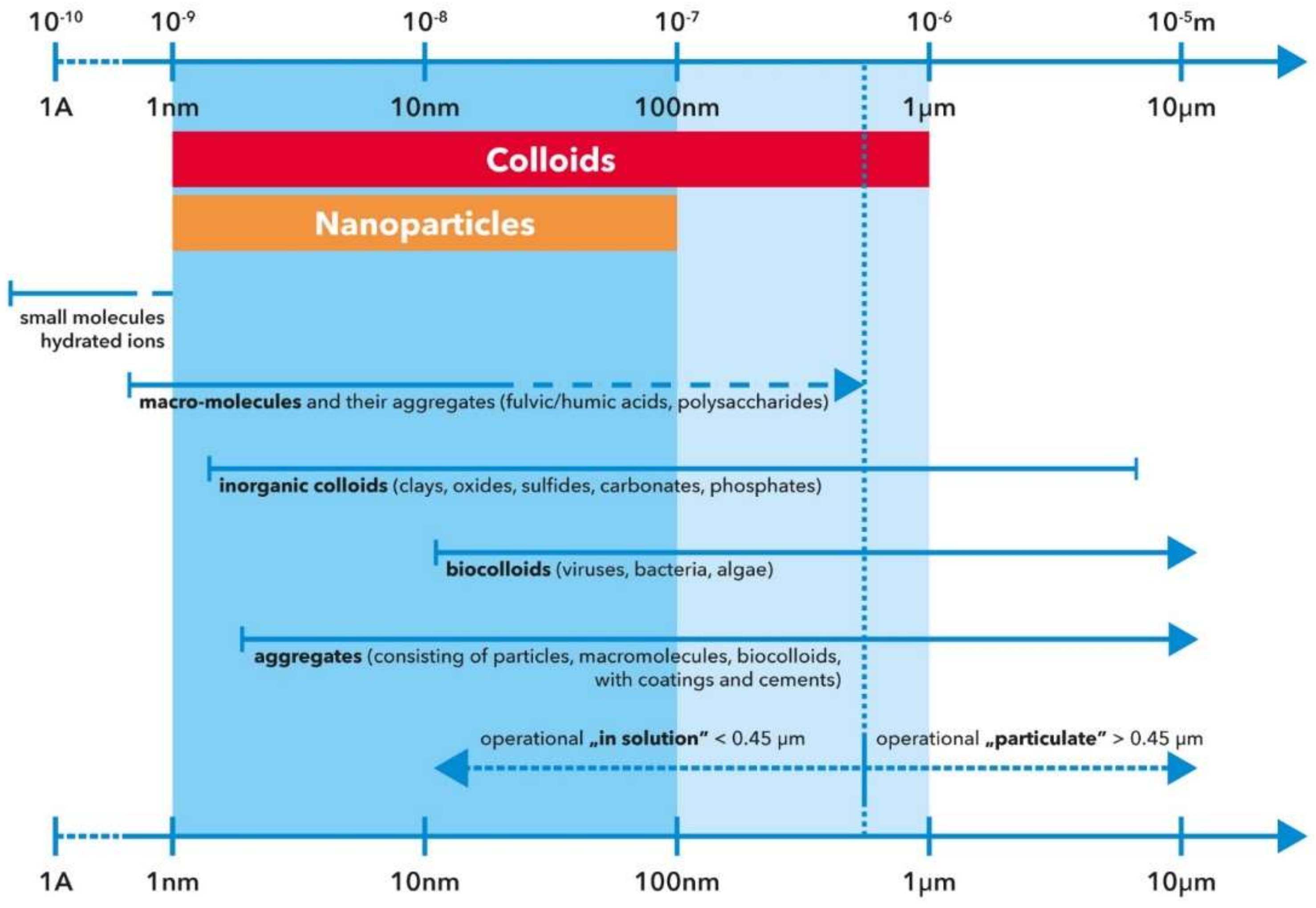
The synthesis and properties of NPs represent a fundamental issue to understand their fate in the waters and sediments of estuary environments. These particles can be considered as complex mixtures where different compounds can be adsorbed or exchanged [20]. As already stated, NPs are particles with at least one dimension less than 100 nm and are defined by the International Union of Pure and Applied Chemistry (IUPAC) [23] as a sub-fraction of colloids (Figure 1). Colloids can be natural, formed through long-term processes of millions of years, and anthropic. Practical examples of natural colloids include desert dusts, aerosols and metal oxide NPs [15]. Some authors [20,24] further distinguish between different anthropic NPs in terms of unintended by-products from technological processes or specific manufactured products. Christian et al. [20] reported how NPs must be considered as complex mixtures, capable to adsorb light as a dye and to dissolve as a small molecule. They have an exceptionally high surface-area-to-volume ratio and this feature is at the base of their extraordinary properties [20]. This property means that a large part of a NP’s atoms are on the particle’s surface rather than in the core or bulk material [25].
One of the most commonly manufactured NPs is titanium dioxide (TiO2 NPs), which are often found in surface waters, sediments, sludge and wastewaters [26]. Piccinno et al. [27] estimated the overall production of TiO2 at 3000 tons/year and that it will likely increase exponentially until 2025 [28]. They find wide use in water treatment, photocatalysis, solar cells and paints [27]. Products containing TiO2 NPs are UV filter creams, food additives, self-cleaning coated glass, construction materials, paints, biomedical products, toothpaste, wood preservatives and photocatalysts [29]. They are mixtures of two crystalline structures, namely, anatase and rutile, with the former requiring the largest amount of manufacturing processing [30] and with the highest photocatalytic activity [31], and the rutile form is used as white coating pigment [32]. Besides the size and surface area, these metal nanomaterials have unique properties in terms of their shape, crystallinity purity, stability corrosiveness and photocatalytic activity [29], as well as excellent optical and electrical properties [29]. TiO2 represents one of the most used semiconductors for photocatalysis [29]. The textile industry, for example, makes large use of these products to photodegrade dyes in wastewaters [29].
The increased production and use of these products increase their presence in surface waters [33] and coastal seawaters [34]. TiO2 NPs have also been found to interact with many pollutants and this increases their diffusion and toxicity [35]. Some authors estimated a total concentration of TiO2 NPs in swimming pool water of 21–60 µg/L [36]. Some authors made conservative estimates of 4 kg of TiO2 nanoparticles in the water from a touristic Mediterranean beach on a summer’s day [37]. Due to their specific properties, TiO2 NPs are accumulated on the surface microlayer of the water column [38]. Due to this preferential localisation, the release of these NPs in waters causes enormous concern due to their involvement in the photocatalysis and phototransformation processes of organic compounds, with negative effects on organisms [39]. Solar radiation may photoexcite TiO2 NPs from UV filters, producing significant amounts of hydrogen peroxide (H2O2) and generating stress on marine phytoplankton [37].
Other important ENMs in terms of their intrinsic properties, large use and commercialisation are silicon oxide (SiO2), cerium oxide (CeO2), silver (Ag) and zinc oxide (ZnO) [15]. The use of synthetic silica, i.e., SiO2 ENMs, goes back to the middle of the last century. Recently, SiO2 ENMs production has significantly increased for mass applications, such as tires [40]. CeO2 NPs are used in the automotive industry for particulate abatement, as a catalytic converter and fuel additive [41], as glass polisher and heat-resistant plating [42] and for chemical–mechanical planarisation in the fabrication of integrated circuits [43]. Nanosilver (Ag ENM) has been employed in the medical field since the beginning of the last century due to its antibacterial properties [44,45]. It is also commonly used in many consumer products (CPs) in deodorants and socks [46]. Unlike SiO2 ENMs, however, Ag ENMs have been studied broadly [27]. Among the most threatening ENMs are ZnO NPs, with an annual global production of 10 Mt [47]. About ten percent of this production is released into the environment and 95% of it enters the aquatic system, accumulating in sediments [48]. ZnO NPs are also used in fertilisers, paints and animal feeders [15], and much like TiO2 NPs, are used in sunscreens to protect skin from UV irradiation [15]. The use of ZnO NPs may exceed that of TiO2 NPs since these nanoparticles can absorb UV-A and UV-B radiation, while TiO2 NPs only absorb UV-B, offering better protection and improved opaqueness [49]. ZnO NPs are also used in ceramics and rubber processing, wastewater treatment and as a fungicide [49].
1.2. The Sources of ENMs
The major sources of ENMs leading to release into the environment are sewage spills from wastewater treatment plants [50,51]. There are data from Callaghan and MacCormack [6] reporting levels of ENMs in wastewater treatment plants effluent from 10−4 to >101 μg/L.
Another source is urban runoff [52]. This is because ENMs are widely used in urban settings, for instance, the TiO2 used as pigments in paints of white road marking, containing at least 10% by weight of TiO2 pigments [53]. Some studies measured high concentrations of TiO2 in road dust, sludge from storm drains and roadside soils, which were suspected to have an anthropogenic origin. It was predicted that road marking material will be 450,000 tons in 2025, which may result in a significant release of TiO2 pigment to urban runoff [54]. Another source of TiO2 in urban runoff comes from the large use of exterior paints [54]. However, TiO2 NPs can be also incorporated into material surfaces to allow them to self-clean [55]. In fact, since the creation of self-cleaning glass, TiO2 has been used to create self-cleaning nanoparticles, working in a two-stage cleaning process: the first consists of photocatalysis of any fouling matter on the surface, i.e., glass, while the second occurs when the glass becomes super hydrophilic and allows water to wash away the catalysed debris on the surface.
There is an objective difficulty in discriminating natural from engineered TiO2 in all kinds of environmental matrices, especially when they are present at high concentrations [56,57]. The main differences between natural and engineered NPs are the elemental composition, elemental ratios, purity, surface coating, narrow size distribution and morphology [20,33,39]. Another difference is that TiO2 ENMs contain 1–15% by weight of artificial coatings, i.e., oxyhydrates and oxides of Si and Al, except for TiO2 used as a food additive [49].
In the case of TiO2 NPs, a good indicator for discriminating natural or anthropic sources is the Ti/Nb ratio. This is because natural TiO2 NPs are carriers of Nb in rocks [50]. Wang et al. [57] quantified this ratio in real urban runoff, two bridges in Columbia, and Ballona Creek and Los Angeles River, USA, to reveal the presence of TiO2 engineered particles. In urban runoff, they observed an increase in the elemental ratios Ti/Nb relative to natural background values, indicating Ti contamination. The presence of TiO2 engineered particles was confirmed using transmission electron microscopy and energy-dispersive spectroscopy. TiO2 engineered NPs in urban runoff were in the range of 5–150 mg/L.
Besides freshwater effluents, ENMs can also come from the large use of cosmetic products for solar irradiation protection; sanitary products, such as sticky plasters; and deodorants [58], as is the case of Ag NPs. The dissolved Ag can form colloids or remain as NPs. Blaser et al. [59] modelled the presence of Ag levels along the course of the Rhine in Germany and suggested that concentrations in waters and sediment might increase towards the estuary from 4 to 320 ng/L in waters and from 0.04 to 14 µg/g in sediments. Leaving the model approach behind, real measurements of Ag release indicated the formation of colloidal complexes with Fe/Mn oxyhydroxides/sulphides and sedimentation downstream from an industrial plant [60]. Wong et al. [49] estimated an annual release of 250 t of these creams into the marine system.
Once released into the environment, NPs undergo dispersion, transportation, aggregation and sedimentation, which are all processes that influence their reactivity, mobility and toxicity [61]. Many ENMs travel through domestic waste pipes and then reach wastewater treatment plants and, ultimately, the oceans. Based on a model estimation, O’Brien and Cummins [62] reported that WWTPs and water purification facilities can remove 60–70%, even up to 96%, of NPs. Thus, models are widely adopted by researchers due to the difficulty in analysing sampled NPs and the low ENMs released from different industries [15]. The objective difficulty of these models is that their output can be distant from the real release volume in an estuary environment, which might represent an important sink of such engineered particles.
1.3. The Estimation of ENMs Release
The regulation of ENMs and risk estimation is possible with a clear comprehension of the production and release of these emerging contaminants. From a regulatory point of view, data on ENMs’ environmental production, release and exposure are required to estimate the associated risk. The current literature is quite sparse in terms of the data produced. A former estimate from 2004 [63] reported a production rate of nano-metal oxides, namely, TiO2, ZnO and Fe2O3, for skincare products of 103 tonnes/y. There are estimations that at least 25% of sunscreen is washed off during bathing and swimming [64], meaning that around 250 tonnes of these NPs can be discharged into the marine environment. Given ENMs diffuse applications, an analysis of the environmental distribution and concentration of the applied NPs is advisable. Models of exposure of existing or new ENMs, in combination with toxicological data, can contribute to a prospective risk assessment [65]. Thus, there are considerable uncertainties in the estimation of ENMs released into the environment. To date, the quantification of ENMs in environmental samples, especially in solid samples, such as soil and sediment, is not possible [66] and cannot be directly detectable using analytic methods due to their very low concentrations. Even if detected, it is difficult to discriminate between natural and engineered nanomaterials [67]. Compared with the toxicological examination of ENMs, few data are available about their actual release [68]. Among ENMs and in terms of release, Ag ENMs are again the most studied nanomaterial so far [69]. A valid tool to produce environmental risk analyses involves modelling predicted environmental concentrations (PECs) [66] on the basis of existing environmental levels of ENMs. Several modelling studies have presented quantitative estimations of the environmental concentrations of ENMs [59,66]. Sun et al. [70] used a stochastic approach to predict the amount of CeO2 ENMs in freshwater and reported a range of 1 pg/L in 2017 to a few hundred ng/L in 2050. Relative to CeO2, the SiO2 ENMs estimates are approximately 1000 times higher, and those for Ag ENMs are 10 times lower [70]. Keller et al. [5] used models to quantify the release of global ENMs production on a world scale base and estimated that most of them were released from coatings, paints, pigments, electronics and optics, personal care products and energy production. ENM wastes amounted to ~318.300 metric tons, with ~10% accumulated in aquatic systems. The framework for estimating emissions can be easily improved as better data becomes available [5].
1.4. ENMs Behaviour in Marine Environments
Due to their low dimensions and electrical properties, ENMs tend to aggregate, which affects their reactivity, behaviour, fate, mobility and risk consequences [71]. One of the main effects of aggregation is the stability reduction; this occurs through homo-aggregation between particles of the same dimension and hetero-aggregation between ENMs and natural colloids [72]. Hetero-aggregation occurs when interacting with particles of different physico-chemical features [73] and depends on the surface chemistry of the particles interacting, as well the solution chemistry [74]. Aggregation occurring through adsorption processes with co-existing colloids influences the mobility and the transference of ENMs in different compartments. This occurs through colloidal bridging and or charge neutralisation or by surface coverage. Both homo- and hetero-aggregation occur in parallel, while at the low electrolyte level, only hetero-aggregation occurs [75]. Mobility and toxicity were found to be a function of aggregate size and while mobility generally decreases, toxicity increases with the dimension [29].
Disaggregation is the opposite process with opening up of the agglomerates and with the obvious increase of the stability of NPs [76]. The stability of the ENMs’ dispersion is critical for the assessment of potential risk [77]. The stabilisation of ENMs also has important consequences on the photocatalytic properties of the particles and the related processes of organic contaminant photodegradation, reuse of ENMs suspension and sedimentation [78].
The processes of aggregation and sedimentation of ENMs occur throughout the waste management process. In wastewater treatment plants (WWTPs), ENMs are removed by activated sludge [79]. There is scant knowledge of the fate of ENMs in wastes and landfills. Waste composition, particle properties and physico-chemical conditions of the landfill condition the fate of the nanoparticles and, hence, the transportation to the open sea [80]. Throughout the disposal steps, NPs may end up in soil, surface waters and marine systems.
1.4.1. Factors Influencing Aggregation
Once released in water, nanoparticles tend to aggregate. The aggregation of ENMs is an important phenomenon in the marine environment because of the attraction between particles, gravity and Brownian effects, which affects their sedimentation [29]. This feature depends on the size, concentration, charge and shape of the particles, as well as the features of the aqueous media, such as pH, ionic strength and natural dissolved organic matter [21,81]. When the sizes of the aggregates increase, the stability decreases, favouring sedimentation [82]. Increasing NPs’ concentration increases the probability of the particles coming together and, thus, interacting and aggregating [83]. However, the presence of impurities of phosphorus, silicon and ions in general affects aggregation due to modification of the charge of the surface [84] Aggregation is in fact also influenced by cationic and anionic charges [85]. Anions act by neutralising the surface charge of ENMs, reducing the electric double layer, which is an effect that increases with the charge of the anions [86]. Aggregation is hindered by electrostatic repulsion and or steric hindrance. Aggregation can be modified by modifying the charge via adding so-called capping agents, namely, citrate, polyvinylpyrrolidone, gum Arabic and oleate [87,88], favouring dispersion [89] in contrast with the natural aggregation effect of the van der Waals’ forces [90] or, in other cases, reducing dissolution. This means that capping can significantly modify the ENMs behaviour in high-ionic-strength sea waters. When the charge is neutralised, the ENMs form large aggregates. In seawater, the salinity, CO32−, SO42−, Mg2+ and Ca2+ affect ENMs’ behaviour. The ionic strength of seawater shields the repulsive forces of the nanoparticles and forces them to aggregate in a few hours [81]. This effect, on the one hand, reduces the exposure of the water column living organisms and, on the other, enhances the risk for benthic species [91].
Another important factor is the presence of natural organic matter (NOM), which is very ubiquitous; is a key encountered compound [92] and, in its dissolved form (DOM), significantly impacts ENMs’ stability. DOM is a very complex cocktail of different compounds with different physical and chemical properties, molecular weights and compositions varying with the originating material and biogeochemical occurring processes [93]. Many authors observed the adsorption of DOM onto nanoparticle’s surfaces, altering the physico-chemical properties and, hence, the stability and transport of ENMS in porous media [94,95,96]. One clear issue is the direct relationship between the DOM concentration and nanoparticle mobility [94,97]. Besides the DOM concentration, other key features are the composition, charge density and molecular weight (Mw) of DOM. Thus, fulvic acids (FA) and humic acids (HA) behave differently, as shown by Vindehal et al. [92] in the interaction with silver nanoparticles. FA were found to impede goethite nanoparticles aggregation, having a more charged moiety per mole of C. In the same way, Li et al. [98] also observed the increased ability of low molecular weight acids on the increased mobility of graphene oxide nanoparticles. Zhang et al. [99] studied how DOM from different sources modifies the stability and mobility of TiO2 NPs in acid-saturated porous media and found that the aromaticity and weight average molecular weight features were the key properties that determine the transport potential of the nanoparticles. Dissolved fulvic acids interact with TiO2 NPs via phenolic and carboxylic ligands [100], while the higher content of aromatic and alkyl groups of humic acids increases the hydrophobicity and adsorption onto TiO2 NPs [29].
One of the main parts of NOM is the exopolymer substances (EPs) produced by bacteria and phytoplankton and made up of polysaccharides and proteins [101]. These biopolymers behave as surfactants and can form marine gels. Authors such as Quigg et al. [102] reported that these biopolymers play an important role in the interaction between marine biota and ENMs, such as CuO NPs and Ag NPs [103,104]. Xu [29] reported how the release of extracellular polymeric substances by marine algae affects ENM aggregation, adsorbing on nanoparticles via chemical bonding and electrostatic interaction, favouring settlement of the particles.
1.4.2. ENMs Behaviour in Estuary Environment
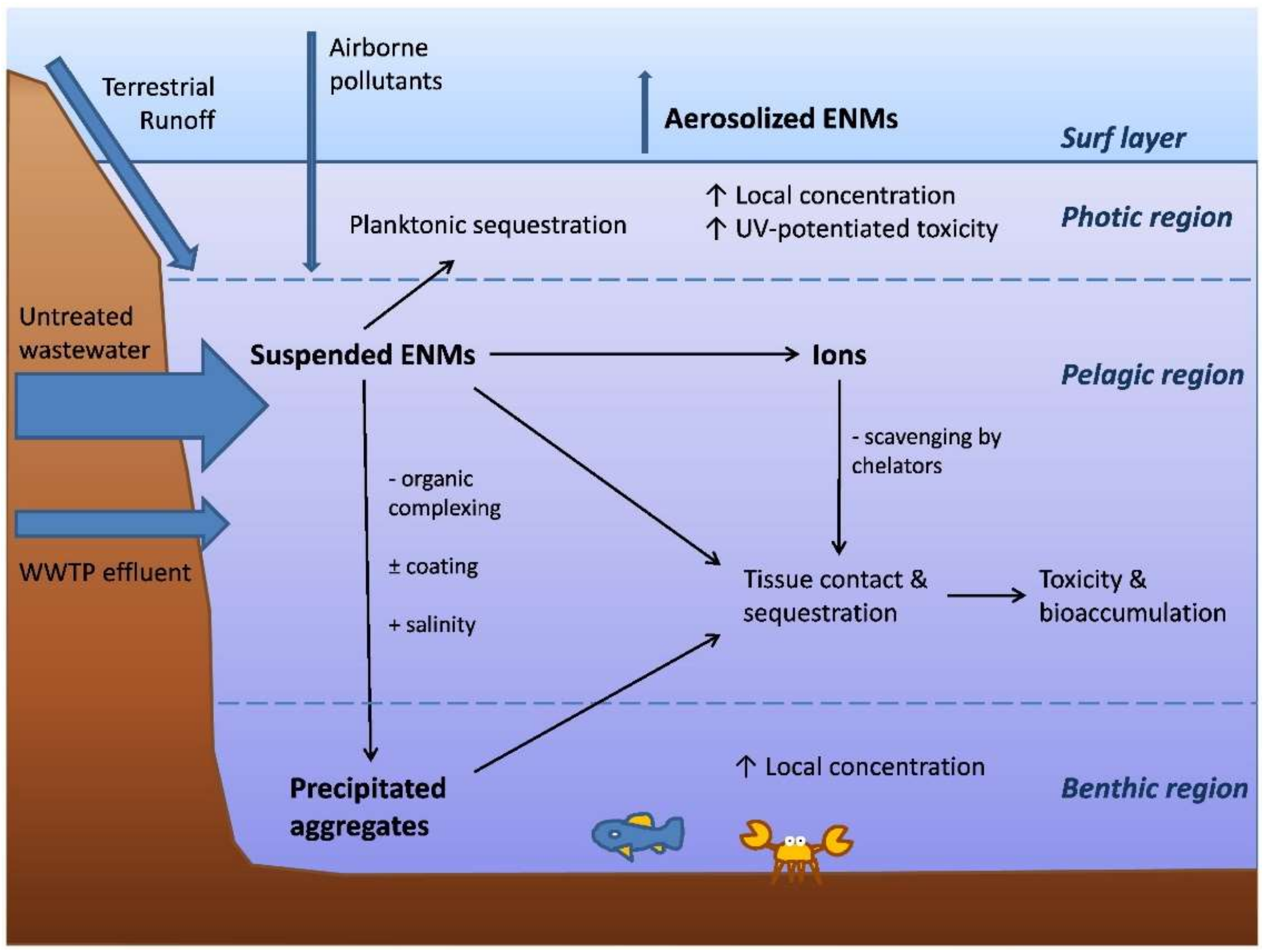
Along river watercourses, ENMs release metals with different kinetics, depending on the hetero-aggregation and interaction with NOMs of different molecular weights. When ENMs reach an estuary, more complex and different processes occur (Figure 2), as an estuary is a very sensitive and critical environment that collects domestic, agricultural, shipping and industrial runoffs [105]. Thus, the significant input of colloids and suspended particulate matter [106] and the increased ionic strength favor aggregation and particles sediment, posing a risk for the benthos. In this context, ENMS go through intense processes of aggregation and sedimentation and sink slowly to the floor, depending on the ionic strength, temperature and NOM levels [107]. Moreover, in the estuary the increased hydrodynamics resuspend the particles making ENMs available for a larger number of organisms, including filter, suspension and deposit feeders [15]. Tappin et al. [108] reported higher loads of Ag+ in the Adriatic Sea than those of rivers because of the high estuary hydrodynamicity that remobilises Ag+ from sediments and increases the overall pool of dissolved metal and the ecological risk for marine biota [15].
These features make these environments particularly susceptible to the potential toxicity risk of ENMs. Besides NOMs and colloids, ENMS can react with various contaminants and cations under different physico-chemical conditions, such as pH, ionic strength and type and concentration of cations. The colloidal behaviour and stability of ENMs depend on the physico-chemical interactions with the environment. They can release metal ions, precipitate, stabilise in the water columns via interaction with NOMs or be taken by algae. At the top of the water, they can be aerosolised and dispersed and cause extra toxicity in the photic region spreading UV energy (Figure 2) [6]. This interaction is driven by NOMs’ surface coating of ENMs, particle aggregation, disaggregation and aggregate structure [20]. For example, the sorption of humic substances (HS) on iron-oxide NPs, which occurs through the formation of a nanoscale surface coating [109], increases the negative charge of an NP’s surface according to the NP’s point of zero charge (PZC) and the pH of the solution. Such a process may produce a dual effect: (i) decreases particle aggregation via a charge stabilisation and steric stabilisation mechanism, and (ii) increases the aggregation through charge neutralisation due to fibrillar attachment [110,111,112]. Another important driving force is the concentration of the ENMs, whose increase produces aggregation [113]. Thus, in aqueous systems, different processes influence NPs’ stability; aggregation; sedimentation; nanoscale film formation; charge enhancement; steric stabilisation by NOMs; ionic strength; binding cations, such as Ca2+; NP properties and their concentration; nature of the organic matter; and the conditions of the aqueous media, such as pH and ionic strength. From the above considerations and reports, it might be concluded that the ecological impact and environmental behaviour of ENMs in estuary depend mainly on the stability of the nanoparticles if they are dispersed or aggregated [114,115].
1.4.3. Interaction of ENMs with Pollutants
Due to the high surface-to-mass ratio, ENMs can adsorb pollutants or/and trap them via aggregation [116]. Due to these properties, the NPs of zero-valent iron are used for remediation purposes of organic contaminants [117]. Sorption is driven by the intrinsic properties of the NPs, such as composition, size, purity, morphology, porosity, aggregation/disaggregation and aggregate structure, as well as the conditions of the medium, such as pH and ionic strength. Pure anatase TiO2 NPs display higher sorption capacity mixtures relative to the rutile form [118]. The particle dimension is of paramount importance, for instance, it was observed that the sorption of Cu2+ on hematite is higher than 7 for 88 nm particles and that the affinity is significantly higher at lower pH for smaller particles [119]. In the same way, the affinity of Pb2+ for TiO2 NPs of 20–33 nm was much higher than that on 520 nm materials [20].
2. Bioavailability and Bioaccumulation of ENMs
When ENMs are released from the source, i.e., WWTPs, they move in freshwaters towards estuaries and find different physico-chemical conditions throughout their trip. In freshwater, the dissolved metals from the ENMs form a complex with colloidal oxyhydroxides of Fe and Al and tend to sediment, as is the case for example of Ag+. In an estuary, nanoparticles aggregate, with a reduction of the surface area and bioavailability. The bioavailability of ENMs is influenced by the NOM’s surface coating, aggregation and disaggregation, and hence, so is the ENMs’ transport in surface waters and sedimentation. The stabilisation of ENMs via surface coating increases their residence time in the water column and their transport distance. In contrast, aggregation induces ENM settling to the sediments and accumulation by benthic organisms as final receptors [15].
In the sea environment, NPs are rarely nano-sized, as they aggregate with the salinity increase, with a consequent reduction in the surface area and dissolution. In salty water, the dissolved metal cations from ENM complexes with free Cl− ions, and hence toxicological effects, are limited to situations where nanoparticles are adsorbed or internalised in biota with the successive dissolution of the metal [15]. Bustamante and Miramand [120] measured high levels of cerium (10.85 µg/g) and silver (61.2 µg/g) accumulation in the digestive gland of Chlamys varia relative to other organs and tissues, such as the gonads, gills, muscle and soft tissues. Finally, disaggregation results in the formation of small aggregates that are resuspended and become mobile in the water column, carrying pollutants and nutrients with them [20].
Bioaccumulation in aquatic organisms depends on many properties of the nanoparticles, and many of them are interrelated. First, in terms of size, the smaller the particles are, the easier they are taken up, with a substantial difference between the smaller particles following the pinocytosis path, and the larger particles taking the phagocytosis route, and yet others that are capable to create their own membrane channels [121].
Bioaccumulation may occur via adsorption on the surface of a cell, organ and body; internalisation in cells; dissolution of ions; and a mechanistic nano effect. Since marine fish drink a consistent amount of water to compensate for water lost through osmosis, ENMs dissolve in the digestive tract, releasing free ions into the organisms [15]. The main targeted organs in biota are the gills, gut and intestine [15]. Some evidence of tissue bioaccumulation was reported for the estuary species sheephead minnow, Cyprinodon variegatus, exposed to Ag NPs. Another tested freshwater organism was the medaka fish, Oryzias melastigma, exposed to ZnO NPs. It was observed that non-oxidative stress occurred as mechanical injury and genotoxicity [49]. Many studies reported how ENMs, such as Ag NPs, can accumulate in biofilms [15,122,123], with direct uptake of Ag+ from the water columns or metals released from marine-discarded consumer products (CPs) and then transferred to bivalves via biofilm ingestion, for example. Thus, the uptake of ENMs depends on the species, as well the delivery method of metals, ions, NPs and CPs; the capping agents’ size; dissolution; and the manufacturing process of CPs [123].
An important factor regulating bioavailability and bioaccumulation is the rate of dissolution of ENMs, which varies with the nature of the metal oxide nanoparticle. For example, CeO2 NPs are insoluble [124], Cu2O NPs are soluble [125] and Ag NPs dissolve Ag+ and form peroxide radicals [21]. For the same nanoparticle, metal dissolution is regulated by the size, coating and medium. In freshwater, the physico-chemical characteristics are different than those in marine water and, hence, so are the bioaccumulation and toxicity. An interesting case is that of ZnO NPs, which dissolve high levels of Zn2+ up to levels of 3.2–4.8 mg/L in a marine system [49]; this case is particularly important on beaches, where bathers make large use of ZnO NPs in UV filter creams. Dissolution and, hence, bioaccumulation and potential toxicity, are also affected by the particle shape, with rods being more soluble than spheres and causing additional damage due to sharp edges, piercing cell membranes [126].
3. Toxicity of ENMs in Marine Biota
One of the main factors limiting the knowledge of ENMs’ toxicity in water environments is the lack of ENM quality standard regulations for freshwater and saltwater. The existing ones released by the US EPA deal with the presence of free metal ions and are based on the application of the biotic free ligand model (BLM) and the free ion active model (FIAM) [6], which have limited applications to evaluating ENM toxicity [127].
ENM toxicity is regulated by water quality parameters and physico-chemical and colloidal properties of ENM formulation. According to some authors, ENMs pose a relatively low risk for most environmental compartments [44]. Of the same opinion are Callaghan and MacCormack [6], who reported that ENMs generally cause mild acute toxicity to adult fish and crustaceans, with a sensitivity that may be significant for a limited number of species and life stages.
Other authors, such as Baker et al. [15] and Roma et al. [128], considered that once ENMs release their vehiculated metals, these ions can be extremely toxic for marine fauna and this depends on their speciation and physical and chemical forms. The metal present in ENMs and that in free ionic form differs depending on the charge. For instance, ionic silver exists as Ag+, while Ag NPs have a negative charge [129]. Authors attribute the toxicity of these ENMs to ion shedding [130], while others report additional toxicity mechanisms [131]. Johari et al. [132] studied the toxicity of Ag NPs and ionic silver on marine microalgae Dunaliella salina, evidencing higher toxicity of soluble ionic silver than Ag NPs. In the same way, Oukarroum et al. [133] and Miao et al. [134] attributed the Ag NPs’ toxicity to the direct interaction of NPs on the marine microalgae cell’s surface and the consequent formation of cell aggregates, the release of Ag+, formation of ROS and lipid peroxidation injury. The induced aggregation of NPs on the algal cells might cause a reduction in accessibility of irradiation with consequent inhibition of growth and nutrient adsorption [135]. In this context, high salinity levels can cause a reduction in the toxicity of Ag NPs. In fact, an increased salinity may lead to variation in the silver bioavailability, with a dominance of aggregation processes of NPs forming large agglomerates with different surface area, charge and size parameters [136]. In estuary waters, Ag+ toxicity is related to the ionic strength and presence of ligands, such as Cl−, I− and Br−, with AgCl insoluble salt formation competing with the different halogenated complexes [137]. At higher salinity levels, it is likely that excessive Cl− could react with the AgCl precipitate and form soluble forms of AgClm−1 [132].
This means that ENMs behave differently from their ionic counterparts in terms of ecological risk [128]. The direct interaction of ENMs or their dissolved metals with the organism proteins may induce ion regulatory stress and/or developmental toxicity [6]. A deeper discussion on the ENMs’ biological effects is given in Section 3.2.
The literature provides only a few studies reporting on specific cases of direct comparison between the toxicity effects of ENMs and their metal ions [49,138,139,140], and the results are quite contrasting.
Some papers [106] report the toxicity of ENMs similar to that of the corresponding ions released. This was tested through an in vitro test based on neutral red retention time (NRRT) applied to the genus Mytilus and taking copper, chromium, cobalt, gold and titanium oxide/dioxides into consideration. Mussels are filter feeder organisms and, hence, represent the ideal selective sentinel for probing the environmental fate of ENMs since they can bioconcentrate significant levels of NPs [141,142]. The NRRT assay detects decreased lysosomal membrane stability in haemocytes from bivalves exposed to pollutants [105] and, hence, represents an indication of organism health status because animals exposed to pollutants have compromised lysosomal stability [105]. In some cases, abnormal lysosomal accumulation of toxic pollutants leads to oxidative damage and cell death [143]. The test showed how copper, chromium and cobalt were toxic and matched with other studies reporting the cytotoxic effects of the former metal oxides. Cong et al. [144] studied the embryotoxicity of both ZnO NPs and ionic Zn2+ to marine medaka Oryzias melastigma. They reported how the biological effect of ZnO NPs was significantly higher than those of aqueous Zn2+, attributing this pattern to particulate or aggregate forms with increased mortality of embryos, decreased percentage of total hatching success and increased malformation and edema of newly hatched larvae. The same authors concluded that the Zn speciation from ZnO NPs released in marine seawater may play an important role in the dissolution and toxicity processes [144].
Other studies reported on more toxic effects from the ionic form. Some examples are the case of zinc oxide for the crustaceous Tigriopus japonicus and Elasmopus rapax. Noor et al. [145] reported how Zn2+ caused greater metabolic damages than ZnO NPs, inhibiting the production of ATP in mitochondria and modifying the profile of free amino acids in the blue mussel Mytilus edulis. A similar trend was observed by Cong et al. [146], exposing the polichete Hediste diversicolor to Ag NPs and ionic Ag+, showing a genotoxicity effect, even though there was no bioaccumulation difference between the two forms. In the same way, the experiments conducted by Mouneyrac et al. [138] on the H. diversicolor and the bivalve Scrobicularia plana reported similar oxidative stress response, apoptosis and genotoxicity, but silver nanoparticles produced more elevated levels of DNA damage. Different toxicity effects were observed by McCarthy et al. [139] in the oyster Crassostrea virginica exposed to ionic silver, showing cellular damage in gills, and nanosilver particles producing hepatopancreas function damage. D’Agata et al. [140] found a greater accumulation of TiO2 NPs in Mytilus galloprovincialis and greater toxicity of titanium dioxide, compromising metallothionein gene expression and producing histological impairments. Gomes et al. [147] reported different mechanisms of toxicity from silver nanoparticles and the relative ionic counterpart, as well as different sets of proteins expressed in gills and digestive glands, for the same mussel. Other organisms, including anellids such as Polychaeta, feeding on sediments also ingest ENMs and are exposed to metal dissolution or particle clogging in the gills, where they can internalise the nanoparticles with sublethal effects not related to dissolution [15]. Garcia Alonso et al. [148] exposed the polychaete Nereis diversicolor to Ag NPs capped with citrate and found particle aggregation in association with the villi and in the plasma membranes and endocytotic pits. In these pits, Ag NPs were internalised with particle sequestration in the lysosomes. The study also evidenced the association of Ag+ with metallothionein proteins and Ag NPs associated with organelles and metal-rich granules, suggesting a double route of uptake, namely, aqueous ions and particulate uptake. Similarly, another study [146] with coelomocytes exposed to Ag NPs reported genotoxic effects that did not depend on Ag+ dissolution. Sub-lethal effects of TiO2 NPs exposed to Arenicola marina were attributed to the particle association in the gut epithelium [149]. Fabrega et al. [150] exposed the shrimp Corophium volutator to ZnO NPs and found high levels of Zn in the hepatopancreas in insoluble sulphur-rich sphaerites.
The higher toxicity of the ionic form relative to the bound metal in NPs was reported by Sendra et al. [151]. The authors tested the toxicological response of the unicellular microalgae Chlorella autotrophica, which possesses a typical cellulosic cell wall, and Dunaniella salina, which lacks a cell wall, to Ag and CeO2 NPs. Being at the bottom of the marine phytoplankton food web, these microalgae are sensitive to NPs, with effects on reproduction and metabolic functions [151]. Exposure of two algae to Ag and Ce in both NP and ionic forms affected the reproductive, structural and physiological mechanisms of both algae. For both species, treatments with silver were more toxic than those with cerium and, for both metals, the ionic form was more toxic than the NP. The absence of a cell wall in D. salina did not influence its sensitivity, where it was more tolerant than C. autotrophica. Authors attributed the higher tolerance to the production of extracellular polymeric substances and the elimination of the compounds to the external environment.
What emerges from the literature survey is that an important role is also given by the kind of exposed organism. Hanna et al. [152] exposed the amphipod Leptocherius plumulosus to CuO NPs and ZnO NPs for 10 d. The authors highlighted that, while for ZnO NPs, the main drive of toxicity was found to be dissolution, for CuO NPs, toxicity did not depend on metal dissolution [152]. The polychaete worm Nereodes diversicolor exposed to CuO NPs generated the accumulation of Cu and production of ROS and highlighted a different uptake route between NP and ionic metals. Burić et al. [153] assayed the species-specific effects of low concentrations of 60 nm Ag NPs in the range of 1–100 µg/L on the embryonal development in Mediterranean urchins Arbacia lixula, Paracentrotus lividus and Sphaerechinus granularis, with times of exposure from 30 min to 24 h. The most sensitive species was A. lixula, with a negative influence on embryonal development and even arrested development. The greatest impact was observed for embryos exposed to NPs at 6 and 24 h post fertilisation. Similar effects were observed for the other tested organisms but at higher concentrations of first exposure. The results indicated that the toxic effects of Ag NP were species-specific and that the time of exposure of embryos was an important factor in the development of abnormalities. Thus, the sea urchin embryo development test could be used for nanoparticle toxicity testing [153].
While the toxicity potential of ENMs in an aquatic environment is still a matter of debate, it is rather clear that ENMs have a unique toxicity mechanism and pathway: being nano-sized, they can easily enter cells, organelles and nuclei and cause oxidative stress [154]. Metals vehiculated by NPs have different paths of penetration depending on the shape, structure, surface charge, chemical composition, solubility, aggregation, presence of functional groups and reactivity with other compounds of NPs [128]. All these features influence how nanoparticles enter the cell via endocytosis, which is the conventional cellular route of entrance of a particle ≤100 nm due to the presence of protein particle binding sites, production of oxygen reactive species and inflammatory action [155]. The interaction of ENMs with proteins elicits the response of the immunological system and localised and systematic inflammations negatively impact the hatching and embryo growth and integrity [6].
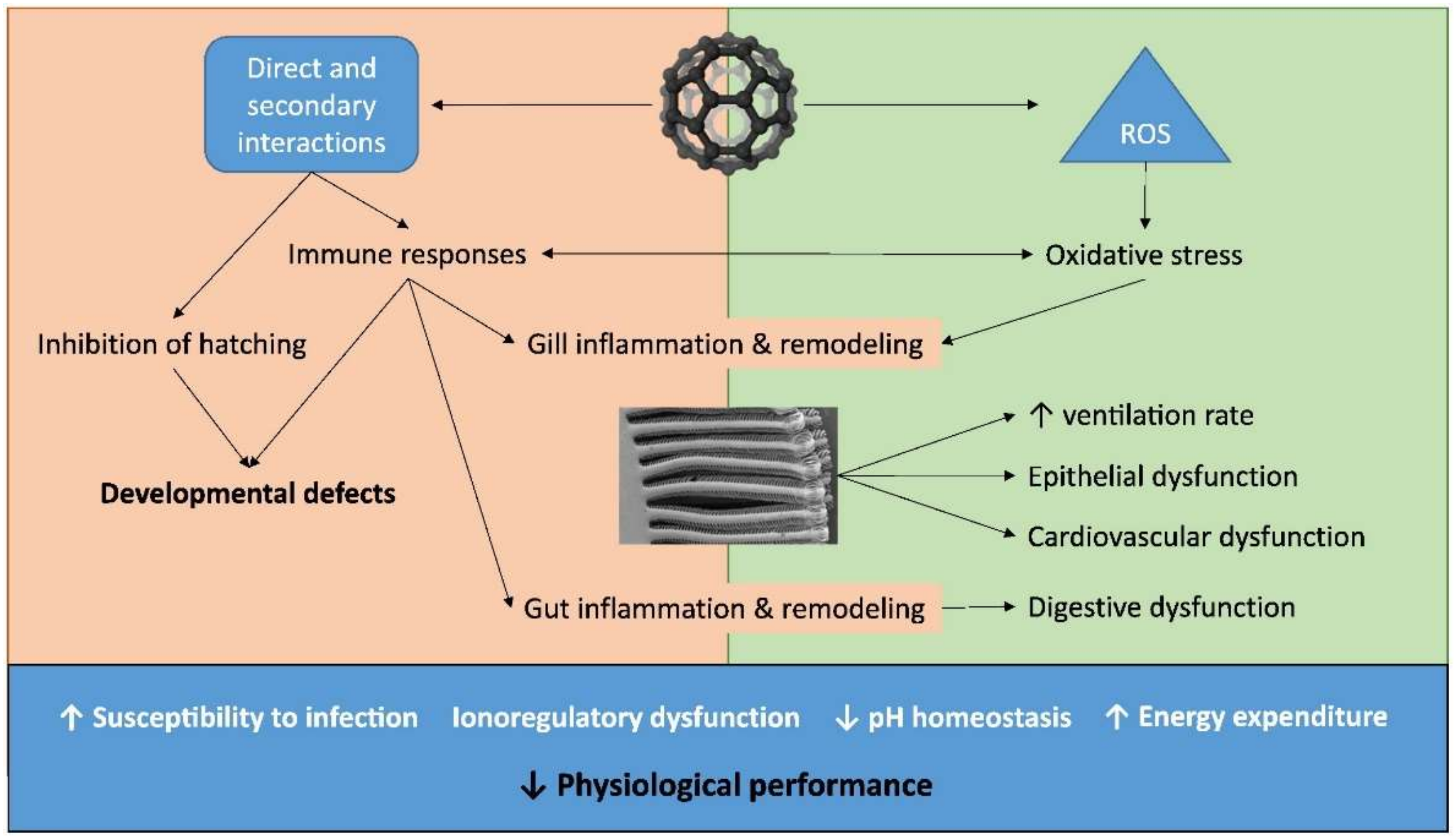
Figure 3 summarizes the different routes of toxicity of ENMs. Regarding the toxicity target, they can cause inflammation of respiratory and digestive epithelia, compromise cardiovascular activity and produce hyper responses of the immune system [6]. They cause multiple forms of damage, such as blockage of cell channels, hindering membrane functions and DNA integrity, oxidation of proteins and release of reactive oxygen species (ROS) [15]. The generation of ROS can be direct or because of the immune system’s response in impacted organisms. This yields tissue damage and/or epithelial remodelling, such as in the gills and gut. The oxidative stress can occur on the surface of the nanoparticles via radicals, transition metals and interaction of the nanoparticles with cellular parts, or via macrophages activation [156].
Since marine water and sediments of estuaries are the final sinks of ENMs, there are a plethora of organisms that are potentially exposed, including crustaceans, molluscs and algae [107]. However, as already seen, ENMs undergo an intense process of sedimentation and aggregation in seawater and, thus, benthic organisms are significantly exposed. Although the literature is quite sparse in terms of toxicity data on marine organisms, below is a summary of the most significant data found. Starting from the most primitive prokaryotic organisms, i.e., bacteria organised in biofilms, it was reported that Ag NPs inhibit the growth of specific species, and the process increases the availability of the nanoparticles for biofilms grazing organisms as gastropods [15]. At a higher trophic level, i.e., algae, ENMs may interfere with the adsorption of nutrients, as was the case observed by Peng et al. [157], who reported the inhibition growth of diatoms in the presence of ZnO NPs. Other studies [158] considered the exposure of macroalgae to Ag NPs and found a toxicity relationship with the progressive dissolution of Ag+, with surface adsorption being the main bioaccumulation path. This might represent a risk for surface grazers. In the case of other microscopic marine organisms, such as rotifers, ingesting particles from the water column brought about the accumulation of smaller-sized NPs in the stomach and intestine, with evidence of toxicity from intracellular dissolution [159]. These mechanisms of bioaccumulation and adsorption internalisation cause ENMs to be available for macro-vertebrate filter feeders. In all cases, ion dissolution is then responsible for toxicity, even though NOM complexation might attenuate toxicity. Wong et al. [48] reported how ZnO NPs were more toxic to the first stage of larvae of the copepod Tigriopus japonicus than ionic Zn2+, hypothesising mechanical inhibition of the nanoparticles besides Zn2+ dissolution.
Some marine organisms in coastal waters can be used as a way to monitor the presence of nanoparticles [160]. This is, for example, the case of TiO2 NPs, which were found to be toxic for fishes, invertebrates, bacteria [161,162,163] and phytoplankton [164]. Matranga and Corsi [107] proposed the use of model organisms and molecular approaches to evaluate the ecotoxicological impact of ENMs on the marine environment. Other organisms can be rather tolerant. The response of the marine organisms varies with the species, the particle’s physico-chemical properties, such as nature and size, photocatalytic activity, shape, ionic strength and pH of the medium, and UV irradiation. Important marine bioindicators of ENMs’ presence in estuary systems are microalgae, which are very important in the primary production of marine ecosystems and may transfer the metals along the marine food chain [102]. Barhoumi and Dewez [165] studied the hypersaline unicellular green algae Dunaliella salina as a pollution indicator of ENMs in a marine ecosystem. This was because the algae possess an elevated rate of bioaccumulation and sensitivity. Morelli et al. [166] exposed the green alga Dunaliella tertiolecta to TiO2 NPs and reported neither inhibition of growth nor alteration of cellular chlorophyll concentration. They observed a transient increase in extracellular ROS production and that the protein moiety of exopolymeric substances excreted by the algae significantly affected the biological activity of the NPs. Antizar-Ladsilso [167] studied the impact of Ag NPs on benthic prokaryotes in a heavily polluted Hooghly estuary in India and observed that microbial communities can be an indicator of sediment functioning. Authors found that exposure to NPs caused an increase in the number of bacteria in a consortium in sediments, highlighting that microbial communities may be resistant to the antimicrobial effects of commercial Ag-NPs. Key microorganisms, such as Pelobacter propionicus, disappeared, indicating that Ag-NPs have the potential to shape estuarine sediment bacterial community structure. Among the most used indicators of ENMs presence in a marine environment, there are the bivalve molluscs, which are filter feeders and able to capture particles from the water column [44]. They may internalise nano- and microparticles through endocytosis and, hence, used as indicators of ENMs [44]. Johnnson et al. [44] exposed eastern oysters Crassostrea virginica and hepatopancreas tissues to a range of TiO2-NP concentration (5–5000 mg/L) under natural sunlight. Authors reported a dose-dependent decrease in lysosomal stability at exposures as low as 50 mg/L. ENMs accumulation caused lysosomal destabilisation and the absence of oxidative stress. Particle size screening indicated a concentration effect over agglomeration, with small agglomerates and individual particles are more easily biologically taken up. Authors concluded that low TiO2-NPs levels might produce sublethal toxicity on oysters, especially under natural sunlight conditions.
3.1. Biological ENMs Effects
In a marine environment, the metals from ENMs can quite easily accumulate in organisms beyond the metabolic need and cause damage [168]. This depends on the metal speciation, method of exposure and specific organism characteristics [98]. Metals can bind to the surface of the organism or enter the cells [169] via endocytosis in the digestive system [170]. If the concentration of the metal is excessive, the excretion mechanisms are hampered [171]. When ENMs go through the membranes, they enter the lysosome and release a high amount of ions, generating ROS via Fenton-type reactions and increasing the antioxidant response [172]. Thus, marine organisms may respond to ENM exposure by altering their biological status, which can be measured through different enzymatic and non-enzymatic biomarkers. The former regard the upregulation of the phase II antioxidant enzymes, i.e., catalase, superoxide dismutase, glutathione peroxidase, glutathione reductase and transferase. These biomarkers serve to scavenge or counter ROS, avoiding oxidative damage during chronic exposure [173]. Another enzymatic biomarker that can be measured is acetylcholinesterase, which regulates many physiological functions [174]. The excessive production of ROS becomes problematic for organisms [175] and ENMs and ROS can move to deeper tissues through blood flows [55]. Organisms can respond to the excess of ROS, producing antioxidant enzymes and non-enzymes, such as metallothioneins [128]. Dosages of such protein levels are used as biomarkers of ENM toxicity. Another often-used biomarker is the thiobarbituric acid reactive substances (TBRAS) assay, which measures the lipid peroxidation of polyunsaturated fatty acids caused by ROS [176]. Furthermore, the lysosomal membrane stability is a stress biomarker, and, in this case, the neutral red activation retention assay is used to measure the increased lysosomal contents in the cytosol as a response to the increased ROS amount [128]. ROS can also cause DNA damage, including strand breaks, mutations, chromosomal aberration [177] and even cell death [178]. DNA strand breaks and mutagenicity endpoints are determined using chromosome aberrations and micronucleus assays [179].
3.2. Factors Influencing Marine ENM Effects
The factors that can influence the biological responses to ENM exposure are temperature, feeding, reproductive status and salinity [131,180,181,182]. These influences have only been studied to a limited extent and there is a limited comprehensive understanding of EMPs’ impact on estuary environments and their risk assessment [183]. Salinity is the major parameter that is responsible for the stress of estuarine organisms and influences the toxicological effects of ENMs. Noor et al. [145] investigated the role of ZnO NPs on intermediate metabolite homeostasis in the blue mussel Mytilus edulis under different salinity conditions, finding that fluctuating salinity conditions were less stressful than low constant hypo-osmotic stress. A period of 24 h was found to be sufficient to restore the metabolic functions of this euryhaline species. The biological effects measured through different biomarkers decreased as the salinity stress increased.
A similar study was carried out by Wu et al. [131], who investigated the interactive effects of salinity (normal 15, low 5 and fluctuating, 5–15‰) and of ZnO NPs and ionic Zn2+ on the immune system of Mytilus edulis from a brackish area of the Baltic Sea. Exposure to ZnO NPs caused hemocyte mortality, phagocytosis and lysosomal volume increases. At a salinity of 15‰, ZnO NPs suppressed the mRNA expression of the Toll-like receptors involved in pathogen recognition. With fluctuating salinity, ZnO NPs increased the expression of multiple immune-related genes in hemocytes, whereas low salinity had immune suppressive effects that overshadowed those of ZnO NPs. This variable immune system response was attributed not to aggregation or solubility of ZnO NPs but to the interactive toxic effects of nanoparticles and the physiological effects of the osmotic stress.
3.3. Measurements of ENMs in Aquatic System
As already stated, the main difficulty in evaluating ENM toxicity arises from the objective difficulty in measuring nanoparticle levels in a water environment. Nano-sized metal particles in an aquatic environment are difficult to measure, i.e., in terms of concentration, particle size distribution, surface area and electromagnetic behaviour [184]. Cell membranes having pores larger than the size of the nanoparticles trap them with difficulty, impairing detection [185]. Manufacturing membranes of the dimension of nanomaterials can, in some cases, resolve this problem, allowing for multiple studies of ENMs release measurements and modelling ENMs’ release and life cycles [128].
A stochastic approach was developed by Dumont et al. [186], who estimated an average level of silver nanoparticles of 0.002 ng/L and zinc oxide of 1.5 ng/L for a representative portion (~50%) of the European rivers. In ~10% of the rivers, the model estimated higher concentrations of 0.18 ng/L and 150 ng/L, respectively. One of the major problems of these models is that they consider only the initial form of the ENMs, ignoring the modification throughout the lifespan of the nanoparticle [5]. Another common problem of model approaches is that they too often consider riverine scenarios with few estimations for estuarine environments: Gottschalk et al. [47] estimated levels of TiO2 at 0.004–0.099 pg/kg in seawater and 4.3–120 pg/g in sediments, ZnO at 0.006–0.4 pg/L in seawater and 6–220 pg/kg in sediment, CuO at 0.02–0.07 pg/L in seawater and 25–83 pg/kg in sediment, Ag at 0–0.06 in seawater and 0–0.7 pg/kg in sediment, and CeO2 at 0.03–2 in seawater and 0.04–2 pg/kg in sediment.
4. Conclusions
This review highlighted how the available information on the toxicological effects of ENMs on marine organisms is very limited. The review outlined the existing gap in knowledge on the toxicological effects of ENMs on marine biota, with most of the current literature focusing mainly on bacteria and molluscs due to larger measurable endpoints or friendliness. Moreover, their final biological effect is rather ambiguous, either from metal dissolution and/or a nano-effect. It is likely that in an estuarine environment, the high variability of the salinity condition causes a significant variation in the immune system response of marine biota, where the interactive toxic effects of nanoparticles and the physiological effects of the osmotic stress play a more significant role rather than nanoparticle aggregation or solubility of nanoengineered metals.
A low amount of knowledge exists on the role of pH, temperature, nutrient availability and salinity, and often these factors induce different responses depending on the specific involved marine species and age. This means that for each specific marine environment and specific sentinel population, specific multi-biomarkers sensitive to ENMs can be set to measure different stress for a range of environmental marine conditions. Moreover, these emerging contaminants, which are present at very low levels, even pico levels, relative to other categories of new pollutants, are difficult to measure. One of the major efforts of future research will involve setting up in situ reliable, reproducible and sensitive detection procedures in complex matrices. Potential acute exposure risk for estuary biota may possibly be limited to localised areas, i.e., beaches, as well as near ENMs “point sources”, e.g., production plant outfalls and waste treatment plants.
The results from different surveys highlight the need to design an effective set of biomarkers for a set of key estuary fauna species that are capable of giving information on the impact of ENMs. It will also be important to define suitable stabilisation techniques to have better control of the release of nanoparticles in estuary and coastal environments and their overall fate and toxicological effects. It is also important to develop effective approaches that can discriminate natural and engineered NPs, their sources and factors, and mechanisms that determine these ENMs’ behaviours under the complex framework of the transient marine environment. Another issue that needs to be clarified is the influence of light irradiation on the release of toxic soluble metal from ENMs under different ionic strength, hardness and dissolved organic matter concentrations.
Author Contributions
Writing—original draft preparation, conceptualisation and data curation, M.A.; writing—review and editing, L.F. All authors read and agreed to the published version of the manuscript.
Funding
This research received no external funding.
Institutional Review Board Statement
Not applicable.
Informed Consent Statement
Not applicable.
Data Availability Statement
Not applicable.
Conflicts of Interest
The authors declare no conflict of interest.
References
- Emergen Research. Nanotechnology Market Size, Share, Trends, by Type (Nanomaterials, Nanocomposites, Nano Devices, Nano Tools), by Industry (Food and Agriculture, Healthcare, Information and Technology, Environment, Energy, Cosmetics), and by Region, Forecast to 2028. 2021, p. 250. Available online: https://www.emergenresearch.com/press-releases (accessed on 21 January 2022).
- Liu, Y.; Tourbin, M.; Lachaize, S.; Guiraud, P. Nanoparticles in wastewaters: Hazards, fate and remediation. Powder Technol. 2014, 255, 149–156. [Google Scholar] [CrossRef] [Green Version]
- European Commission. 2011/696/EU Commission Recommendation of 18 October 2011 on the definition of nanomaterial. Off. J. Eur. Union 2011, L275, 8. [Google Scholar]
- Project on Emerging Nanotechnologies, 2013. Consumer Products Inventory [WWWDocument]. Available online: http://www.nanotechproject.org/cpi (accessed on 21 January 2022).
- Keller, A.A.; McFerran, S.; Lazareva, A.; Suh, S. Global life cycle releases of engineered nanomaterials. J. Nanopart. Res. 2013, 15, 1692. [Google Scholar] [CrossRef]
- Callaghan, N.I.; MacCormack, T.J. Ecophysiological perspectives on engineered nanomaterial toxicity in fish and crustaceans. Comp. Biochem. Physiol. Part C Toxicol. Pharmacol. 2017, 193, 30–41. [Google Scholar] [CrossRef]
- Williams, R.J.; Harrison, S.; Keller, V.; Kuenen, J.; Lofts, S.; Praetorius, A.; Svendsen, C.; Vermeulen, L.C.; van Wijnen, J. Models for assessing engineered nanomaterial fate and behaviour in the aquatic environment. Curr. Opin. Environ. Sustain. 2019, 36, 105–115. [Google Scholar] [CrossRef]
- Markus, A.A.; Parsons, J.R.; Roex, E.W.M.; de Voogt, P.; Laane, R.W.P.M. Modelling the transport of engineered metallic nanoparticles in the river Rhine. Water Res. 2016, 91, 214–224. [Google Scholar] [CrossRef]
- Corsi, I.; Cherr, G.N.; Lenihan, H.S.; Labille, J.; Hassellov, M.; Canesi, L.; Pondero, F.; Frenzilli, G.; Hristozov, D.; Puntes, V.; et al. Common strategies and technologies for the ecosafety assessment and design of nanomaterials entering the marine environment. ACS Nano 2014, 8, 9694–9709. [Google Scholar] [CrossRef] [Green Version]
- Daughton, C.G. Non-regulated water contaminants: Emerging research. Environ. Impact Assess. Rev. 2004, 24, 711–732. [Google Scholar] [CrossRef]
- Moore, M.N. Do nanoparticles present ecotoxicological risks for the health of the aquatic environment? Environ. Int. 2006, 32, 967–976. [Google Scholar] [CrossRef]
- Minetto, D.; Libralato, G.; Volpi Ghirardini, A. Ecotoxicity of engineered TiO2 nanoparticles to saltwater organisms: An overview. Environ. Int. 2014, 66, 18–27. [Google Scholar] [CrossRef]
- Brown, D.M.; Wilson, M.R.; MacNee, W.; Stone, V.; Donaldson, K. Size-dependent proinflammatory effects of ultrafine polystyrene particles: A role for surface area and oxidative stress in the enhanced activity of ultrafines. Toxicol. Appl. Pharmacol. 2001, 175, 191–199. [Google Scholar] [CrossRef] [Green Version]
- Tedesco, S.; Sheehan, D. Nanomaterials as emerging environmental threats. Curr. Chem. Biol. 2010, 4, 151–160. [Google Scholar]
- Baker, T.J.; Tyler, C.R.; Galloway, T.S. Impacts of metal oxide nanoparticles on marine organisms. Environ. Pollut. 2014, 186, 257–271. [Google Scholar] [CrossRef]
- Rosenkranz, P.; Chaudhry, Q.; Stone, V.; Fernandes, T.F. A comparison of nanoparticle and fine particle uptake by Daphnia magna. Environ. Toxicol. Chem. 2009, 28, 2142–2149. [Google Scholar] [CrossRef]
- Croteau, M.N.; Dybowska, A.D.; Luoma, S.N.; Valsami-Jones, E. A novel approach reveals that zinc oxide nanoparticles are bioavailable and toxic after dietary exposures. Nanotoxicology 2011, 5, 79–90. [Google Scholar] [CrossRef]
- Zhao, C.M.; Wang, W.X. Comparison of acute and chronic toxicity of silver nanoparticles and silver nitrate to Daphnia magna. Environ. Toxicol. Chem. 2011, 30, 885–892. [Google Scholar] [CrossRef]
- Asghari, S.; Johari, S.A.; Lee, J.H.; Kim, Y.S.; Jeon, Y.B.; Choi, H.J.; Moon, M.C.; Yu, I.J. Toxicity of various silver nanoparticles compared to silver ions in Daphnia magna. J. Nanobiotechnol. 2012, 10, 14. [Google Scholar] [CrossRef] [Green Version]
- Christian, P.; Von der Kammer, F.; Baalousha, M.; Hofmann, T. Nanoparticles: Structure, properties, preparation and behaviour in environmental media. Environ. Toxicol. Chem. 2008, 17, 326–343. [Google Scholar] [CrossRef]
- Batley, G.E.; Kirby, J.K.; McLaughlin, M.J. Fate and risks of nanomaterials in aquatic and terrestrial environments. Acc. Chem. Res. 2013, 46, 854–862. [Google Scholar] [CrossRef]
- French, R.A.; Jacobson, A.R.; Kim, B.; Isley, S.L.; Penn, R.L.; Baveye, P.C. Influence of ionic strength, pH, and cation valence on aggregation kinetics of titanium dioxide nanoparticles. Environ. Sci. Technol. 2009, 43, 1354–1359. [Google Scholar] [CrossRef]
- IUPAC. IUPAC Compendium of Chemical Terminology, 2nd ed.; McNaught, A.D., Wilkinson, A., Eds.; Blackwell Science: Hoboken, NJ, USA, 1997; p. 446. [Google Scholar]
- Nowack, B.; Bucheli, T.D. Occurrence, behavior and effects of nanoparticles in the environment. Environ. Pollut. 2007, 150, 5–22. [Google Scholar] [CrossRef] [PubMed]
- Nel, A.; Xia, T.; Meng, H.; Wang, X.; Lin, S.J.; Ji, Z.X.; Zhang, H.Y. Nanomaterial testing in the 21st century: Use of a predictive toxicological approach and high through put screening. Acc. Chem. Res. 2013, 46, 607–621. [Google Scholar] [CrossRef] [PubMed]
- Wang, Y.; Zhu, X.; Lao, Y.; Lv, X.; Tao, Y.; Huang, B.; Wang, J.; Zhou, J.; Cai, Z. TiO2 nanoparticles in the marine environment: Physical effects responsible for the toxicity on algae Phaeodactylum tricornutum. Sci. Total Environ. 2016, 565, 818–826. [Google Scholar] [CrossRef] [PubMed]
- Piccinno, F.; Gottschalk, F.; Seeger, S.; Nowack, B. Industrial production quantities and uses of ten engineered nanomaterials in Europe and the world. J. Nanopart. Res. 2012, 14, 1109. [Google Scholar] [CrossRef] [Green Version]
- Jovanović, B. Critical review of public health regulations of titanium dioxide, a human food additive. Integr. Environ. Assess. Manag. 2015, 11, 10–20. [Google Scholar] [CrossRef] [Green Version]
- Xu, F. Review of analytical studies on TiO2 nanoparticles and particle aggregation, coagulation, flocculation, sedimentation, stabilization. Chemosphere 2018, 212, 662–677. [Google Scholar] [CrossRef]
- Hanaor, D.A.H.; Sorrell, C.C. Review of the anatase to rutile phase transformation. J. Mater. Sci. 2011, 46, 855–874. [Google Scholar] [CrossRef] [Green Version]
- Choi, Y.; Umebayashi, T.; Yoshikawa, M. Fabrication and characterization of C-doped anatase TiO2 photocatalysts. J. Mater. Sci. 2004, 39, 1837–1839. [Google Scholar] [CrossRef]
- Menard, A.; Drobne, D.; Jemec, A. Ecotoxicity of nanosized TiO2. Review of in vivo data. Environ. Pollut. 2011, 159, 677–684. [Google Scholar] [CrossRef]
- Gondikas, A.P.; von der Kammer, F.; Reed, R.B.; Wagner, S.; Ranville, J.F.; Hofmann, T. Release of TiO2 nanoparticles from sunscreens into surface waters: A one-year survey at the Old Danube recreational lake. Environ. Sci. Technol. 2014, 48, 5415–5422. [Google Scholar] [CrossRef]
- Botta, C.; Labille, J.; Auffan, M.; Borschneck, D.; Miche, H.; Cabié, M.; Masion, A.; Rose, J.; Bottero, J.Y. TiO2-based nanoparticles released in water from commercialized sunscreens in a life-cycle perspective: Structures and quantities. Environ. Pollut. 2011, 159, 1543–1550. [Google Scholar] [CrossRef]
- Yang, W.W.; Wang, Y.; Huang, B.; Wang, N.X.; Wei, Z.B.; Luo, J.; Miao, A.J.; Yang, L.Y. TiO2 nanoparticles act as a carrier of Cd bioaccumulation in the ciliate Tetrahymena thermophila. Environ. Sci. Technol. 2014, 48, 7568–7575. [Google Scholar] [CrossRef]
- Holbrook, R.D.; Motabar, D.; Quiñones, O.; Stanford, B.; Vanderford, B.; Moss, D. Titanium distribution in swimming pool water is dominated by dissolved species. Environ. Pollut. 2013, 181, 68–74. [Google Scholar] [CrossRef]
- Sánchez-Quiles, D.; Tovar-Sánchez, A. Sunscreens as a source of hydrogen peroxide production in coastal waters. Environ. Sci. Technol. 2014, 48, 9037–9042. [Google Scholar] [CrossRef] [Green Version]
- Guo, X.; Yin, Y.; Tan, Z.; Zhang, Z.; Chen, Y.; Liu, J. Significant enrichment of engineered nanoparticles in water surface microlayer. Environ. Sci. Technol. 2016, 3, 381–385. [Google Scholar] [CrossRef]
- Tong, T.; Fang, K.; Thomas, S.A.; Kelly, J.J.; Gray, K.A.; Gaillard, J.-F. Chemical interactions between nano-ZnO and nano-TiO2 in a natural aqueous medium. Environ. Sci. Technol. 2014, 48, 7924–7932. [Google Scholar] [CrossRef]
- Sun, T.Y.; Gottschalk, F.; Hungerbühler, K.; Nowack, B. Comprehensive probabilistic modelling of environmental emissions of engineered nanomaterials. Environ. Pollut. 2014, 185, 69–76. [Google Scholar] [CrossRef]
- Müller, E.; Hilty, L.M.; Widmer, R.; Schluep, M.; Faulstich, M. Modeling Metal Stocks and Flows: A Review of Dynamic Material Flow Analysis Methods. Environ. Sci. Technol. 2014, 48, 2102–2113. [Google Scholar] [CrossRef] [PubMed]
- EPA 2009. Toxicological Review of Cerium Oxide and Cerium Compounds (2009) EPA/635/R-08/002F. Available online: https://cfpub.epa.gov/ncea/iris/iris_documents/documents/toxreviews/1018tr.pdf (accessed on 21 January 2022).
- Giese, B.; Klaessig, F.; Park, B.; Kaegi, R.; Steinfeldt, M.; Wigger, H.; von Gleich, A.; Gottschalk, F. Risks, Release and Concentrations of Engineered Nanomaterial in the Environment. Sci. Rep. 2018, 8, 1565. [Google Scholar] [CrossRef]
- Johnson, B.D.; Gilbert, S.L.; Khan, B.; Carrol, D.L.; Ringwood, A.H. Cellular responses of eastern oysters, Crassostrea virginica, to titanium dioxide nanoparticles. Mar. Environ. Res. 2015, 111, 135–143. [Google Scholar] [CrossRef]
- Shrivastava, S.; Bera, T.; Roy, A.; Singh, G.; Ramachandrarao, P.; Dash, D. Characterization of enhanced antibacterial effects of novel silver nanoparticles. Nanotechnology 2007, 18, 225103. [Google Scholar] [CrossRef]
- Luoma, S.N. PEN 15-Silver Nanotechnologies and the Environment: Old Problems or New Challenges? 2008. Available online: https://www.researchgate.net/publication/221720677_Silver_Nanotechnologies_and_the_Environment (accessed on 21 January 2022).
- Stoller, M.; Ochando-Pulido, J. ZnO Nano-particles production intensification by means of a spinning disk reactor. Nanomaterials 2020, 10, 1321. [Google Scholar] [CrossRef] [PubMed]
- Gottschalk, F.; Lassen, C.; Kjoelholt, J.; Christensen, F.; Nowack, B. Modeling flows and concentrations of nine engineered nanomaterials in the Danish environment. Int. J. Environ. Res. Public Health 2015, 12, 5581–5602. [Google Scholar] [CrossRef] [PubMed] [Green Version]
- Wong, S.W.; Leung, P.T.; Djurišić, A.B.; Leung, K.M. Toxicities of nano zinc oxide to five marine organisms: Influences of aggregate size and ion solubility. Anal. Bioanal. Chem. 2010, 396, 609–618. [Google Scholar] [CrossRef]
- Loosli, F.; Wang, J.; Rothenberg, S.; Bizimis, M.; Winkler, C.; Borovinskaya, O.; Flamigni, L.; Baalousha, M. Sewage spills are a major source of engineered titanium dioxide release into the environment. Environ. Sci. Nano 2019, 6, 763–777. [Google Scholar] [CrossRef]
- Saharia, A.M.; Zhu, Z.; Aich, N.; Baalousha, M.; Atkinson, J.F. Modeling the transport of titanium dioxide nanomaterials from combined sewer overflows in an urban river. Sci. Total Environ. 2019, 696, 133904. [Google Scholar] [CrossRef]
- Baalousha, M.; Yang, Y.; Vance, M.E.; Colman, B.P.; McNeal, S.; Xu, J.; Blaszczak, J.; Steele, M.; Bernhardt, E.; Hochella, M.F., Jr. Outdoor urban nanomaterials: The emergence of a new, integrated, and critical field of study. Sci. Total Environ. 2016, 557–558, 740–753. [Google Scholar] [CrossRef] [Green Version]
- ASTM D7942 15; Standard Specification for Thermoplastic Pavement Markings in Non Snow Plow Areas. ASTM: West Conshohocken, PA, USA, 2015.
- Coatingsworld. U.S. Demand for Paint & Coatings to Reach 1.4 Billion Gallons in 2019. 2019. Available online: https://www.coatingsworld.com/contents/view_breaking-news/2015-09-07/us-demand-for-paint-coatings-to-reach-14-billion-gallons-in-2019/ (accessed on 9 December 2021).
- Wang, T.; Wen, X.; Hu, Y.; Zhang, X.; Wang, D.; Yin, S. Copper nanoparticles induced oxidation stress, cell apoptosis and immune response in the liver of juvenile Takifugu fasciatus. Fish Shellfish Immunol. 2019, 84, 648–655. [Google Scholar] [CrossRef]
- Adamiec, E. Road environments: Impact of metals on human health in heavily congested cities of Poland. Int. J. Environ. Res. Public Health 2017, 14, 697. [Google Scholar] [CrossRef] [Green Version]
- Wang, J.; Nabi, M.M.; Mohanty, S.K.; Nabiul Afrooz, A.R.M.; Cantando, E.; Aich, N.; Baalousha, M. Detection and quantification of engineered particles in urban runoff Jingjing. Chemosphere 2020, 248, 126070. [Google Scholar] [CrossRef]
- Yu, S.-J.; Yin, Y.-G.; Liu, J.-F. Silver nanoparticles in the environment. Environ. Sci. Process. Impacts 2012, 15, 78–92. [Google Scholar] [CrossRef]
- Blaser, S.; Scheringer, M.; MacLeod, M.; Hungerbühler, K. Estimation of cumulative aquatic exposure and risk due to silver: Contribution of nano-functionalized plastics and textiles. Sci. Total Environ. 2008, 390, 396–409. [Google Scholar] [CrossRef]
- Wen, L.S.; Santschi, P.H.; Gill, G.A.; Tang, D.G. Silver concentrations in Colorado, USA, watersheds using improved methodology. Environ. Toxicol. Chem. 2002, 21, 2040–2051. [Google Scholar] [CrossRef]
- Gao, X.; Lowry, G.V. Progress towards standardized and validated characterizations for measuring physicochemical properties of manufactured nanomaterials relevant to nano health and safety risks. NanoImpact 2018, 9, 14–30. [Google Scholar] [CrossRef]
- O’Brien, N.; Cummins, E. Ranking initial environmental and human health risk resulting from environmentally relevant nanomaterials. J. Environ. Sci. Health Part A Toxic Hazard. Subst. Environ. Eng. 2010, 45, 992–1007. [Google Scholar] [CrossRef]
- Pikethly, M.J. Nanomaterials—The driving force. Mater. Today 2004, 7, 20–29. [Google Scholar] [CrossRef]
- Danovaro, R.; Bongiorni, L.; Corinaldesi, C.; Giovannelli, D.; Damiani, E.; Astolfi, P.; Greci, L.; Pusceddu, A. Sunscreens cause coral bleaching by promoting viral infections. Environ. Health Perspect. 2008, 116, 441–447. [Google Scholar] [CrossRef] [Green Version]
- Hischier, R.; Nowack, B.; Gottschalk, F.; Hincapie, I.; Steinfeldt, M.; Som, C. Life cycle assessment of façade coating systems containing manufactured nanomaterials. J. Nanopart. Res. 2015, 17, 68. [Google Scholar] [CrossRef]
- Gottschalk, F.; Sun, T.; Nowack, B. Environmental concentrations of engineered nanomaterials: Review of modeling and analytical studies. Environ. Pollut. 2013, 181, 287–300. [Google Scholar] [CrossRef]
- Mueller, N.C.; Nowack, B. Exposure modeling of engineered nanoparticles in the environment. Environ. Sci. Technol. 2008, 42, 4447–4453. [Google Scholar] [CrossRef]
- Bornhöft, N.A.; Sun, T.Y.; Hilty, L.M.; Nowack, B. A dynamic probabilistic material flow modeling method. Environ. Model. Softw. 2016, 76, 69–80. [Google Scholar] [CrossRef] [Green Version]
- Caballero-Guzman, A.; Sun, T.; Nowack, B. Flows of engineered nanomaterials through the recycling process in Switzerland. Waste Manag. 2015, 36, 33–43. [Google Scholar] [CrossRef] [PubMed]
- Sun, T.Y.; Bornhöft, N.A.; Hungerbühler, K.; Nowack, B. Dynamic Probabilistic Modeling of Environmental Emissions of Engineered Nanomaterials. Environ. Sci. Technol. 2016, 50, 4701–4711. [Google Scholar] [CrossRef] [PubMed]
- Hotze, E.M.; Phenrat, T.; Lowry, G.V. Nanoparticle aggregation: Challenges to understanding transport and reactivity in the environment. J. Environ. Qual. 2010, 39, 1909–1924. [Google Scholar] [CrossRef] [Green Version]
- Sendra, M.; Yeste, M.P.; Gatica, J.M.; Moreno-Garrido, I.; Blasco, J. Homo agglomeration and hetero agglomeration of TiO2 in nanoparticle and bulk form, onto freshwater and marine microalgae. Sci. Total Environ. 2017, 592, 403–411. [Google Scholar] [CrossRef]
- Bansal, P.; Deshpande, A.P.; Basavaraj, M.G. Hetero aggregation of oppositely charged nanoparticles. J. Colloid Interface Sci. 2017, 492, 92–100. [Google Scholar] [CrossRef]
- Praetorius, A.; Labille, J.; Scheringer, M.; Thill, A.; Hungerbühler, K.; Bottero, J.-Y. Hetero aggregation of titanium dioxide nanoparticles with model natural colloids under environmentally relevant conditions. Environ. Sci. Technol. 2014, 48, 10690–10698. [Google Scholar] [CrossRef] [Green Version]
- Wang, Z.Y.; Zhang, L.; Zhao, J.; Xing, B.S. Environmental processes and toxicity of metallic nanoparticles in aquatic systems as affected by natural organic matter. Environ. Sci. Nano 2016, 3, 240–255. [Google Scholar] [CrossRef]
- Bennett, S.W.; Zhou, D.; Mielke, R.; Keller, A.A. Photoinduced disaggregation of TiO2 nanoparticles enables transdermal penetration. PLoS ONE 2012, 7, e48719. [Google Scholar]
- Jiang, C.; Cao, Y.; Xiao, G.; Zhu, R.; Lu, Y. A review on the application of inorganic nanoparticles in chemical surface coatings on metallic substrates. RSC Adv. 2017, 7, 7531–7539. [Google Scholar] [CrossRef] [Green Version]
- Fernández-Ibáñez, P.; Blanco, J.; Malato, S.; de las Nieves, F.J. Application of the colloidal stability of TiO2 particles for recovery and reuse in solar photocatalysis. Water Res. 2003, 37, 3180–3188. [Google Scholar] [CrossRef]
- Qiu, G.; Au, M.-J.; Ting, Y.-P. Impacts of nano-TiO2 on system performance and bacterial community and their removal during biological treatment of wastewater. Water Air Soil Pollut. 2016, 227, 386. [Google Scholar] [CrossRef]
- Boldrin, A.; Hansen, S.F.; Baun, A.; Hartmann, N.I.B.; Astrup, T.F. Environmental exposure assessment framework for nanoparticles in solid waste. J. Nanopart. Res. 2014, 16, 2394. [Google Scholar] [CrossRef] [Green Version]
- Keller, A.A.; Wang, H.; Zhou, D.; Lenihan, H.S.; Cherr, G.; Cardinale, B.J.; Miller, R.; Ji, Z. Stability and aggregation of metal oxide nanoparticles in natural aqueous matrices. Environ. Sci. Technol. 2010, 44, 1962–1967. [Google Scholar] [CrossRef]
- Jayalath, S.; Wu, H.; Larsen, S.C.; Grassian, V.H. Surface adsorption of Suwannee River humic acid on TiO2 nanoparticles: A study of pH and particle size. Langmuir 2018, 34, 3136–3145. [Google Scholar] [CrossRef]
- Miller, R.J.; Lenihan, H.S.; Muller, E.B.; Tseng, N.; Hanna, S.K.; Keller, A.A. Impacts of metal oxide nanoparticles on marine phytoplankton. Environ. Sci. Technol. 2010, 44, 7329–7334. [Google Scholar] [CrossRef]
- Liu, X.; Chen, G.; Su, C. Effects of material properties on sedimentation and aggregation of titanium dioxide nanoparticles of anatase and rutile in the aqueous phase. J. Colloid Interface Sci. 2011, 363, 84–91. [Google Scholar] [CrossRef]
- Domingos, R.F.; Peyrot, C.; Wilkinson, K.J. Aggregation of titanium dioxide nanoparticles: Role of calcium and phosphate. Environ. Chem. 2010, 7, 61–66. [Google Scholar] [CrossRef]
- He, X.; Mitrano, D.M.; Nowack, B.; Bahk, Y.K.; Figi, R.; Schreiner, C.; Bürki, M.; Wang, J. Agglomeration potential of TiO2 in synthetic leachates made from the fly ash of different incinerated wastes. Environ. Pollut. 2017, 223, 616–623. [Google Scholar] [CrossRef]
- Li, J.; Schiavo, S.; Xiangli, D.; Rametta, G.; Miglietta, M.L.; Oliviero, M.; Changwen, W.; Manzo, S. Early ecotoxic effects of ZnO nanoparticle chronic exposure in Mytilus galloprovincialis revealed by transcription of apoptosis and antioxidant-related genes. Ecotoxicology 2018, 27, 369–384. [Google Scholar] [CrossRef]
- Yang, X.; Gondikas, A.P.; Marinakos, S.M.; Auffan, M.; Liu, J.; Hsu-Kim, H.; Meyer, J.N. Mechanism of silver nanoparticle toxicity is dependent on dissolved silver and surface coating in Caenorhabditis elegans. Environ. Sci. Technol. 2012, 46, 1119–1127. [Google Scholar] [CrossRef] [PubMed]
- Bhatt, I.; Tripathi, B.N. Interaction of engineered nanoparticles with various components of the environment and possible strategies for their risk assessment. Chemosphere 2011, 82, 308–317. [Google Scholar] [CrossRef] [PubMed]
- Studart, A.R.; Amstad, E.; Gauckler, L.J. Colloidal stabilization of nanoparticles in concentrated suspensions. Langmuir 2007, 23, 1081–1090. [Google Scholar] [CrossRef] [PubMed]
- Zhou, C.; Vitiello, V.; Pellegrini, D.; Wu, C.; Morelli, E.; Buttino, I. Toxicological effects of CdSe/ZnS quantum dots on marine planktonic organisms. Ecotoxicol. Environ. Saf. 2016, 123, 26–31. [Google Scholar] [CrossRef]
- Vindedahl, A.M.; Stemig, M.S.; Arnold, W.A.; Penn, R.L. Character of humic substances as a predictor for goethite nanoparticle reactivity and aggregation. Environ. Sci. Technol. 2016, 50, 1200–1208. [Google Scholar] [CrossRef]
- Li, H.; Qiu, Y.; Wang, X.; Liu, W.; Chen, G.; Ma, Y.; Xing, B. Suspension stability and aggregation of multi-walled carbon nanotubes as affected by dissolved organic matters extracted from agricultural wastes. Environ. Pollut. 2016, 210, 323–329. [Google Scholar] [CrossRef] [Green Version]
- Chen, G.X.; Liu, X.Y.; Su, C.M. Distinct effects of humic acid on transport and retention of TiO2 rutile nanoparticles in saturated sand columns. Environ. Sci. Technol. 2012, 46, 7142–7150. [Google Scholar] [CrossRef]
- Louie, S.M.; Spielmansun, E.R.; Small, M.J.; Tilton, R.D.; Lowry, G.V. Correlation of the physicochemical properties of natural organic matter samples from different sources to their effects on gold nanoparticle aggregation in monovalent electrolyte. Environ. Sci. Technol. 2015, 49, 2188–2198. [Google Scholar] [CrossRef]
- Wang, M.; Gao, B.; Tang, D. Review of key factors controlling engineered nanoparticle transport in porous media. J. Hazard. Mater. 2016, 318, 233–246. [Google Scholar] [CrossRef] [Green Version]
- Chen, M.; Xu, N.; Christodoulatos, C.; Wang, D. Synergistic effects of phosphorus and humic acid on the transport of anatase titanium dioxide nanoparticles in water-saturated porous media. Environ. Pollut. 2018, 243, 1368–1375. [Google Scholar] [CrossRef]
- Li, J.; Chen, J.; Lu, T.; Wang, Y.; Zhang, H.; Shang, Z.; Li, D.; Zhou, Y.; Qi, Z. Effects of low-molecular weight organic acids on the transport of graphene oxide nanoparticles in saturated sand columns. Sci. Total Environ. 2019, 666, 94–102. [Google Scholar] [CrossRef]
- Zhang, R.; Zhang, H.; Tu, C.; Luo, Y. The limited facilitating effect of dissolved organic matter extracted from organic wastes on the transport of titanium dioxide nanoparticles in acidic saturated porous media. Chemosphere 2019, 237, 124529. [Google Scholar] [CrossRef]
- Wu, F.; Falfushynska, H.; Dellwig, O.; Piontkivska, H.; Sokolova, I.M. Photocatalytic effects of titanium dioxide nanoparticles on aquatic organisms—Current knowledge and suggestions for future research. Aquat. Toxicol. 2017, 185, 138–148. [Google Scholar]
- Verdugo, P.; Alldredge, A.L.; Azam, F.; Kirchman, D.L.; Passow, U.; Santschi, P.H. The oceanic gel phase: A bridge in the DOM-POM continuum. Mar. Chem. 2004, 92, 67–85. [Google Scholar] [CrossRef]
- Quigg, A.; Chin, W.C.; Chen, C.S.; Zhang, S.; Jiang, Y.; Miao, A.J.; Schwehr, K.A.; Xu, C.; Santschi, P.H. Direct and indirect toxic effects of engineered nanoparticles on algae: Role of natural organic matter. ACS Sustain. Chem. Eng. 2013, 1, 686–702. [Google Scholar] [CrossRef]
- Zhang, S.; Jiang, Y.; Chen, C.; Spurgin, J.; Schwehr, K.A.; Quigg, A.; Chin, W.; Santschi, P.H. Aggregation, dissolution, and stability of quantum dots in marine environments: Importance of extracellular polymeric substances. Environ. Sci. Technol. 2012, 46, 8764–8772. [Google Scholar] [CrossRef]
- Adeleye, A.S.; Conway, J.R.; Perez, T.; Rutten, P.; Keller, A.A. Influence of extracellular polymeric substances on the long-term fate, dissolution, and speciation of copper-based nanoparticles. Environ. Sci. Technol. 2014, 48, 12561–12568. [Google Scholar] [CrossRef] [Green Version]
- Hu, W.; Culloty, S.; Darmody, G.; Lynch, S.; Davenport, J.; Ramirez-Garcia, S.; Dawson, K.; Lynch, I.; Doyle, H.; Sheehan, D. Neutral red retention time assay in determination of toxicity of nanoparticles. Mar. Environ. Res. 2015, 111, 158–161. [Google Scholar] [CrossRef] [Green Version]
- Xie, B.; Xu, Z.; Guo, W.; Li, Q. Impact of natural organic matter on the physicochemical properties of aqueous C60 nanoparticles. Environ. Sci. Technol. 2008, 42, 2853–2859. [Google Scholar] [CrossRef]
- Matranga, V.; Corsi, I. Toxic effects of engineered nanoparticles in the marine environment: Model organisms and molecular approaches. Mar. Environ. Res. 2012, 76, 32–40. [Google Scholar] [CrossRef]
- Tappin, A.D.; Barriada, J.L.; Braungardt, C.B.; Evans, E.H.; Patey, M.D.; Achterberg, E.P. Dissolved silver in European estuarine and coastal waters. Water Res. 2010, 44, 4204–4216. [Google Scholar] [CrossRef] [PubMed]
- Baalousha, M.; Lead, J.R. Characterization of natural aquatic colloids (<5 nm) by flow-field flow fractionation and atomic force microscopy. Environ. Sci. Technol. 2007, 41, 1111–1117. [Google Scholar] [CrossRef] [PubMed]
- Tipping, E.; Higgins, D.C. The effect of adsorbed humic substances on the colloid stability of haematite particles. Colloids Surf. 1982, 5, 85–92. [Google Scholar] [CrossRef]
- Jekel, M.R. The stabilization of dispersed mineral particles by adsorption of humic substances. Water Res. 1986, 20, 1543–1554. [Google Scholar] [CrossRef]
- Buffle, J.; Wilkinson, K.J.; Stoll, S.; Filella, M.; Zhang, J. A generalized description of aquatic colloidal interactions: The three-colloidal component approach. Environ. Sci Technol. 1998, 32, 2887–2899. [Google Scholar] [CrossRef]
- Phenrat, T.; Saleh, N.; Sirk, K.; Tilton, R.D.; Lowry, G.V. Aggregation and sedimentation of aqueous nanoscale zerovalent iron dispersions. Environ. Sci. Technol. 2007, 41, 284–290. [Google Scholar] [CrossRef]
- Zhang, R.C.; Zhang, H.B.; Tu, C.; Hu, X.F.; Li, L.Z.; Luo, Y.M.; Christie, P. Facilitated transport of titanium dioxide nanoparticles by humic substances in saturated porous media under acidic conditions. J. Nanopart. Res. 2015, 17, 165. [Google Scholar] [CrossRef]
- Louie, S.M.; Tilton, R.D.; Lowry, G.V. Critical review: Impacts of macromolecular coatings on critical physicochemical processes controlling environmental fate of nanomaterials. Environ. Sci. Nano 2016, 3, 283–310. [Google Scholar] [CrossRef]
- Lyven, B.; Hassellov, M.; Turner, D.R.; Haraldsson, C.; Andersson, K. Competition between iron- and carbon-based colloidal carriers for trace metals in a freshwater assessed using flow field flow fractionation coupled to ICPMS. Geochim. Cosmochim. Acta 2003, 67, 3791–3802. [Google Scholar] [CrossRef]
- Obare, S.O.; Meyer, G.J. Nanostructured materials for environmental remediation of organic contaminants in water. J. Environ. Sci. Health Part A Toxic Hazard. Subst. Environ. Eng. 2004, 39, 2549–2582. [Google Scholar] [CrossRef]
- Giammar, D.E.; Maus, C.J.; Xie, L. Effects of particle size and crystalline phase on lead adsorption to titanium dioxide nanoparticles. Environ. Eng. Sci. 2007, 24, 85–95. [Google Scholar] [CrossRef]
- Madden, A.S.; Hochella, J.; Luxton, T.P. Insights for size dependent reactivity of hematite nanomineral surfaces through Cu2+ sorption. Geochim. Cosmochim. Acta 2006, 70, 4095–4104. [Google Scholar] [CrossRef]
- Bustamante, P.; Miramand, P. Subcellular and body distributions of 17 trace elements in the variegated scallop Chlamys varia from the French coast of the Bay of Biscay. Sci. Total Environ. 2005, 337, 59–73. [Google Scholar] [CrossRef] [Green Version]
- Wang, T.; Bai, J.; Jiang, X.; Nienhaus, G.U. Cellular uptake of nanoparticles by membrane penetration: A study combining confocal microscopy with FTIR Spectro electrochemistry. ACS Nano 2012, 6, 1251–1259. [Google Scholar] [CrossRef]
- Ferry, J.L.; Craig, P.; Hexel, C.; Sisco, P.; Frey, R.; Pennington, P.L.; Fulton, M.H.; Scott, I.G.; Decho, A.W.; Kashiwada, S.; et al. Transfer of gold nanoparticles from the water column to the estuarine food web. Nat. Nanotechnol. 2009, 4, 441–444. [Google Scholar] [CrossRef]
- Cleveland, D.; Long, S.E.; Pennington, P.L.; Cooper, E.; Fulton, M.H.; Scott, G.I.; Brewer, T.; Davis, J.; Petersen, E.J.; Wood, L. Pilot estuarine mesocosm study on the environmental fate of silver nanomaterials leached from consumer products. Sci. Total Environ. 2012, 421–422, 267–272. [Google Scholar] [CrossRef]
- Zhang, H.; Ji, Z.; Xia, T.; Meng, H.; Low-Kam, C.; Liu, R.; Pokhrel, S.; Lin, S.; Wang, X.; Liao, Y.P.; et al. Use of metal oxide nanoparticle band gap to develop a predictive paradigm for oxidative stress and acute pulmonary inflammation. ACS Nano 2012, 6, 4349–4368. [Google Scholar] [CrossRef]
- Bielmyer, G.K.; Grosell, M.; Brix, K.V. Toxicity of silver, zinc, copper, and nickel to the copepod Acartia tonsa exposed via a phytoplankton diet. Environ. Sci. Technol. 2006, 40, 2063–2068. [Google Scholar] [CrossRef]
- Baalousha, M.; Ju-Nam, Y.; Cole, P.A.; Hriljac, J.A.; Jones, I.P.; Tyler, C.R.; Stone, V.; Fernandes, T.F.; Jepson, M.A.; Lead, J.R. Characterization of cerium oxide nanoparticles—Part 2: Non size measurements. Environ. Toxicol. Chem. 2012, 31, 994–1003. [Google Scholar] [CrossRef]
- Choi, O.; Clevenger, T.E.; Deng, B.; Surampalli, R.Y.; Ross, L., Jr.; Hu, Z. Role of sulfide and ligand strength in controlling nanosilver toxicity. Water Res. 2009, 43, 1879–1886. [Google Scholar] [CrossRef]
- Roma, J.; Matos, A.R.; Vinagre, C.; Duarte, B. Engineered metal nanoparticles in the marine environment: A review of the effects on marine fauna. Mar. Environ. Res. 2020, 161, 105110. [Google Scholar] [CrossRef] [PubMed]
- Liu, J.; Hurt, R.H. Ion release kinetics and particle persistence in aqueous nano-silver colloids. Environ. Sci. Technol. 2010, 44, 2169–2175. [Google Scholar] [CrossRef] [PubMed]
- U.S. EPA. National Recommended Water Quality Criteria; EPA-822-R-02-047; Office of Science and Technology: Washington, DC, USA, 2002.
- Wu, F.; Falfushynska, H.; Dellwig, O.; Piontkivska, H.; Sokolova, I.M. Interactive effects of salinity variation and exposure to ZnO nanoparticles on the innate immune system of a sentinel marine bivalve, Mytilus edulis. Sci. Total Environ. 2020, 712, 136473. [Google Scholar] [CrossRef] [PubMed]
- Johari, S.A.; Sarkheil, M.; Tayemeh, M.B.; Veisi, S. Influence of salinity on the toxicity of silver nanoparticles (AgNPs) and silver nitrate (AgNO3) in halophilic microalgae, Dunaliella salina. Chemosphere 2018, 209, 156–162. [Google Scholar] [CrossRef] [PubMed]
- Oukarroum, A.; Bras, S.; Perreault, F.; Popovic, R. Inhibitory effects of silver nanoparticles in two green algae, Chlorella vulgaris and Dunaliella tertiolecta. Ecotoxicol. Environ. Saf. 2012, 78, 80–85. [Google Scholar] [CrossRef] [PubMed]
- Miao, A.-J.; Luo, Z.; Chen, C.-S.; Chin, W.-C.; Santschi, P.-H.; Quigg, A. Intracellular uptake: A possible mechanism for silver engineered nanoparticle toxicity to a freshwater alga Ochromonas danica. PLoS ONE 2010, 5, e8613. [Google Scholar] [CrossRef] [PubMed] [Green Version]
- Wei, C.; Zhang, Y.; Guo, J.; Han, B.; Yang, X.; Yuan, J. Effects of silica nanoparticles on growth and photosynthetic pigment contents of Scenedesmus obliquus. J. Environ. Sci. 2010, 22, 155–160. [Google Scholar] [CrossRef]
- Truong, L.; Zaikova, T.; Richman, E.K.; Hutchison, J.E.; Tanguay, R.L. Media ionic strength impacts embryonic responses to engineered nanoparticle exposure. Nanotoxicology 2012, 6, 691–699. [Google Scholar] [CrossRef] [Green Version]
- Ward, T.J.; Kramer, J.R. Silver speciation during chronic toxicity tests with the mysid, Americamysis bahia. Comp. Biochem. Physiol. Part C Toxicol. Pharmacol. 2002, 133, 75–86. [Google Scholar] [CrossRef]
- Mouneyrac, C.; Buffet, P.-E.; Poirier, L.; Zalouk-Vergnoux, A.; Guibbolini, M.; Risso-de Faverney, C.; Gilliland, D.; Berhanu, D.; Dybowska, A.; Châtel, A.; et al. Fate and effects of metal-based nanoparticles in two marine invertebrates, the bivalve mollusc Scrobicularia plana and the annelid polychaete Hediste diversicolor. Environ. Sci. Pollut. Res. 2014, 21, 7899–7912. [Google Scholar] [CrossRef]
- McCarthy, M.P.; Carroll, D.L.; Ringwood, A.H. Tissue specific responses of oysters, Crassostrea virginica, to silver nanoparticles. Aquat. Toxicol. 2013, 138, 123–128. [Google Scholar] [CrossRef]
- D’Agata, A.; Fasulo, S.; Dallas, L.J.; Fisher, A.S.; Maisano, M.; Readman, J.W.; Jha, A.N. Enhanced toxicity of ‘bulk’ titanium dioxide compared to ‘fresh’ and ‘aged’ nano-TiO2 in marine mussels (Mytilus galloprovincialis). Nanotoxicology 2014, 8, 549–558. [Google Scholar] [CrossRef]
- Ward, J.E.; Kach, D.J. Marine aggregates facilitate ingestion of nanoparticles by suspension-feeding bivalves. Mar. Environ. Res. 2009, 68, 137–142. [Google Scholar] [CrossRef] [Green Version]
- Canesi, L.; Ciacci, C.; Fabbri, R.; Marcornini, A.; Pojano, G.; Gallo, G. Bivalve molluscs as a unique target group for nanoparticle toxicity. Mar. Environ. Res. 2012, 76, 16–21. [Google Scholar] [CrossRef]
- Moore, M.N.; Viarengo, A.; Donkin, P.; Hawkins, A.J.S. Autophagic and lysosomal reactions to stress in the hepatopancreas of blue mussels. Aquat. Toxicol. 2007, 84, 80–91. [Google Scholar] [CrossRef]
- Cong, Y.; Jin, F.; Wanga, J.; Mu, J. The embryotoxicity of ZnO nanoparticles to marine medaka, Oryzias melastigma. Aquat. Toxicol. 2017, 185, 11–18. [Google Scholar] [CrossRef]
- Noor, M.N.; Wu, F.; Sokolov, E.P.; Falfushynska, H.; Timm, S.; Haider, F.; Sokolova, I.M. Salinity-dependent effects of ZnO nanoparticles on bioenergetics and intermediate metabolite homeostasis in a euryhaline marine bivalve, Mytilus edulis. Sci. Total Environ. 2021, 774, 145195. [Google Scholar] [CrossRef]
- Cong, Y.; Banta, G.T.; Selck, H.; Berhanu, D.; Valsami-Jones, E.; Forbes, V.E. Toxic effects and bioaccumulation of nano-, micron- and ionic-Ag in the polychaete, Nereis diversicolor. Aquat. Toxicol. 2011, 105, 403–411. [Google Scholar] [CrossRef] [Green Version]
- Gomes, T.; Pereira, C.G.; Cardoso, C.; Bebianno, M.J. Differential protein expression in mussels Mytilus galloprovincialis exposed to nano and ionic Ag. Aquat. Toxicol. 2013, 136, 79–90. [Google Scholar] [CrossRef]
- García-Alonso, J.; Khan, F.R.; Misra, S.K.; Turmaine, M.; Smith, B.D.; Rainbow, P.S.; Luoma, S.N.; Valsami-Jones, E. Cellular Internalization of Silver Nanoparticles in Gut Epithelia of the Estuarine Polychaete Nereis diversicolor. Environ. Sci. Technol. 2011, 45, 4630–4636. [Google Scholar] [CrossRef]
- Galloway, T.S.; Cipelli, R.; Guralnik, J.; Ferrucci, L.; Bandinelli, S.; Corsi, A.M.; Money, C.; Mccormack, C.; Melzer, D. Daily bisphenol A excretion and associations with sex hormone concentrations: Results from the InCHIANTI adult population study. Environ. Health Perspect. 2010, 118, 1603–1608. [Google Scholar] [CrossRef] [PubMed] [Green Version]
- Fabrega, J.; Tantra, R.; Amer, A.; Björn, S. Sequestration of Zinc from Zinc Oxide Nanoparticles and Life Cycle Effects in the Sediment Dweller Amphipod Corophium volutator. Environ. Sci. Technol. 2012, 46, 1128–1135. [Google Scholar] [CrossRef] [PubMed]
- Sendra, M.; Blasco, J.; Araújo, C.V.M. Is the cell wall of marine phytoplankton a protective barrier or a nanoparticle interaction site? Toxicological responses of Chlorella autotrophica and Dunaliella salina to Ag and CeO2 nanoparticles. Ecol. Indic. 2018, 95, 1053–1067. [Google Scholar] [CrossRef]
- Hanna, S.K.; Miller, R.J.; Zhou, D.; Keller, A.A.; Lenihan, H.S. Accumulation and toxicity of metal oxide nanoparticles in a soft-sediment estuarine amphipod. Aquat. Toxicol. 2013, 142–143, 441–446. [Google Scholar] [CrossRef] [PubMed] [Green Version]
- Burić, P.; Jakšić, Z.; Štajner, L.; Sikirić, M.D.; Jurašin, D.; Cascio, C.; Calzolai, L.; Mark Lyons, D. Effect of silver nanoparticles on Mediterranean Sea urchin embryonal development is species specific and depends on moment of first exposure. Mar. Environ. Res. 2015, 111, 50–59. [Google Scholar] [CrossRef] [PubMed] [Green Version]
- Kohen, R.; Nyska, A. Invited review: Oxidation of biological systems: Oxidative stress phenomena, antioxidants, redox reactions, and methods for their quantification. Toxicol. Pathol. 2002, 30, 620–650. [Google Scholar] [CrossRef] [Green Version]
- Khan, I.; Saeed, K.; Khan, I. Nanoparticles: Properties, applications and toxicities. Arab. J. Chem. 2019, 12, 908–931. [Google Scholar] [CrossRef]
- Nel, A.; Xia, T.; Mädler, L.; Li, N. Toxic potential of materials at the nanolevel. Science 2006, 311, 622–627. [Google Scholar] [CrossRef] [Green Version]
- Peng, L.; Peng, Q.; Li, Y. Controlled synthesis and self-assembly of highly monodisperse Ag and Ag2S nanocrystals. Chemistry 2011, 17, 941–946. [Google Scholar]
- Turner, A.; Brice, B.; Brown, M.T. Interactions of silver nanoparticles with the marine macroalga, Ulva lactuca. Ecotoxicology 2012, 21, 148–154. [Google Scholar] [CrossRef]
- Snell, T.W.; Hicks, D.G. Assessing toxicity of nanoparticles using Brachionus manjavacas (Rotifera). Environ. Toxicol. 2011, 26, 146–152. [Google Scholar] [CrossRef]
- Amiard-Triquet, C.; Amiard, J.C.; Mouneyrac, C. Aquatic Ecotoxicology. Advancing Tools for Dealing with Emerging Risks; Academic Press: London, UK, 2015; p. 504. [Google Scholar]
- Paterson, G.; Ataria, J.M.; Hoque, M.E.; Burns, D.C.; Metcalfe, C.D. The toxicity of titanium dioxide nanopowder to early life stages of the Japanese medaka (Oryzias latipes). Chemosphere 2011, 82, 1002–1009. [Google Scholar] [CrossRef]
- Canesi, L.; Corsi, I. Effects of nanomaterials on marine invertebrates. Sci. Total Environ. 2016, 565, 933–940. [Google Scholar] [CrossRef]
- Cherchi, C.; Gu, A.Z. Impact of titanium dioxide nanomaterials on nitrogen fixation rate and intracellular nitron storage in Anabaena variabilis. Environ. Sci. Technol. 2010, 44, 8302–8307. [Google Scholar] [CrossRef]
- Sendra, M.; Sánchez-Quiles, D.; Blasco, J.; Moreno-Garrido, I.; Lubián, L.M.; Pérez-García, S.; Tovar-Sánchez, A. Effects of TiO2 nanoparticles and sunscreens on coastal marine microalgae: Ultraviolet radiation is key variable for toxicity assessment. Environ. Int. 2017, 98, 62–68. [Google Scholar] [CrossRef]
- Barhoumi, L.; Dewez, D. Toxicity of superparamagnetic iron oxide nanoparticles on green alga Chlorella vulgaris. BioMed Res. Int. 2013, 2013, 647974. [Google Scholar] [CrossRef] [Green Version]
- Morelli, E.; Gabellieri, E.; Bonomini, A.; Tognotti, D.; Grassi, G.; Corsi, I. TiO2 nanoparticles in seawater: Aggregation and interactions with the green alga Dunaliella tertiolecta. Ecotoxicol. Environ. Saf. 2018, 148, 184–193. [Google Scholar] [CrossRef]
- Antizar-Ladisla, B.; Bhattacharya, B.D.; Ray Chaudhuri, S.; Sarkar, S.K. Impact of silver nanoparticles on benthic prokaryotes in heavy metal-contaminated estuarine sediments in a tropical environment. Mar. Pollut. Bull. 2015, 99, 104–111. [Google Scholar] [CrossRef]
- Yang, L.; Wang, W.X. Comparative contributions of copper nanoparticles and ions to copper bioaccumulation and toxicity in barnacle larvae. Environ. Pollut. 2019, 249, 116–124. [Google Scholar] [CrossRef]
- Rainbow, P.S. Trace metal accumulation in marine invertebrates: Marine biology or marine chemistry? J. Mar. Biol. Assoc. U. K. 1997, 77, 195–210. [Google Scholar] [CrossRef]
- Iversen, T.G.; Skotland, T.; Sandvig, K. Endocytosis and intracellular transport of nanoparticles: Present knowledge and need for future studies. Nano Today 2011, 6, 176–185. [Google Scholar] [CrossRef]
- Rainbow, P.S. Trace metal concentrations in aquatic invertebrates: Why and so what? Environ. Pollut. 2002, 120, 497–507. [Google Scholar] [CrossRef]
- Ploetz, E.; Zimpel, A.; Cauda, V.; Bauer, D.; Lamb, D.C.; Haisch, C.; Zahler, S.; Vollmar, A.M.; Wuttke, S.; Engelke, H. Metal–Organic Framework Nanoparticles Induce Pyroptosis in Cells Controlled by the Extracellular pH. Adv. Mater. 2020, 32, 1907267. [Google Scholar] [CrossRef] [PubMed] [Green Version]
- Volland, M.; Hampel, M.; Martos-Sitcha, J.A.; Trombini, C.; Martínez-Rodríguez, G.; Blasco, J. Citrate gold nanoparticle exposure in the marine bivalve Ruditapes philippinarum: Uptake, elimination and oxidative stress response. Environ. Sci. Pollut. Res. 2015, 22, 17414–17424. [Google Scholar] [CrossRef]
- Jebali, J.; Khedher, S.B.; Sabbagh, M.; Kamel, N.; Banni, M.; Boussetta, H. Cholinesterase activity as biomarker of neurotoxicity: Utility in the assessment of aquatic environment contamination. Rev. Gest. Costeira Integr.—J. Integr. Coast. Zone Manag. 2013, 13, 525–537. [Google Scholar] [CrossRef] [Green Version]
- Finkel, T. Signal transduction by reactive oxygen species. J. Cell Biol. 2011, 194, 7–15. [Google Scholar] [CrossRef] [Green Version]
- Pryor, W.A.; Stanley, J.P. Letter: A suggested mechanism for the production of malondialdehyde during the autoxidation of polyunsaturated fatty acids. Nonenzymatic production of prostaglandin endoperoxides during autoxidation. J. Org. Chem. 1975, 40, 3615–3617. [Google Scholar] [CrossRef]
- Ng, C.T.; Li, J.J.; Bay, B.H.; Yung, L.Y.L. Current studies into the genotoxic effects of nanomaterials. J. Nucleic Acids 2010, 2010, 947859. [Google Scholar] [CrossRef] [Green Version]
- Sharma, N.; Rather, M.A.; Ajima, M.N.; Gireesh-Babu, P.; Kumar, K.; Sharma, R. Assessment of DNA damage and molecular responses in Labeo rohita (Hamilton, 1822) following short-term exposure to silver nanoparticles. Food Chem. Toxicol. 2016, 96, 122–132. [Google Scholar] [CrossRef]
- Mahaye, N.; Thwala, M.; Cowan, D.A.; Musee, N. Genotoxicity of metal based engineered nanoparticles in aquatic organisms: A review. Mutat. Res. Rev. Mutat. Res. 2017, 773, 134–160. [Google Scholar] [CrossRef]
- Falfushynska, H.I.; Gnatyshyna, L.L.; Ivanina, A.V.; Khoma, V.V.; Stoliar, O.B.; Sokolova, I.M. Bioenergetic responses of freshwater mussels Unio tumidus to the combined effects of nano-ZnO and temperature regime. Sci. Total Environ. 2019, 650, 1440–1450. [Google Scholar] [CrossRef]
- Blanco-Rayón, E.; Ivanina, A.V.; Sokolova, I.M.; Marigómez, I.; Izagirre, U. Food-type may jeopardize biomarker interpretation in mussels used in aquatic toxicological experimentation. PLoS ONE 2019, 14, e0220661. [Google Scholar] [CrossRef] [Green Version]
- Roznere, I.; Watters, G.T.; Wolfe, B.A.; Daly, M. Nontargeted metabolomics reveals biochemical pathways altered in response to captivity and food limitation in the freshwater mussel Amblema plicata. Comp. Biochem. Physiol. Part D Genom. Proteom. 2014, 12, 53–60. [Google Scholar] [CrossRef]
- Cabral, H.; Fonseca, V.; Sousa, T.; Costa Leal, M. Synergistic effects of climate change and marine pollution: An overlooked interaction in coastal and estuarine areas. Int. J. Environ. Res. Public Health 2019, 16, 2737. [Google Scholar] [CrossRef] [Green Version]
- Heithmar, E.M. Screening Methods for Metal-Containing Nanoparticles in Water; EPA/600/R-11/096; U.S. Environmental Protection Agency: Washington, DC, USA, 2011.
- Sadik, O.A.; Du, N.; Yazgan, I.; Okello, V. Nanotechnology Applications for Clean Water Nanostructured. In Membranes for Water Purification, 2nd ed.; William Andrew: Norwich, NY, USA, 2014; pp. 95–108. [Google Scholar]
- Dumont, E.; Johnson, A.C.; Keller, V.D.; Williams, R.J. Nano silver and nano zinc-oxide in surface waters—Exposure estimation for Europe at high spatial and temporal resolution. Environ. Pollut. 2015, 196, 341–349. [Google Scholar] [CrossRef] [Green Version]
Figure 1.
Dimensional classes of natural colloids and NPs. Adapted from Christian et al. [20].
Figure 1.
Dimensional classes of natural colloids and NPs. Adapted from Christian et al. [20].

Figure 2.
Destiny of engineered nanometals ENMs in an estuary environment. The relative abundance is indicated by the thickness of the arrows. Image used with the permission of Elsevier [6].
Figure 2.
Destiny of engineered nanometals ENMs in an estuary environment. The relative abundance is indicated by the thickness of the arrows. Image used with the permission of Elsevier [6].

Figure 3.
ENMs toxicity routes in an aquatic environment. Image used with the permission of Elsevier [6].
Figure 3.
ENMs toxicity routes in an aquatic environment. Image used with the permission of Elsevier [6].

Publisher’s Note: MDPI stays neutral with regard to jurisdictional claims in published maps and institutional affiliations. |
© 2022 by the authors. Licensee MDPI, Basel, Switzerland. This article is an open access article distributed under the terms and conditions of the Creative Commons Attribution (CC BY) license (https://creativecommons.org/licenses/by/4.0/).
Share and Cite
MDPI and ACS Style
Arienzo, M.; Ferrara, L. Environmental Fate of Metal Nanoparticles in Estuarine Environments. Water 2022, 14, 1297. https://doi.org/10.3390/w14081297
AMA Style
Arienzo M, Ferrara L. Environmental Fate of Metal Nanoparticles in Estuarine Environments. Water. 2022; 14(8):1297. https://doi.org/10.3390/w14081297
Chicago/Turabian StyleArienzo, Michele, and Luciano Ferrara. 2022. "Environmental Fate of Metal Nanoparticles in Estuarine Environments" Water 14, no. 8: 1297. https://doi.org/10.3390/w14081297
Note that from the first issue of 2016, this journal uses article numbers instead of page numbers. See further details here.