Article

Microbial Community Structure and Bacterial Lineages Associated with Sulfonamides Resistance in Anthropogenic Impacted Larut River

1
Institute of Biological Sciences, Universiti Malaya, Kuala Lumpur 50603, Malaysia
2
Institute of Ocean and Earth Sciences (IOES), Universiti Malaya, Kuala Lumpur 50603, Malaysia
3
Leading Academia in Marine and Environment Pollution Research (LaMer), Center for Marine Environmental Studies (CMES), Ehime University, Matsuyama 790-8577, Japan
*
Authors to whom correspondence should be addressed.
Water 2022, 14(7), 1018; https://doi.org/10.3390/w14071018
Submission received: 10 December 2021 / Revised: 14 March 2022 / Accepted: 16 March 2022 / Published: 23 March 2022
(This article belongs to the Special Issue Antibiotic Resistance in Environmental Waters)

Abstract

:
Anthropogenic activities often contribute to antibiotic resistance in aquatic environments. Larut River Malaysia is polluted with both organic and inorganic pollutants from domestic and industrial wastewater that are probably treated inadequately. The river is characterized by high biochemical oxygen demand, chemical oxygen demand, total suspended solids, ammonia, and heavy metals. In our previous study, sulfonamides (SAs) and sulfonamide resistance genes (sul) were detected in the Larut River. Hence, in this study, we further examined the microbial community structure, diversity of sulfonamide-resistant bacteria (SARB), and their resistance genes. The study also aimed at identifying cultivable bacteria potential carriers of sul genes in the aquatic environment. Proteobacteria (22.4–66.0%), Firmicutes (0.8–41.6%), Bacteroidetes (2.0–29.4%), and Actinobacteria (5.5–27.9%) were the most dominant phyla in both the effluents and river waters. SARB isolated consisted only 4.7% of the total genera identified, with SAR Klebsiella as the most dominant (38.0–61.3%) followed by SAR Escherichia (0–22.2%) and Acinetobacter (3.2–16.0%). The majority of the SAR Klebsiella isolated from the effluents and middle downstream were positive for sul genes. Sul genes-negative SAR Escherichia and Acinetobacter were low (<20%). Canonical-correlation analysis (CCA) showed that SAs residues and inorganic nutrients exerted significant impacts on microbial community and total sul genes. Network analysis identified 11 SARB as potential sul genes bacterial carriers. These findings indicated that anthropogenic activities exerted impacts on the microbial community structure and SAs resistance in the Larut River.

1. Introduction

The increasing community-acquired infections of antibiotic-resistant bacteria (ARB) and occurrence of environmental ARB and their resistance genes (ARGs) have raised concern about antibiotic resistance in natural environments [1,2,3,4]. Aquatic environments are suffering from different types and levels of anthropogenic antibiotic pollution, including discharge of untreated and crudely treated municipal, hospital, agricultural, livestock, and pharmaceutical industries wastewaters [5]. Subsequently, aquatic environments may represent the origin, amplifier, and/or reservoir of the environmental, human, and/or animals ARB and ARGs as well as act as a bioreactor facilitating the emergence and dissemination of ARGs transfer between the pathogenic and non-pathogenic bacteria [6,7,8].
There are three possible types of selection for antibiotic resistance in aquatic environments [9]: (i) natural selection for antibiotic resistance through the production of antibiotics by aquatic organisms (fungi, bacteria) [10]; (ii) anthropogenic selection of ARB and ARGs through anthropogenic activities [11]; and (iii) introduction of ARB and ARGs from anthropogenic sources [12]. Studies have shown that exposure of bacteria to a sub-inhibitory concentration of antibiotics may play a significant role in the selection of antibiotic resistance [13,14]. However, ARB with resistance genes on plasmids or genomes could remain in the environments with or without selective pressures [15,16]. These discrepancies could be due to more complicated mechanisms of development and preservation of ARB and ARGs in the environment. Thus, understanding the survival of ARB from anthropogenic sources as well as the persistence of ARGs in the natural environment is essential to more accurately assess the role of aquatic environments as reservoirs of resistance.
Most studies to date on ARGs in the aquatic environment are those that confer resistance to beta-lactam antibiotics, aminoglycosides, fluoroquinolones, tetracyclines, and macrolides [17]. In fact, studies related to sulfonamide resistance genes (sul) in surface waters were rarely reported, particularly for anthropogenic polluted rivers. There are numerous investigations on the composition of the bacterial assemblage and ARGs in rivers indicating that external pollution and environmental factors affect the bacterial community assembly [18,19]. However, most studies reported data either on the types and detection frequency of ARG or ARB. Despite metagenomic analysis that has been conducted in some studies to characterize the acquired resistome in aquatic environments, understanding the water quality, variation of bacterial communities, and anthropogenic factors that affect the fate of ARGs in the aquatic environment remain scarce.
Sulfonamides (SAs) have been applied in humans and veterinary as antibiotic prophylaxis for almost a century [20]. SAs possess a high excretion rate, high solubility, and persistence in the environment [21,22]. Bacterial SA resistance is mediated by the horizontal transfer of foreign dhps gene or a part of it or via plasmids that carry sul genes encoding alternative drug-resistant variants of the dihydropteroate synthase (DHPS) enzyme [23]. SA binds to DHPS to inhibit dihydrofolic acid formation and thus halts bacterial growth. Distribution profiles of sul genes vary in aquatic bacterial assemblages [24]. However, whether the ARGs carriers are potential harmful pathogens or harmless non-pathogens remains unresolved while the emergence and spread of ARGs have been a rising threat in recent decades [25,26].
In our recent monitoring work, we revealed that sulfamethoxazole (SMX), sulfadimethoxine (SDM), and sulfadiazine (SDZ) are the most prevalent SAs residues detected in the Larut River. Our findings have shown that SMX-resistant (SMXr) heterotrophic bacteria and SMXr-enteric bacteria, as well as the ratio of SMXr bacteria to total bacteria, increased at sites receiving wastewater effluents [14]. As SAs residues are also prevalent here, we hypothesized that environmental bacterial assemblage, SARB, and sul genes are similarly influenced by the anthropogenic influence. To better understand the impact of environmental bacterial assemblage on the SAs resistance genes and the potential sul gene bacterial carriers in anthropogenically impacted Larut River, we used the culture-dependent method to identify and examine the diversity of culturable SAs-resistant (SAR) isolates and their sul genes. A comparison of culturable to total microbial community profile structure was carried out to identify the potential sul gene bacterial carriers. We also determined the factors affecting the bacterial community composition and the fate of sul genes in this river. The results from this study will be useful to strategize effective mitigating strategies for combating environmental antibiotic resistance dissemination and transmission of resistant genes through the water system.

2. Materials and Methods
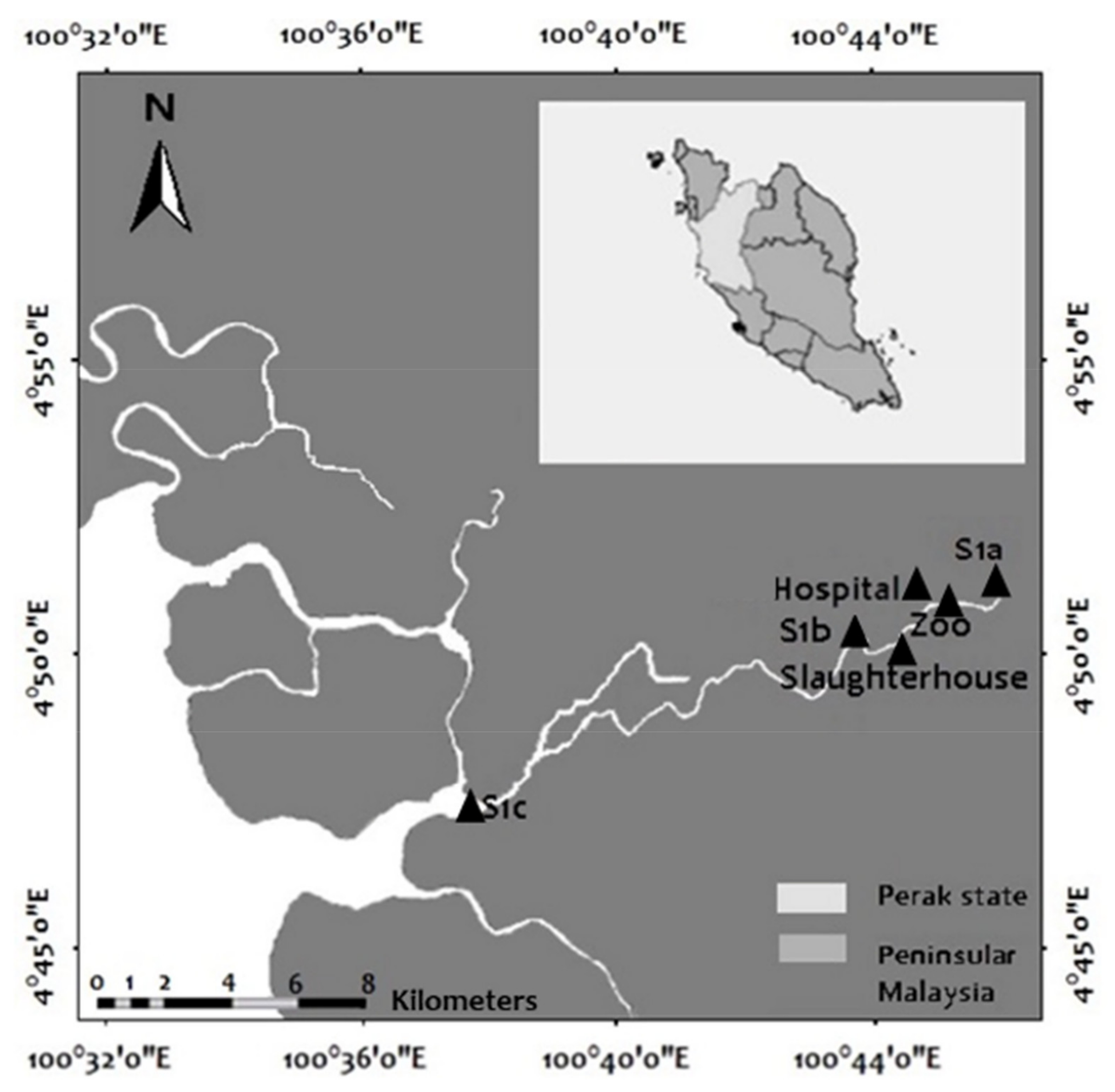
2.1. Sampling Sites

The samples were collected from six sampling sites located upstream (S1a) and downstream (S1b and S1c) of Larut River, Taiping, Perak, including wastewater effluents from zoos, hospitals, and slaughterhouses (Figure 1). These wastewaters were collected from the major outlet flowing into the adjacent river, and they were mainly greywater. Larut River is the only river from the Larut hill flowing through the city, receiving the wastewater effluents that are eventually discharged into the Straits of Malacca. Thus, this river was selected as a model to study the anthropogenic impacts on SAs resistance in the aquatic environment [14]. Grab sampling was conducted for three consecutive months to collect the water samples with an acid-washed bucket. Although each grab sample represents a snapshot of the environment, the average of the three different months should adequately reflect the environmental condition. All samples were placed in sterilized sampling bottles and kept on ice until analysis in the laboratory. A concurrent study to measure the physicochemical (temperature, pH, salinity, dissolved oxygen) and inorganic nutrient parameters (ammonium (NH4), nitrite (NO2), nitrate (NO3), phosphate (PO4)) was conducted, and the results have been published [14].

2.2. Metagenomics DNA Extraction

A total of 500 mL of water samples were filtered through a 0.22 µM Millipore GTTP (Merck, Kenilworth, NJ, USA) membrane filter. Total genomic DNA was extracted using PowerWater® DNA Isolation Kit (MoBio Laboratories, Carlsbad, CA, USA) according to the manufacturer’s instructions. DNA with concentration >30 ng μL−1 was sent for 16S metagenomics analysis.

2.3. Microbial Community Structure

The microbial community structure was analyzed using the Illumina MiSeq platform (Majorbio, China). The marker region of the bacteria was amplified using 338F and 806R primers with unique barcodes. PCR amplification was conducted at 95 °C for 2 min, followed by 25 cycles at 95 °C for 30 s, 55 °C for 30 s, and 72 °C for 30 s and a final extension at 72 °C for 5 min. The libraries were constructed using the Illumina MiSeq PE250/300 Platform according to standard protocols. Sample libraries were pooled in equimolar and paired-end sequenced (2 × 250/300 bp) on an Illumina MiSeq platform. The raw “Fastq” files were demultiplexed and quality filtered using Quantitative Insights into Microbial Ecology (QIIME, version 1.9.1). Operational taxonomic units (OTUs) were clustered with 97% similarity cut-off using the UPARSE in the USEARCH software (version 7.1, http://drive5.com/uparse/, accessed on 24 January 2019) [11]. All quality sequence “Fasta” files were checked and removed for chimeric sequences by the UCHIME reference algorithm [7]. The taxonomic information of each 16S rRNA gene sequence was analyzed by Ribosomal Database Project Classifier (RDP Classifier 11.1, http://rdp.cme.msu.edu/, accessed on 31 December 2019) [16] against the SILVA rRNA databases 123 (SSU123) using a confidence threshold of 0.7 [27]. The diversity indices (Chao1 and Shannon indices) were calculated using Mothur (version v.1.30.1, https://mothur.org/wiki/, accessed on 31 December 2019) [28]. The microbial community heat map was drawn using Microsoft Excel. Bray-Curtis distance matrix was calculated using PAST software [29], and hierarchical cluster analysis was presented as a dendrogram using the Unweighted Pair Group Method with Arithmetic Mean (UPGMA) [30].

2.4. Identification of Culturable Sulfonamides-Resistant Bacteria

A total of 278 culturable SAR isolates were isolated from water samples collected from the six sampling sites. The culturable SAR bacterial isolation was carried out using tryptic soy agar (TSA), and CHROMagarTM Orientation (COR) supplemented with 60 µg mL−1 SMX in triplicates [31]. The culturable SAR isolates were cultured in nutrient broth supplemented with 60 μg mL−1 SMX at 37 °C for 18–24 h prior to DNA extraction using the boiling method [32]. The 16S rDNA genes were amplified using PCR (Applied Biosystem, CA, United States) with 27F and 1492R primers (Table 1) [4] and subjected to 16S rRNA Sanger sequencing. Basic Local Alignment Search Tool (BLAST) was used for bacterial identification [8].

2.5. Detection of SAR Genes (sul1, sul2 and sul3) in the Sulfonamides-Resistant Bacteria

The presence of sul genes was determined using PCR (Applied Biosystem, California, United States) with gene-specific primers (Table 1). Amplification conditions for the sul genes were as follows: pre-denaturation at 95 °C for 5 min, 35 cycles of denaturation at 95 °C for 30 s, annealing at 68 °C (for sul1 and sul2) [33] and 63 °C (sul3) [34] for 30 s and extension 72 °C for 30 s, followed by a final extension at 72 °C for 7 min. Gel electrophoresis was performed on 1.5% agarose gel.

2.6. Statistical Analyses

Canonical-correspondence analysis (CCA) was used to explore the impacts’ probable links among SAs residues, environmental physicochemical parameters, microbial community structure, and total sul genes in the Larut River [14]. The relationship between the categories was represented in a two-dimensional graph. Network visualization was performed based on the logarithm-transformed culturable SARB population and the abundance of their sul genes using Gephi 0.9.2 to measure the associations between these parameters.

3. Results and Discussion

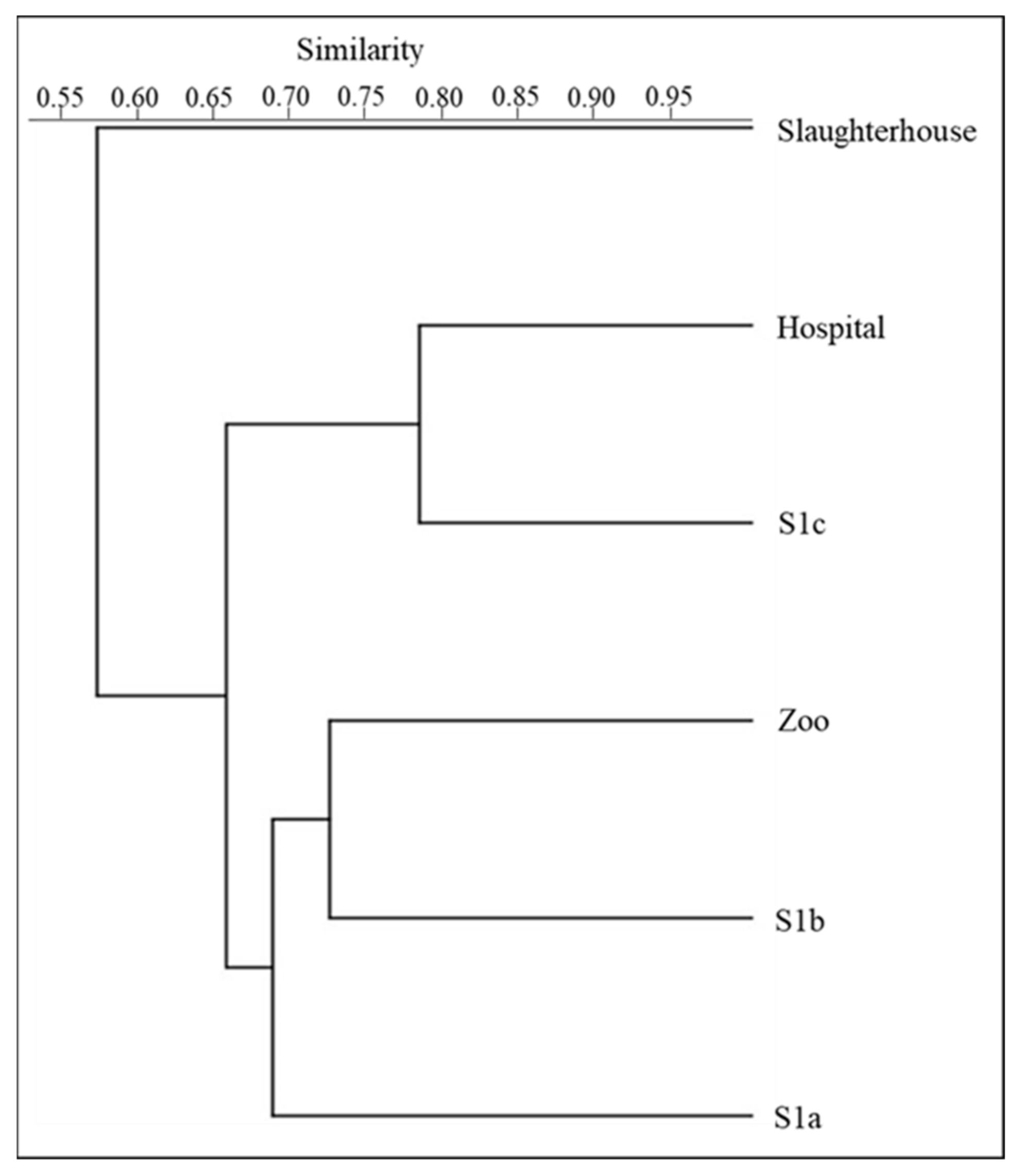
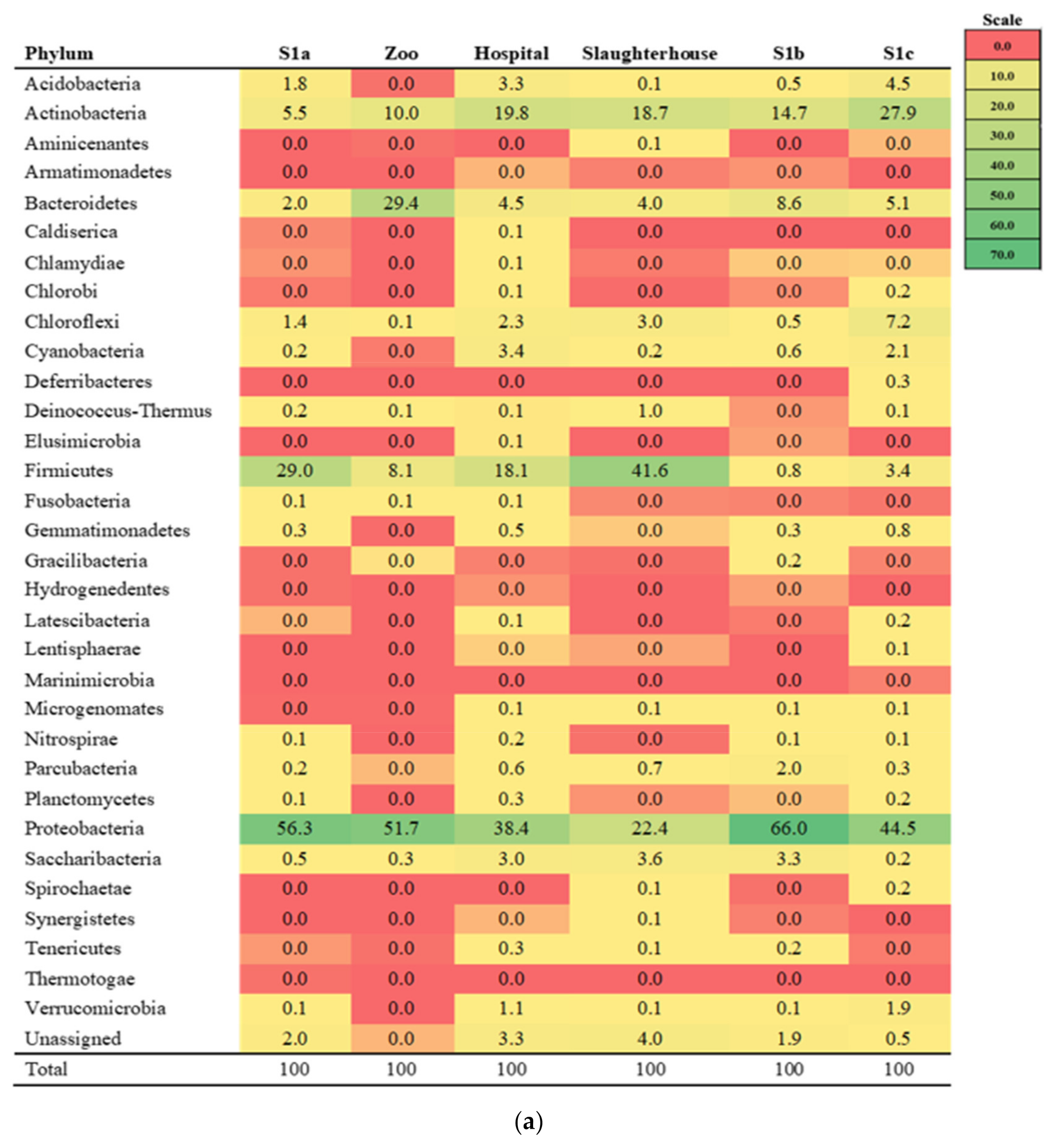
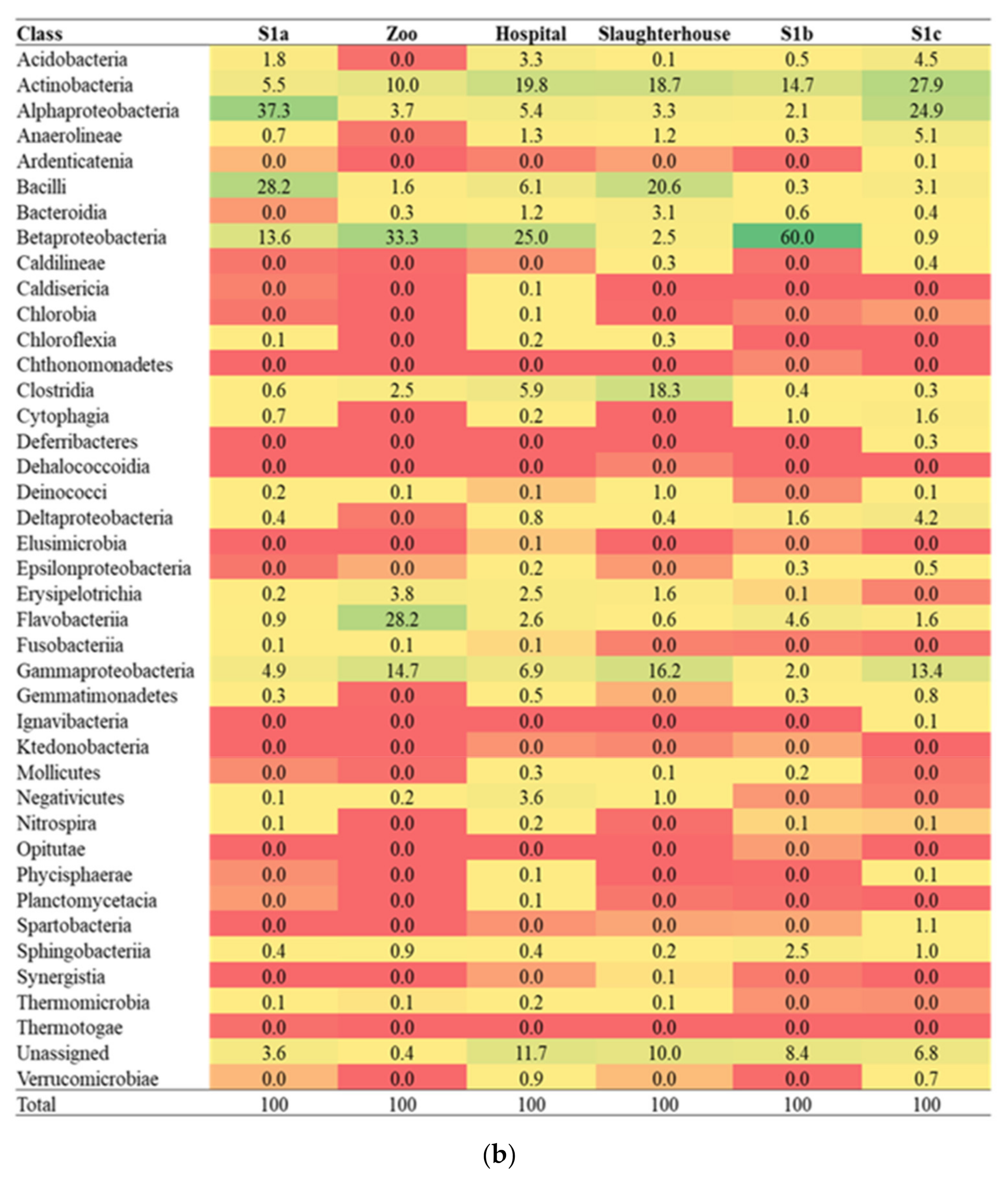
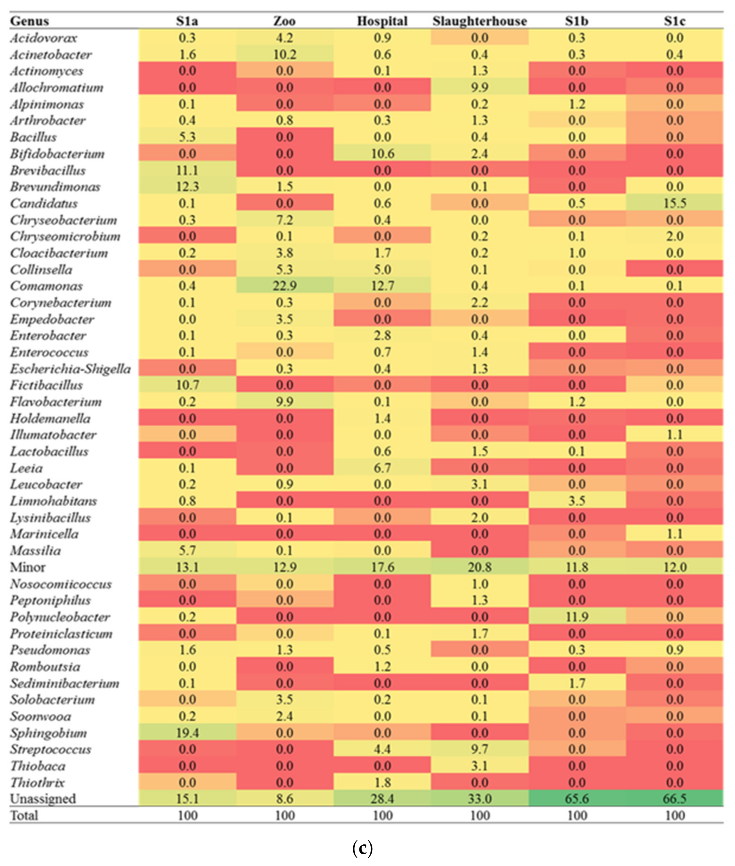
3.1. Diversity of the Microbial Community

Metagenomics sequencing from six samples obtained 50997 to 68919 reads (Supplementary Table S1). These reads were clustered into 499 to 1227 OTUs (>97% sequence similarity). The rarefaction curves of OTUs were saturated for all of the sampling sites (Supplementary Figure S1). The microbial community composition varied among the sites. Two outcomes corresponding to anthropogenic inputs were observed: (i) wastewater effluents supported the microbial community diversity in which the bacterial diversity and species richness were pronouncedly increased in effluents from hospital (Chao index (Chao 1):1278; Shannon index (H): 4.91) and slaughterhouse (Chao 1:1254; H: 5.24); compared to zoos (Chao 1, 633; H, 3.87) as well as river water. (ii) Lower bacterial diversity and species richness by which Comamonas was found to be present predominantly in effluent from the zoo.
Cluster analysis indicated that the microbial community composition in zoo effluent and S1b, which received all wastewater effluents from the zoo, hospital, and slaughterhouse, were more similar compared to S1a (Figure 2). This could be due to the influence of effluent discharge that contained more animal waste as Lye et al. [14] detected SMX and SDM among the commonly used SAs in these wastewaters. In contrast, at S1c, which is further downstream and near the river mouth, salinity may be the important factor in shaping the bacterial community structure to be similar to the microbial community in hospital wastewater, which is characterized by high TDS [35]. Studies have shown shifts in aquatic bacterial diversity according to salinity gradient [36,37]. However, further research is needed to confirm this. The distinct bacterial community profile observed in slaughterhouses may be due to poultry waste, and the distinctly lower DO levels (<LOQ) [31] as DO is an important environmental driver that influences bacterial communities in various marine and freshwater ecosystems [38,39].
In this study, we identified 32 phyla, 40 classes, and 509 genera. Proteobacteria, Firmicutes, Bacteroidetes, and Actinobacteria dominated in both effluents and river waters (Figure 3a–c). These four phyla were followed by six other major phyla, including Acidobacteria, Cyanobacteria, Parcubacteria, Verrucomicrobia (except for zoo effluent), Chloroflexi, and Saccharibacteria. The dominant phyla detected in this study are reportedly predominant in soil and various aquatic environments [40,41,42,43,44]. However, the dominant bacterial phyla in the aquatic ecosystem generally varied depending on the type of water samples and anthropogenic pollutants [41,45,46]. Within the Proteobacteria phylum, Alphaproteobacteria was enriched in S1a (37.3%) and S1c (24.9%), Betaproteobacteria in zoo effluent (33.3%) and S1b (60.0%) while Gammaproteobacteria in zoo (14.7%) and slaughterhouse (16.2%) effluents and S1c (13.4%). These findings concurred with previous studies that reported the survival of the Proteobacteria in wastewater as members of this phylum are known to have versatile metabolic pathways and intense biodegradative activity [47,48,49,50]. For Firmicutes, Bacilli was enriched in S1a (28.2%) and slaughterhouse effluent (20.6%), while Clostridia was only in the slaughterhouse effluent (18.3%). The lower DO conditions in the slaughterhouse effluent might have provided more suitable conditions for the growth of these sporeformers [51]. In Bacteroidetes, Flavobacteriia predominated in the zoo effluent. A similar finding has been reported by Bondarczuk and Piotrowska-Seget [45] in a lake receiving wastewater, and the presence of Flavobacteriia is frequently associated with the high availability of organic matter [45,52]. Actinobacteria were enriched to >10% abundance in all of the effluents and river waters except in S1a. The members of this phylum are well known for tolerance to low levels of organic carbon, and thus, they can survive in a wide range of habitats [53,54,55].

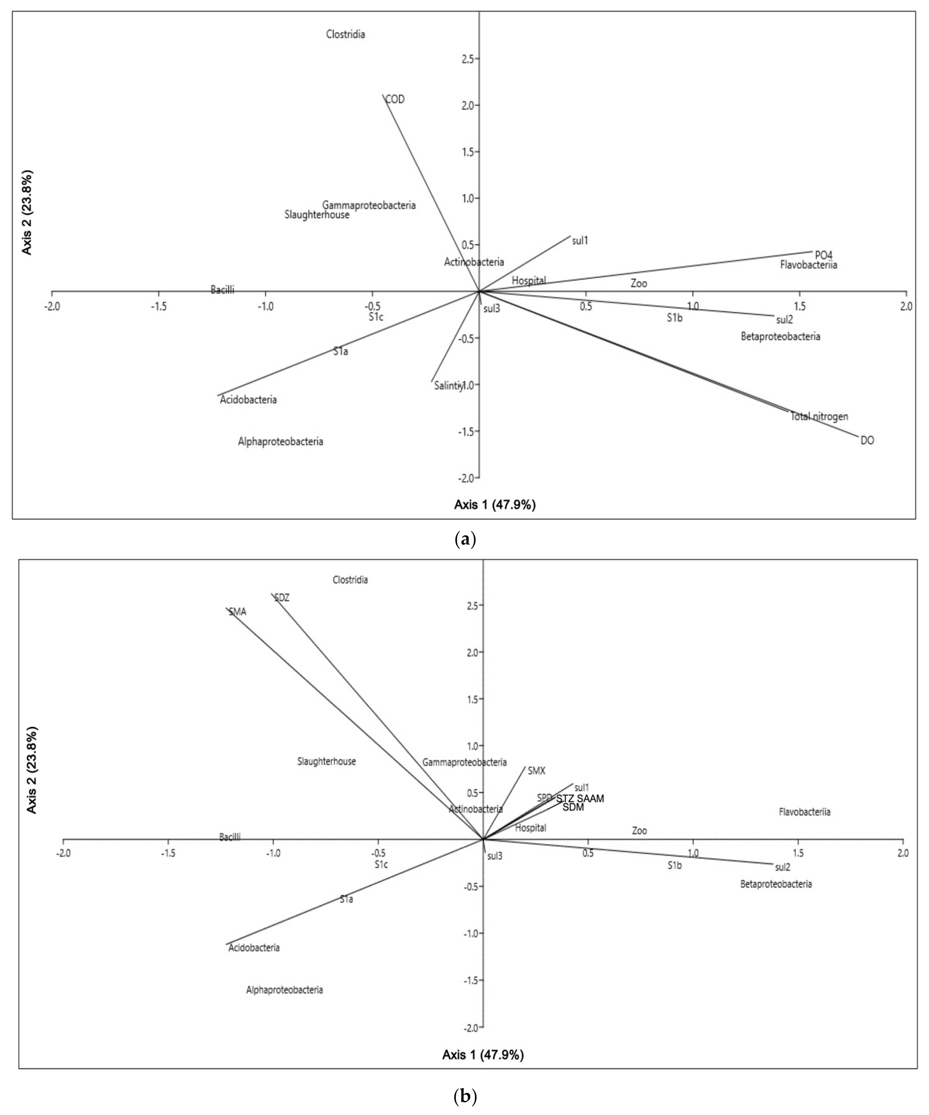
3.2. Environmental Physicochemical Parameters, Sulfonamides Residues and Their Relations to Microbial Community

CCA results (Figure 4a) showed that the microbial community was significantly shaped by salinity, DO, chemical oxygen demand (COD), phosphate (PO4), and total nitrogen (TN). The results are in agreement with the findings of previous studies that the changes in the abundance and composition of the microbial bacterial community can be attributed to the in situ physicochemical parameters [56,57,58]. Salinity and DO are important drivers of bacterial diversity and composition [59,60,61]. In the riverine waters, Betaproteobacteria and total sul2 genes showed a positive association with DO, while Acidobacteria was associated with salinity. On the contrary, a negative correlation was observed between DO and Clostridia and Bacilli. Moreover, COD also had a significant influence on Actinobacteria, Proteobacteria (Gammaproteobacteria), and Firmicutes (Bacilli and Clostridia) from the slaughterhouse effluent. Similar observations have been reported by Liu et al. [62]. The relationship between bacteria and PO4 is rarely reported [63,64]. In the present study, we found that Flavobacteriia and total sul1 genes in the zoo and hospital effluents were associated positively with PO4. This correlation could be attributed to phosphorus sources. Zheng et al. [64] have revealed the role of phosphorus in shaping the microbial composition and function in activated sludges. TN has been associated with Betaproteobactria concurring with Guo et al. [65], who reported that a higher nitrogen source could enrich Betaproteobacteria.
Our results also showed that microbial community was significantly shaped by SMA, SDZ, SMX, SPD, SDM, STZ, and SAAM, in which 47.9% of the variations could be explained by CCA1 and 23.7% by CCA2 (Figure 4b). The key antibiotics detected in the hospital (SPD, SMX, SDM, STZ, and SAAM) exhibited significant correlations with the microbial community and total sul1 genes. In the slaughterhouse effluent, Actinobacteria and Gammaproteobacteria showed positive associations with SDZ and SMA. These observed correlations were similar to the works of Xiong et al. [43], Guan et al. [66], Visca et al. [67], and Xu et al. [44]. Differences in the response by the observed microbial community to SA were probably due to the development of antibiotic resistance mechanisms in indigenous bacteria under antibiotic selective pressure [68].

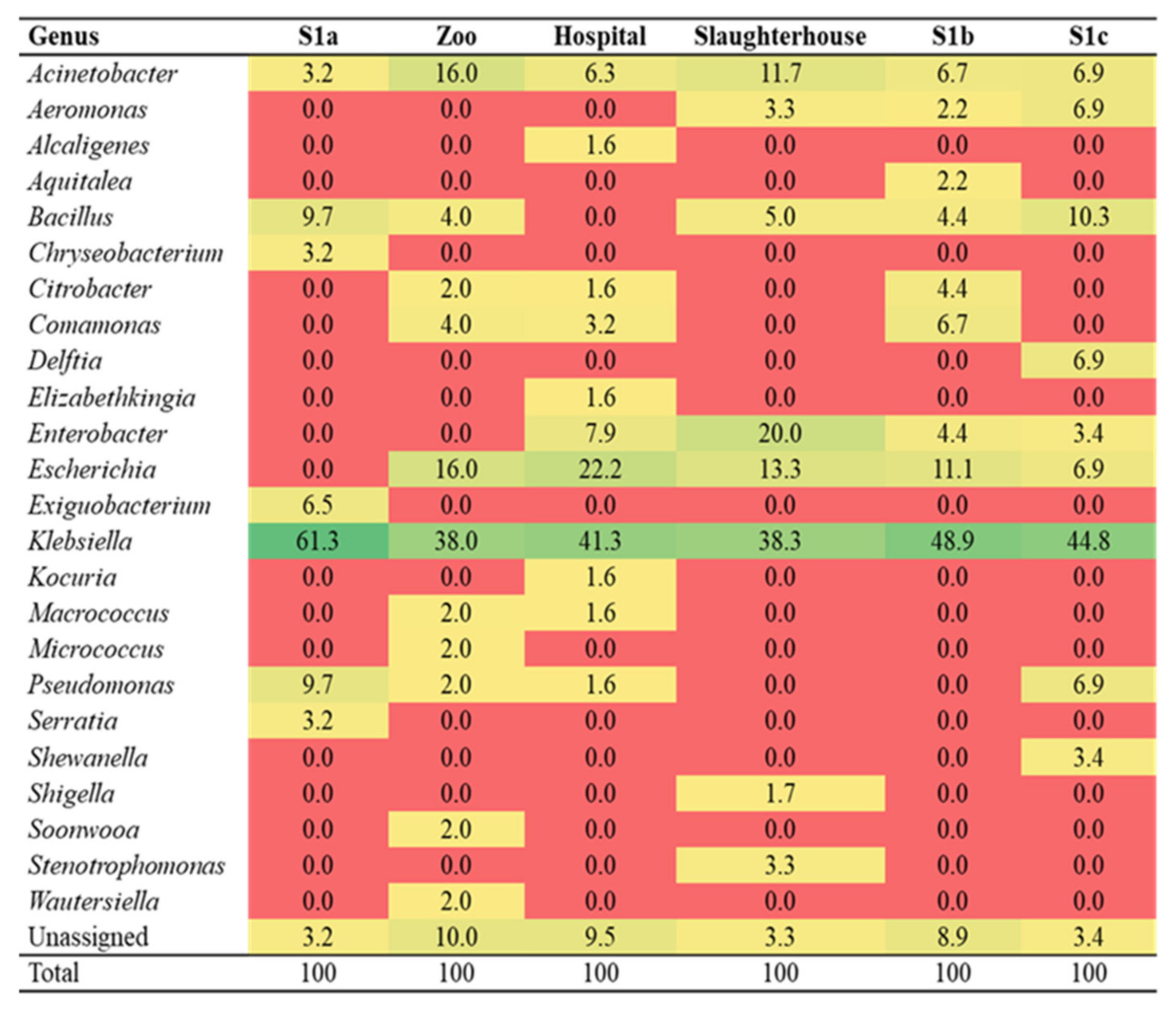
3.3. Identification of Culturable Sulfonamide-Resistant Bacteria

We had isolated a total of 278 SARB that belonged to 24 genera, which consisted of only 4.7% of the total number of genera identified with the metagenomics approach (Figure 5). SAR Klebsiella (38.0%–61.3%) were predominant in both the effluents and the Larut River. The majority of the SAR Klebsiella isolated was mainly K. pneumoniae (Supplementary Table S2). Our results concurred with previous studies that reported K. pneumoniae is able to survive a broad ecological range, which could be attributed to their highly diverse genome [25,69,70,71,72]. The SAR Escherichia (0–22.2%) and Acinetobacter (3.2–16.0%) were more frequently isolated from the effluents than the Larut River. Among these SARB, Acinetobacter sp., Bacillus sp., and Pseudomonas sp. are common bacterial genera intrinsically resistant to SAs, whereas Serratia sp., Alcaligens sp., Aquitalea sp., and Delftia sp. are poorly characterized in terms of SAR resistance. In this study, approximately half of the SARB found in the effluents from the zoo, hospital, and slaughterhouse were not able to persist in the river water. This was evident in the reduction in SARB isolated from wastewater effluents and S1b to S1c [14]. The reduction in SAR could be attributed to environmental factors and reduced antibiotic selection pressure [10,31,73,74] that may have limited their distribution and persistence along the Larut River.

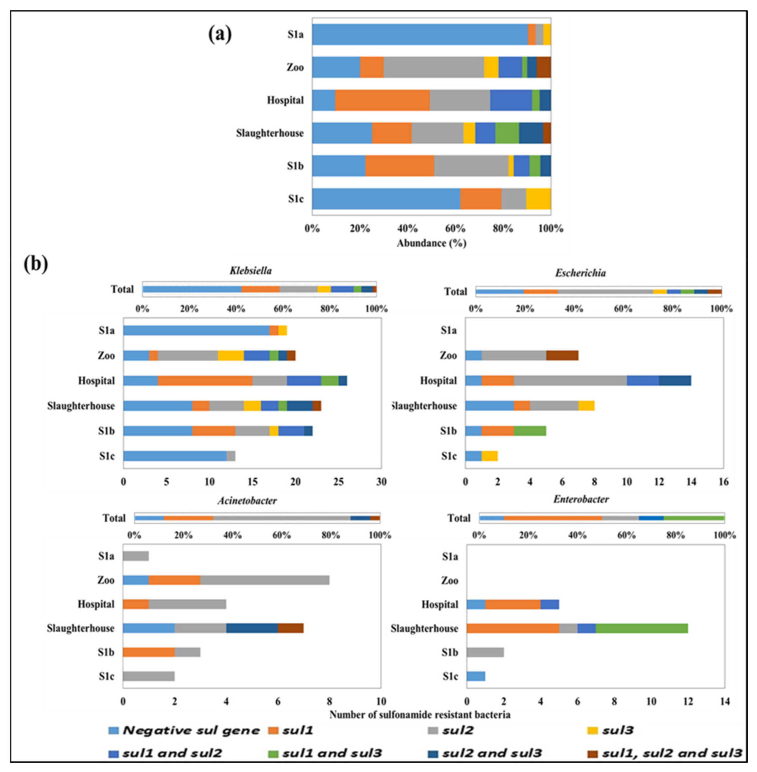
3.4. Detection of sul1, sul2, and sul3 Genes in the Culturable Sulfonamide-Resistant Isolates

The sul genes were detected in all sites with a distribution pattern of sul2 (24.5%) > sul1 (21.2%) > sul3 (4.0%) (Figure 6a). The majority of the SARB harboring sul genes were found in the zoo (80.0%), hospital (90.5%), and slaughterhouse (75.0%) effluents. SARB isolated from the zoo and slaughterhouse effluents harbored sul2 gene, whereas the hospital wastewater effluent was dominated by the SARB with sul1 gene. These findings indicated that SAR is common in the aquatic environment of Larut River and is in agreement with Suzuki et al. [75], who showed that sul1 and sul2 are ubiquitous in aquatic bacteria. Notably, the SARB in these wastewater effluents was found to harbor multiple sul genes. Contrarily, SARB that harbored no sul gene in their genome was more prevalent in the upstream and further downstream river, suggesting that the SARB carrying sul genes was associated with anthropogenic activities. Furthermore, a decreasing trend was observed in SARB harboring multiple sul genes from the immediate downstream (S1b) to further downstream (S1c). The decreased abundance of SARB that possessed sul genes and the sul genotypes at S1c might be due to the reduction in SA selection pressure where Björkman and Andersson [76] reported that resistance is associated with metabolic cost suggesting a decline of ARB with lower antibiotic use. Schulz zur Wiesch et al. [77] reported that gene-based resistance in bacteria strains is often costly, and the fitness loss may be reflected in a reduced growth rate in vivo [78] or in vitro [79,80] and transmission rate [81] in the absence of antibiotics. Studies also revealed that the cost of resistance is among the most important factor determining the rate and extent of resistance emergence [9,79,82,83,84].
Interestingly, all the combinations of sul genes in the culturable SARB were found in the wastewater effluents. In this study, ~40% of the SAR Klebsiella did not carry sul genes and were mainly detected in S1a and S1c while the rest were harboring sul genes, i.e., mainly sul1 and sul2 (~32%) (Figure 6b). These results indicated that SAR in Klebsiella in wastewater environment did not rely on sul genes but other SAR mechanisms. With the rising cases of community-acquired infections of resistant bacteria, it is interesting to look into the SAR mechanisms, including folate synthesis and efflux systems in Klebsiella in the aquatic environments, to predict and counteract the emergence and future evolution of antibiotic resistance in these bacteria.
We observed different sul genotypes and sul genes distribution patterns in SAR Acinetobacter, Escherichia, and Enterobacter. However, the majority (>80%) of these SARB harbored sul genes suggesting that SAR of these bacteria were mostly dependent on resistance genes. The SAR Acinetobacter that dominated in the zoo (16.0%) and slaughterhouse (11.7%) effluents were consistent with previous studies that reported Acinetobacter as a potential environmental sul genes reservoir from animal sources (pig slurry and manured agricultural soils). Our findings also suggested a potential link for dissemination of sul genes between the natural environments, clinical, and agricultural settings. However, direct evidence of ARGs dissemination between the environmental and clinical resistome is rare as these ARGs may undergo several rounds of evolutions between the natural and clinical environments [85,86].

3.5. Identification of Potential sul Genes Bacterial Carrier

A total of 19 nodes (8 sul genotypes and 11 SARB) and 43 edges were obtained through the network analysis. Acinetobacter, Aeromonas, Bacillus, Comamonas, Citrobacter, Enterobacter, Escherichia, Klebsiella, Macrococcus, Pseudomonas, and Stenotrophomonas were identified as potential sul genes carriers and found associated with more than one sul genotype except for SAR Macrococcus and Pseudomonas that were associated with only one sul genotype (Figure 7).
In this study, a comparison of culturable SARB population to total microbial community profile structure was carried out to identify the potential sul gene bacterial carriers in this river. The culturable method employed has recovered most of the same dominant bacterial phyla in the total microbial community (Proteobacteria, Actinobacteria, Firmicutes, and Bacteroidetes). However, the results showed that the most prevalent SAR bacterial genera isolated from the culturable SARB were different from the total community. Klebsiella and Citrobacter were not detected in the total microbial community, whereas Macrococcus and Stenotrophomonas were detected at <1% across the sampling sites (Supplementary Figure S2). These results are in agreement with Suzuki et al. [75] that reported culturable bacteria are not a major component of the total bacteria community and are selected by the culture method causing biases in bacterial diversity. Furthermore, conventional culture methods for monitoring ARB and ARGs only revealed ≤0.1% of the true bacterial community [87,88]. Thus, the relationships between ARGs and bacteria species cultivated found are limited to a fraction of the overall river bacterial community.
Notably, nine out of eleven (81.8%) identified potential sul genes bacterial carriers belonged to Proteobacteria while the other two carriers belonged to Firmicutes. Those carrying sul genes were dominated by culturable SAR Klebsiella, Escherichia, Acinetobacter, and Enterobacter and might serve as reservoirs of sul genes and disseminate these genes among the microbial community. It is noted that the abundance and distribution of sul genotypes in the culturable SARB could be largely affected by the sul genotypes in SAR Klebsiella, which were present as the most dominant culturable SARB. The potential environmental sul genes carriers in Proteobacteria, as well as reservoirs, includes Acinetobacter, Aeromonas, Enterobacter, Escherichia, Klebsiella, and Pseudomonas, and their positive correlation with sul1 has been reported [66,89,90,91,92]. For Comamonadaceae and Aeromonadaceae, strong correlations with sul1 and sul2 have also been reported [19].
A correlation was also reported between Firmicutes, particularly Bacillus and Clostridia, and sul1 and sul2 [50,66,92]. Interestingly, this present study had identified SAR Macrococcus that belonged to Firmicutes as one of the sul gene bacterial carriers associated with sul1. Generally, animal origin Macrococcus has been reported to have resistance to methicillin (mecB gene) and TMP (cfr gene) [93,94]. Therefore, this is the first study to show Macrococcus resistance to SA. Further studies are needed to confirm the emergence and dissemination of the sul genes in this genus in the aquatic environment.
Aeromonas, Comamonas, Pseudomonas, and Stenotrophomonas were also detected in the Larut River by both culturable and metagenomics approaches suggesting that these bacteria are the common sul genes carriers residing in Larut River. Furthermore, studies have also revealed the ability of Acinetobacter, Bacillus, Brevundimonas, Comamonas, Escherichia, Klebsiella, Pseudomonas, and Stenotrophomonas for the degradation of SA in various environments and acclimated membrane reactors [26,95,96]. The catabolism of SAs in these bacteria is important not only for antibiotic degradation to remove pollutants in the environment but also for antibiotic resistance as the enzymes involved in degradation are a potential resistance mechanism [97]. Kim et al. [98] revealed that SA metabolism may have evolved in SARB, which has already acquired the class 1 integron under SA selection pressures.
Masco et al. [99] reported that Bifidobacterium is intrinsically resistant to SMX. There have been reports of resistance of SA in Sphingobium, Brevibacillus, Candidatus, and Polynucleobacter [100,101]. Correlations between Candidatus and sul1 and sul2 and Polynucleobacter with sul2 have also been reported [102,103]. Thus, even though these bacteria were not isolated in the present study, they could be the important sul genes bacterial carriers in the Larut River. Therefore, further studies using more variety of culture media and targeting more diverse bacteria are needed to study the antibiotic-resistant profile.
Martínez [3] reported that natural environments represent reservoirs of ARGs, and changes in these ecosystems might be relevant for the emergence of previously unknown resistance determinants in bacterial pathogens. However, the effect of environmental changes on the dynamics of the bacteria population and their ARGs have received less attention [104]. Past research has focused on antibiotic resistance in pathogenic bacteria, where the information about the extent to which commensal and non-pathogenic bacteria can act as antibiotic resistance reservoirs is lacking [105]. Therefore, more studies on whether the anthropogenic activities might enrich the ARB population in the natural environments and facilitate the transfer of ARGs will be important to address in the future. Moreover, Narciso-da-Rocha and Manaia [106] reported that there is still a significant difference between the ARB profile through culture-dependent and metagenomics analysis. The ARGs and their affiliated taxa are still very unclear [107]. Therefore, the combination of correlation analysis, culture-dependent, and metagenomics should be performed for further confirmation of the relationships between the ARGs and their bacterial hosts to the taxa level [92].

4. Conclusions

The zoo, hospital, and slaughterhouse wastewater effluents had exerted impacts on the bacterial microbial community and culturable SAR bacteria population in the Larut River. Our findings showed that the zoo, hospital, and slaughterhouse were potential sources of high diversity and number of potentially pathogenic and clinically important bacteria into the receiving river. The SARB isolated from the wastewater effluents harbored higher sul genes and displayed more diverse sul genotypes. The absence of sul genes was detected in ~40% of the culturable SAR Klebsiella, and more study is needed to understand their resistance mechanisms in the emergence and evolution of SAs resistance in aquatic environments. We have identified 11 sul genes carriers (Acinetobacter, Aeromonas, Bacillus, Comamonas, Citrobacter, Enterobacter, Escherichia, Klebsiella, Macrococcus, Pseudomonas, and Stenotrophomonas) that could act as reservoirs of antibiotic resistance. CCA revealed that SAs residues and inorganic nutrients exerted significant impacts on microbial community and total sul genes. However, further studies are required to understand the development of resistance mechanisms and the relationships between opportunistic pathogens and ARGs.

Supplementary Materials

The following are available online at https://www.mdpi.com/article/10.3390/w14071018/s1, Table S1: Summary of diversity and richness of microbial community. Table S2: Sulfonamides resistant bacteria identified to species level. Figure S1: Rarefaction curves estimated at 97% of microbial community. Figure S2: Bacterial genera detected by 16S rDNA metagenomics.

Author Contributions

Conceptualization, Y.-L.L., L.-C.C. and C.-W.B.; methodology, Y.-L.L., L.-C.C., C.-W.L. and C.-W.B.; validation, Y.-L.L. and C.-W.B.; formal analysis, Y.-L.L., C.-W.L. and C.-W.B.; investigation, Y.-L.L., L.-C.C. and C.-W.B.; resources, L.-C.C., S.S., C.-W.L. and C.-W.B.; data curation, Y.-L.L. and C.-W.B.; writing-original draft preparation, Y.-L.L., L.-C.C. and C.-W.B.; writing-review and editing, Y.-L.L., L.-C.C., C.-W.L., S.S. and C.-W.B.; visualization, Y.-L.L. and C.-W.B.; supervision, L.-C.C., C.-W.L. and C.-W.B.; project administration, L.-C.C. and C.-W.B.; funding acquisition, L.-C.C., S.S., C.-W.L. and C.-W.B. All authors have read and agreed to the published version of the manuscript.

Funding

This work was supported by the Institution Centre of Excellence (HICoE) Phase II Fund, Ministry of Higher Education (IOES-2014D); University of Malaya (High-Impact Research Grant UM.C/625/1/HIR/MOHE/SC/20 (UM.S/P/HIR/MOHE/24, RU009D-2015, IF030A-2017); private funding (PV009-2019) awarded to Chai Lay Ching and partly supported by the Ministry of Education, Culture, Sports, Science and Technology, Japan (MEXT) to a project on Joint Usage/Research Centre, Leading Academia in Marine and Environmental Research (LaMer).

Institutional Review Board Statement

Not applicable.

Informed Consent Statement

Not applicable.

Data Availability Statement

Data is contained within the article and Supplementary Materials.

Acknowledgments

Thank you to the academic editors and the four anonymous reviewers for their valuable comments.

Conflicts of Interest

The authors declare no conflict of interest.

References

  1. Forsberg, K.J.; Reyes, A.; Wang, B.; Selleck, E.M.; Sommer, M.O.A.; Dantas, G. The Shared Antibiotic Resistome of Soil Bacteria and Human Pathogens. Science 2012, 337, 1107–1111. [Google Scholar] [CrossRef] [PubMed] [Green Version]
  2. Gibson, M.K.; Forsberg, K.; Dantas, G. Improved annotation of antibiotic resistance determinants reveals microbial resistomes cluster by ecology. ISME J. 2014, 9, 207–216. [Google Scholar] [CrossRef] [PubMed]
  3. Martínez, J.L. Antibiotics and Antibiotic Resistance Genes in Natural Environments. Science 2008, 321, 365–367. [Google Scholar] [CrossRef] [PubMed]
  4. Suzuki, M.T.; Giovannoni, S.J. Bias caused by template annealing in the amplification of mixtures of 16S rRNA genes by PCR. Appl. Environ. Microbiol. 1996, 62, 625–630. [Google Scholar] [CrossRef] [PubMed] [Green Version]
  5. Li, D.; Qi, R.; Yang, M.; Zhang, Y.; Yu, T. Bacterial community characteristics under long-term antibiotic selection pressures. Water Res. 2011, 45, 6063–6073. [Google Scholar] [CrossRef] [PubMed]
  6. Baquero, F.; Martinez, J.L.; Cantón, R. Antibiotics and antibiotic resistance in water environments. Curr. Opin. Biotechnol. 2008, 19, 260–265. [Google Scholar] [CrossRef] [PubMed]
  7. Poirel, L.; Rodriguez-Martinez, J.-M.; Mammeri, H.; Liard, A.; Nordmann, P. Origin of Plasmid-Mediated Quinolone Resistance Determinant QnrA. Antimicrob. Agents Chemother. 2005, 49, 3523–3525. [Google Scholar] [CrossRef] [Green Version]
  8. Rizzo, L.; Manaia, C.; Merlin, C.; Schwartz, T.; Dagot, C.; Ploy, M.C.; Michael, I.; Fatta-Kassinos, D. Urban wastewater treatment plants as hotspots for antibiotic resistant bacteria and genes spread into the environment: A review. Sci. Total Environ. 2013, 447, 345–360. [Google Scholar] [CrossRef] [Green Version]
  9. Handel, A.; Regoes, R.R.; Antia, R. The Role of Compensatory Mutations in the Emergence of Drug Resistance. PLoS Comput. Biol. 2006, 2, e137. [Google Scholar] [CrossRef] [Green Version]
  10. Gutiérrez, I.R.; Watanabe, N.; Harter, T.; Glaser, B.; Radke, M. Effect of sulfonamide antibiotics on microbial diversity and activity in a Californian Mollic Haploxeralf. J. Soils Sediments 2010, 10, 537–544. [Google Scholar] [CrossRef]
  11. Economou, V.; Gousia, P. Agriculture and food animals as a source of antimicrobial-resistant bacteria. Infect. Drug Resist. 2015, 8, 49–61. [Google Scholar] [CrossRef] [Green Version]
  12. Laffite, A.; Kilunga, P.I.; Kayembe, J.M.; Devarajan, N.; Mulaji, C.; Giuliani, G.; Slaveykova, V.; Poté, J. Hospital Effluents Are One of Several Sources of Metal, Antibiotic Resistance Genes, and Bacterial Markers Disseminated in Sub-Saharan Urban Rivers. Front. Microbiol. 2016, 7, 1128. [Google Scholar] [CrossRef] [Green Version]
  13. Jørgensen, K.M.; Wassermann, T.; Jensen, P.Ø.; Hengzuang, W.; Molin, S.; Høiby, N.; Ciofu, O. Sublethal Ciprofloxacin Treatment Leads to Rapid Development of High-Level Ciprofloxacin Resistance during Long-Term Experimental Evolution of Pseudomonas aeruginosa. Antimicrob. Agents Chemother. 2013, 57, 4215–4221. [Google Scholar] [CrossRef] [Green Version]
  14. Lye, Y.L.; Bong, C.W.; Lee, C.W.; Zhang, R.J.; Zhang, G.; Suzuki, S.; Chai, L.C. Anthropogenic impacts on sulfonamide residues and sulfonamide resistant bacteria and genes in Larut and Sangga Besar River, Perak. Sci. Total Environ. 2019, 688, 1335–1347. [Google Scholar] [CrossRef]
  15. Bien, T.L.T.; Sato-Takabe, Y.; Ogo, M.; Usui, M.; Suzuki, S. Persistence of Multi-Drug Resistance Plasmids in Sterile Water under Very Low Concentrations of Tetracycline. Microbes Environ. 2015, 30, 339–343. [Google Scholar] [CrossRef] [Green Version]
  16. Gullberg, E.; Cao, S.; Berg, O.G.; Ilbäck, C.; Sandegren, L.; Hughes, D.; Andersson, D.I. Selection of Resistant Bacteria at Very Low Antibiotic Concentrations. PLoS Pathog. 2011, 7, e1002158. [Google Scholar] [CrossRef] [Green Version]
  17. Sánchez-Baena, A.M.; Caicedo-Bejarano, L.D.; Chávez-Vivas, M. Structure of Bacterial Community with Resistance to Anti-biotics in Aquatic Environments. A Systematic Review. Int. J. Environ. Res. Public Health 2021, 18, 2348. [Google Scholar] [CrossRef]
  18. Girijan, S.K.; Paul, R.; Rejish Kumar, V.J.; Pillai, D. Investigating the impact of hospital antibiotic usage on aquatic environment and aquaculture systems: A molecular study of quinolone resistance in Escherichia coli. Sci. Total Environ. 2020, 748, 141538. [Google Scholar] [CrossRef]
  19. Narciso-Da-Rocha, C.; Rocha, J.; Vaz-Moreira, I.; Lira, F.; Tamames, J.; Henriques, I.; Martinez, J.L.; Manaia, C.M. Bacterial lineages putatively associated with the dissemination of antibiotic resistance genes in a full-scale urban wastewater treatment plant. Environ. Int. 2018, 118, 179–188. [Google Scholar] [CrossRef]
  20. Sarmah, A.K.; Meyer, M.; Boxall, A.B. A global perspective on the use, sales, exposure pathways, occurrence, fate and effects of veterinary antibiotics (VAs) in the environment. Chemosphere 2006, 65, 725–759. [Google Scholar] [CrossRef]
  21. Lamshöft, M.; Sukul, P.; Zühlke, S.; Spiteller, M. Metabolism of 14C-labelled and non-labelled sulfadiazine after administration to pigs. Anal. Bioanal. Chem. 2007, 388, 1733–1745. [Google Scholar] [CrossRef]
  22. Manzetti, S.; Ghisi, R. The environmental release and fate of antibiotics. Mar. Pollut. Bull. 2014, 79, 7–15. [Google Scholar] [CrossRef]
  23. Sköld, O. Resistance to trimethoprim and sulfonamides. Vet. Res. 2001, 32, 261–273. [Google Scholar] [CrossRef] [Green Version]
  24. Esuzuki, S.; Eogo, M.; Miller, T.W.; Eshimizu, A.; Etakada, H.; Siringan, M.A.T. Who possesses drug resistance genes in the aquatic environment? Sulfamethoxazole (SMX) resistance genes among the bacterial community in water environment of Metro-Manila, Philippines. Front. Microbiol. 2013, 4, 102. [Google Scholar] [CrossRef] [Green Version]
  25. Holt, K.E.; Wertheim, H.; Zadoks, R.N.; Baker, S.; Whitehouse, C.A.; Dance, D.; Jenney, A.; Connor, T.R.; Hsu, L.Y.; Severin, J.; et al. Genomic analysis of diversity, population structure, virulence, and antimicrobial resistance in Klebsiella pneumoniae, an urgent threat to public health. Proc. Natl. Acad. Sci. USA 2015, 112, E3574–E3581. [Google Scholar] [CrossRef] [Green Version]
  26. Islas-Espinoza, M.; Reid, B.J.; Wexler, M.; Bond, P.L. Soil Bacterial Consortia and Previous Exposure Enhance the Biodegradation of Sulfonamides from Pig Manure. Microb. Ecol. 2012, 64, 140–151. [Google Scholar] [CrossRef]
  27. Quast, C.; Pruesse, E.; Yilmaz, P.; Gerken, J.; Schweer, T.; Yarza, P.; Peplies, J.; Glöckner, F.O. The SILVA ribosomal RNA gene database project: Improved data processing and web-based tools. Nucleic Acids Res. 2013, 41, D590–D596. [Google Scholar] [CrossRef]
  28. Schloss, P.D.; Gevers, D.; Westcott, S.L. Reducing the Effects of PCR Amplification and Sequencing Artifacts on 16S rRNA-Based Studies. PLoS ONE 2011, 6, e27310. [Google Scholar] [CrossRef] [PubMed] [Green Version]
  29. Hammer, Ø.; Harper, D.A.T.; Ryan, P.D. PAST: Paleontological statistics software package for education and data analysis. Palaeontol. Electron. 2001, 4, 1–9. [Google Scholar]
  30. Wang, L.; Zhang, J.; Li, H.; Yang, H.; Peng, C.; Peng, Z.; Lu, L. Shift in the microbial community composition of surface water and sediment along an urban river. Sci. Total Environ. 2018, 627, 600–612. [Google Scholar] [CrossRef] [PubMed]
  31. Lu, Z.; Na, G.; Gao, H.; Wang, L.; Bao, C.; Yao, Z. Fate of sulfonamide resistance genes in estuary environment and effect of anthropogenic activities. Sci. Total Environ. 2015, 527–528, 429–438. [Google Scholar] [CrossRef]
  32. Hoa, P.T.P.; Nonaka, L.; Viet, P.H.; Suzuki, S. Detection of the sul1, sul2, and sul3 genes in sulfonamide-resistant bacteria from wastewater and shrimp ponds of north Vietnam. Sci. Total Environ. 2008, 405, 377–384. [Google Scholar] [CrossRef]
  33. Kerrn, M.B.; Klemmensen, T.; Frimodt-Møller, N.; Espersen, F. Susceptibility of Danish Escherichia coli strains isolated from urinary tract infections and bacteraemia, and distribution of sul genes conferring sulphonamide resistance. J. Antimicrob. Chemother. 2002, 50, 513–516. [Google Scholar] [CrossRef] [Green Version]
  34. Na, G.; Zhang, W.; Zhou, S.; Gao, H.; Lu, Z.; Wu, X.; Li, R.; Qiu, L.; Cai, Y.; Yao, Z. Sulfonamide antibiotics in the Northern Yellow Sea are related to resistant bacteria: Implications for antibiotic resistance genes. Mar. Pollut. Bull. 2014, 84, 70–75. [Google Scholar] [CrossRef]
  35. Dianawati, R.I.; Wahyuningsih, N.E.; Nur, M. Treatment of hospital waste water by ozone technology. J. Phys. Conf. Ser. 2018, 1025, 012013. [Google Scholar] [CrossRef] [Green Version]
  36. Silveira, C.; Vieira, R.P.; Cardoso, A.M.; Paranhos, R.; Albano, R.; Martins, O.B. Influence of Salinity on Bacterioplankton Communities from the Brazilian Rain Forest to the Coastal Atlantic Ocean. PLoS ONE 2011, 6, e17789. [Google Scholar] [CrossRef] [Green Version]
  37. Wang, J.; Yang, D.; Zhang, Y.; Shen, J.; van der Gast, C.; Hahn, M.W.; Wu, Q. Do Patterns of Bacterial Diversity along Salinity Gradients Differ from Those Observed for Macroorganisms? PLoS ONE 2011, 6, e27597. [Google Scholar] [CrossRef] [Green Version]
  38. Aldunate, M.; De la Iglesia, R.; Bertagnolli, A.D.; Ulloa, O. Oxygen modulates bacterial community composition in the coastal upwelling waters off central Chile. Deep Sea Res. Part II Top. Stud. Oceanogr. 2018, 156, 68–79. [Google Scholar] [CrossRef]
  39. Spietz, R.; Williams, C.M.; Rocap, G.; Horner-Devine, C. A Dissolved Oxygen Threshold for Shifts in Bacterial Community Structure in a Seasonally Hypoxic Estuary. PLoS ONE 2015, 10, e0135731. [Google Scholar] [CrossRef] [Green Version]
  40. Szekeres, E.; Baricz, A.; Chiriac, C.M.; Farkas, A.; Opris, O.; Soran, M.-L.; Andrei, A.-S.; Rudi, K.; Balcázar, J.L.; Dragos, N.; et al. Abundance of antibiotics, antibiotic resistance genes and bacterial community composition in wastewater effluents from different Romanian hospitals. Environ. Pollut. 2017, 225, 304–315. [Google Scholar] [CrossRef]
  41. Vaz-Moreira, I.; Nunes, O.; Manaia, C.M. Bacterial diversity and antibiotic resistance in water habitats: Searching the links with the human microbiome. FEMS Microbiol. Rev. 2014, 38, 761–778. [Google Scholar] [CrossRef]
  42. Wang, X.; Gu, J.; Gao, H.; Qian, X.; Li, H. Abundances of Clinically Relevant Antibiotic Resistance Genes and Bacterial Community Diversity in the Weihe River, China. Int. J. Environ. Res. Public Health 2018, 15, 708. [Google Scholar] [CrossRef] [Green Version]
  43. Xiong, W.; Sun, Y.; Zhang, T.; Ding, X.; Zhenling, Z.; Wang, M.; Zeng, Z. Antibiotics, Antibiotic Resistance Genes, and Bacterial Community Composition in Fresh Water Aquaculture Environment in China. Microb. Ecol. 2015, 70, 425–432. [Google Scholar] [CrossRef]
  44. Xu, K.; Wang, J.; Gong, H.; Li, Y.; Zhou, L.; Yan, M. Occurrence of antibiotics and their associations with antibiotic resistance genes and bacterial communities in Guangdong coastal areas. Ecotoxicol. Environ. Saf. 2019, 186, 109796. [Google Scholar] [CrossRef]
  45. Bondarczuk, K.; Piotrowska-Seget, Z. Microbial diversity and antibiotic resistance in a final effluent-receiving lake. Sci. Total Environ. 2018, 650, 2951–2961. [Google Scholar] [CrossRef]
  46. Drury, B.; Rosi-Marshall, E.; Kelly, J.J. Wastewater Treatment Effluent Reduces the Abundance and Diversity of Benthic Bacterial Communities in Urban and Suburban Rivers. Appl. Environ. Microbiol. 2013, 79, 1897–1905. [Google Scholar] [CrossRef] [Green Version]
  47. Khandeparker, L.; Kuchi, N.; Kale, D.; Anil, A.C. Microbial community structure of surface sediments from a tropical estuarine environment using next generation sequencing. Ecol. Indic. 2016, 74, 172–181. [Google Scholar] [CrossRef]
  48. Narayan, A.; Patel, V.; Singh, P.; Patel, A.; Jain, K.; Karthikeyan, K.; Shah, A.; Madamwar, D. Response of microbial community structure to seasonal fluctuation on soils of Rann of Kachchh, Gujarat, India: Representing microbial dynamics and functional potential. Ecol. Genet. Genom. 2018, 6, 22–32. [Google Scholar] [CrossRef]
  49. Qiu, W.; Sun, J.; Fang, M.; Luo, S.; Tian, Y.; Dong, P.; Xu, B.; Zheng, C. Occurrence of antibiotics in the main rivers of Shenzhen, China: Association with antibiotic resistance genes and microbial community. Sci. Total Environ. 2019, 653, 334–341. [Google Scholar] [CrossRef]
  50. Szekeres, E.; Chiriac, C.; Baricz, A.; Szőke-Nagy, T.; Lung, I.; Soran, M.-L.; Rudi, K.; Dragos, N.; Coman, C. Investigating antibiotics, antibiotic resistance genes, and microbial contaminants in groundwater in relation to the proximity of urban areas. Environ. Pollut. 2018, 236, 734–744. [Google Scholar] [CrossRef]
  51. Tiquia, S.; Schleibak, M.; Schlaff, J.; Floyd, C.; Benipal, B.; Zakhem, E.; Murray, K. Microbial Community Profiling and Characterization of Some Heterotrophic Bacterial Isolates from River Waters and Shallow Groundwater Wells Along the Rouge River, Southeast Michigan. Environ. Technol. 2008, 29, 651–663. [Google Scholar] [CrossRef] [PubMed]
  52. Jaguś, A. Assessment of the effectiveness of tresna reservoir protection based on the soła river waters contamination. Econ. Eng. 2017, 18, 55–60. [Google Scholar] [CrossRef] [Green Version]
  53. Allgaier, M.; Brückner, S.; Jaspers, E.; Grossart, H.-P. Intra- and inter-lake variability of free-living and particle-associated Actinobacteria communities. Environ. Microbiol. 2007, 9, 2728–2741. [Google Scholar] [CrossRef] [PubMed]
  54. Kurtböke, D.I. Ecology and Habitat Distribution of Actinobacteria. In Biology and Biotechnology of Actinobacteria; Wink, J., Mohammadipanah, F., Hamedi, J., Eds.; Springer: Cham, Switzerland, 2017; pp. 123–149. [Google Scholar] [CrossRef]
  55. Wilhelm, S.W.; LeCleir, G.R.; Bullerjahn, G.S.; McKay, R.M.; Saxton, M.A.; Twiss, M.R.; Bourbonniere, R.A. Seasonal changes in microbial community structure and activity imply winter production is linked to summer hypoxia in a large lake. FEMS Microbiol. Ecol. 2013, 87, 475–485. [Google Scholar] [CrossRef]
  56. Loreau, M.; Naeem, S.; Inchausti, P.; Bengtsson, J.; Grime, J.P.; Hector, A.; Hooper, D.U.; Huston, M.A.; Raffaelli, D.; Schmid, B.; et al. Biodiversity and Ecosystem Functioning: Current Knowledge and Future Challenges. Science 2001, 294, 804–808. [Google Scholar] [CrossRef] [Green Version]
  57. Price, J.; Ledford, S.H.; Ryan, M.O.; Toran, L.; Sales, C.M. Wastewater treatment plant effluent introduces recoverable shifts in microbial community composition in receiving streams. Sci. Total Environ. 2018, 613–614, 1104–1116. [Google Scholar] [CrossRef]
  58. Thomas, C.D.; Cameron, A.; Green, R.E.; Bakkenes, M.; Beaumont, L.J.; Collingham, Y.C.; Erasmus, B.F.N.; de Siqueira, M.F.; Grainger, A.; Hannah, L.; et al. Extinction risk from climate change. Nature 2004, 427, 145–148. [Google Scholar] [CrossRef]
  59. Trick, J.K.; Stuart, M.; Reeder, S. Contaminated groundwater sampling and quality control of water analyses. In Environmental Geochemistry: Site Characterization, Data Analysis and Case Histories. Candice Janco; Elsevier: Amsterdam, The Netherlands, 2018; pp. 25–45. [Google Scholar] [CrossRef] [Green Version]
  60. Logares, R.; Lindström, E.; Langenheder, S.; Logue, J.B.; Paterson, H.; Laybourn-Parry, J.; Rengefors, K.; Tranvik, L.J.; Bertilsson, S. Biogeography of bacterial communities exposed to progressive long-term environmental change. ISME J. 2013, 7, 937–948. [Google Scholar] [CrossRef] [Green Version]
  61. Malele, I.; Nyingilili, H.; Lyaruu, E.; Tauzin, M.; Ollivier, B.B.; Cayol, J.-L.; Fardeau, M.-L.; Geiger, A.; Malele, I.; Nyingilili, H.; et al. Bacterial diversity obtained by culturable approaches in the gut of Glossina pallidipes population from a non sleeping sickness focus in Tanzania: Preliminary results. BMC Microbiol. 2018, 18, 164. [Google Scholar] [CrossRef] [Green Version]
  62. Liu, Z.; Xiang, P.; Duan, Z.; Fu, Z.; Zhang, L.; Zhang, Z. Electricity generation, salinity, COD removal and anodic biofilm microbial community vary with different anode CODs in a microbial desalination cell for high-salinity mustard tuber wastewater treatment. RSC Adv. 2019, 9, 25189–25198. [Google Scholar] [CrossRef] [Green Version]
  63. Dao, T.H.; Guber, A.K.; Sadeghi, A.M.; Karns, J.S.; van Kessel, J.S.; Shelton, D.R.; Pachepsky, Y.A.; McCarty, G. Loss of bioactive phosphorus and enteric bacteria in runoff from dairy manure applied to sod. Soil Sci. 2008, 173, 511–521. [Google Scholar] [CrossRef] [Green Version]
  64. Zheng, L.; Ren, M.; Xie, E.; Ding, A.; Liu, Y.; Deng, S.; Zhang, D. Roles of Phosphorus Sources in Microbial Community Assembly for the Removal of Organic Matters and Ammonia in Activated Sludge. Front. Microbiol. 2019, 10, 1023. [Google Scholar] [CrossRef] [Green Version]
  65. Guo, J.; Zheng, Y.; Teng, J.; Song, J.; Wang, X.; Zhao, Q. The seasonal variation of microbial communities in drinking water sources in Shanghai. J. Clean. Prod. 2020, 265, 121604. [Google Scholar] [CrossRef]
  66. Guan, Y.; Jia, J.; Wu, L.; Xue, X.; Zhang, G.; Wang, Z. Analysis of Bacterial Community Characteristics, Abundance of Antibiotics and Antibiotic Resistance Genes Along a Pollution Gradient of Ba River in Xi’an, China. Front. Microbiol. 2018, 9, 3191. [Google Scholar] [CrossRef] [Green Version]
  67. Visca, A.; Caracciolo, A.B.; Grenni, P.; Rolando, L.; Mariani, L.; Rauseo, J.; Spataro, F.; Monostory, K.; Sperlagh, B.; Patrolecco, L. Legacy and Emerging Pollutants in an Urban River Stretch and Effects on the Bacterioplankton Community. Water 2021, 13, 3402. [Google Scholar] [CrossRef]
  68. Perreten, V.; Vorlet-Fawer, L.; Slickers, P.; Ehricht, R.; Kuhnert, P.; Frey, J. Microarray-Based Detection of 90 Antibiotic Resistance Genes of Gram-Positive Bacteria. J. Clin. Microbiol. 2005, 43, 2291–2302. [Google Scholar] [CrossRef] [Green Version]
  69. Barati, A.; Ghaderpour, A.; Chew, L.L.; Bong, C.W.; Thong, K.L.; Chong, V.C.; Chai, L.C. Isolation and Characterization of Aquatic-Borne Klebsiella pneumoniae from Tropical Estuaries in Malaysia. Int. J. Environ. Res. Public Health 2016, 13, 426. [Google Scholar] [CrossRef] [Green Version]
  70. Ghaderpour, A.; Nasori, K.N.M.; Chew, L.L.; Chong, V.; Thong, K.L.; Chai, L.C. Detection of multiple potentially pathogenic bacteria in Matang mangrove estuaries, Malaysia. Mar. Pollut. Bull. 2014, 83, 324–330. [Google Scholar] [CrossRef]
  71. Siu, L.K.; Fung, C.-P.; Chang, F.-Y.; Lee, N.; Yeh, K.-M.; Koh, T.H.; Ip, M. Molecular Typing and Virulence Analysis of Serotype K1 Klebsiella pneumoniae Strains Isolated from Liver Abscess Patients and Stool Samples from Noninfectious Subjects in Hong Kong, Singapore, and Taiwan. J. Clin. Microbiol. 2011, 49, 3761–3765. [Google Scholar] [CrossRef] [Green Version]
  72. Wyres, K.; Holt, K. Klebsiella pneumoniae as a key trafficker of drug resistance genes from environmental to clinically important bacteria. Curr. Opin. Microbiol. 2018, 45, 131–139. [Google Scholar] [CrossRef]
  73. Flanagan, J.L.; Brodie, E.L.; Weng, L.; Lynch, S.V.; Garcia, O.; Brown, R.; Hugenholtz, P.; DeSantis, T.Z.; Andersen, G.L.; Wiener-Kronish, J.P.; et al. Loss of Bacterial Diversity during Antibiotic Treatment of Intubated Patients Colonized with Pseudomonas aeruginosa. J. Clin. Microbiol. 2007, 45, 1954–1962. [Google Scholar] [CrossRef] [PubMed] [Green Version]
  74. Guo, X.; Pang, W.; Dou, C.; Yin, D. Sulfamethoxazole and COD increase abundance of sulfonamide resistance genes and change bacterial community structures within sequencing batch reactors. Chemosphere 2017, 175, 21–27. [Google Scholar] [CrossRef] [PubMed]
  75. Suzuki, S.; Ogo, M.; Koike, T.; Takada, H.; Newman, B. Sulfonamide and tetracycline resistance genes in total- and culturable-bacterial assemblages in South African aquatic environments. Front. Microbiol. 2015, 6, 796. [Google Scholar] [CrossRef] [PubMed]
  76. Björkman, J.; Andersson, D.I. The cost of antibiotic resistance from a bacterial perspective. Drug Resist. Updates 2000, 3, 237–245. [Google Scholar] [CrossRef] [PubMed]
  77. Schulz zur Wiesch, P.; Engelstädter, J.; Bonhoeffer, S. Compensation of Fitness Costs and Reversibility of Antibiotic Resistance Mutations. Antimicrob. Agents Chemother. 2010, 54, 2085–2095. [Google Scholar] [CrossRef] [PubMed] [Green Version]
  78. Majcherczyk, P.A.; Barblan, J.-L.; Moreillon, P.; Entenza, J.M. Development of glycopeptide-intermediate resistance by Staphylococcus aureus leads to attenuated infectivity in a rat model of endocarditis. Microb. Pathog. 2008, 45, 408–414. [Google Scholar] [CrossRef]
  79. Andersson, D.I. The biological cost of mutational antibiotic resistance: Any practical conclusions? Curr. Opin. Microbiol. 2006, 9, 461–465. [Google Scholar] [CrossRef]
  80. Zhang, Q.; Sahin, O.; McDermott, P.F.; Payot, S. Fitness of antimicrobial-resistant Campylobacter and Salmonella. Microbes Infect. 2006, 8, 1972–1978. [Google Scholar] [CrossRef] [Green Version]
  81. Randall, L.P.; Bagnall, M.C.; Karatzas, K.A.; Coldham, N.C.; Piddock, L.J.V.; Woodward, M.J. Fitness and dissemination of disinfectant-selected multiple-antibiotic-resistant (MAR) strains of Salmonella enterica serovar Typhimurium in chickens. J. Antimicrob. Chemother. 2008, 61, 156–162. [Google Scholar] [CrossRef] [Green Version]
  82. Andersson, D.I. Persistence of antibiotic resistant bacteria. Curr. Opin. Microbiol. 2003, 6, 452–456. [Google Scholar] [CrossRef]
  83. Lipsitch, M. The rise and fall of antimicrobial resistance. Trends Microbiol. 2001, 9, 438–444. [Google Scholar] [CrossRef]
  84. Maisnier-Patin, S.; Andersson, D.I. Adaptation to the deleterious effects of antimicrobial drug resistance mutations by compensatory evolution. Res. Microbiol. 2004, 155, 360–369. [Google Scholar] [CrossRef]
  85. Aminov, R.I.; Mackie, R.I. Evolution and ecology of antibiotic resistance genes. FEMS Microbiol. Lett. 2007, 271, 147–161. [Google Scholar] [CrossRef]
  86. Fitzpatrick, D.; Walsh, F. Antibiotic resistance genes across a wide variety of metagenomes. FEMS Microbiol. Ecol. 2016, 92, fiv168. [Google Scholar] [CrossRef]
  87. Bloomfield, S.F.; Stewart, G.S.A.B.; Dodd, C.E.R.; Booth, I.R.; Power, E.G.M. The viable but non-culturable phenomenon explained? Microbiology 1998, 144, 1–3. [Google Scholar] [CrossRef] [Green Version]
  88. Takami, H.; Inoue, A.; Fuji, F.; Horikoshi, K. Microbial flora in the deepest sea mud of the Mariana Trench. FEMS Microbiol. Lett. 1997, 152, 279–285. [Google Scholar] [CrossRef]
  89. Azizian, M.; Pakzad, I.; Arabi, H.; Nasrollahi, A.; Hosainzadegan, H.; Azizi Jalilian, F.; Taherikalani, M.; Sadeghifard, N.; Samadi, N.; Nasser, A. Prevalence of dfr, int and sul Genes in Cotrimoxazole Resistance Klebsiella pneumoniae Isolated from Two Hospitals of Iran. J. Pure Appl. Microbiol. 2014, 8, 2655–2658. [Google Scholar]
  90. Hoa, P.T.P.; Managaki, S.; Nakada, N.; Takada, H.; Shimizu, A.; Anh, D.H.; Viet, P.H.; Suzuki, S. Antibiotic contamination and occurrence of antibiotic-resistant bacteria in aquatic environments of northern Vietnam. Sci. Total Environ. 2011, 409, 2894–2901. [Google Scholar] [CrossRef]
  91. Khamesipour, F.; Tajbakhsh, E. Analyzed the Genotypic and Phenotypic Antibiotic Resistance Patterns of Klebsiella pneumoniae Isolated from Clinical Samples in Iran. Biomed. Res. 2016, 27, 1017–1026. [Google Scholar]
  92. Liu, Y.; Cheng, D.; Xue, J.; Weaver, L.; Wakelin, S.A.; Feng, Y.; Li, Z. Changes in microbial community structure during pig manure composting and its relationship to the fate of antibiotics and antibiotic resistance genes. J. Hazard. Mater. 2020, 389, 122082. [Google Scholar] [CrossRef]
  93. Argudín, M.A.; Deplano, A.; Meghraoui, A.; Dodémont, M.; Heinrichs, A.; Denis, O.; Nonhoff, C.; Roisin, S. Bacteria from Animals as a Pool of Antimicrobial Resistance Genes. Antibiotics 2017, 6, 12. [Google Scholar] [CrossRef]
  94. Tsubakishita, S.; Kuwahara-Arai, K.; Baba, T.; Hiramatsu, K. Staphylococcal Cassette Chromosome mec -Like Element in Macrococcus caseolyticus. Antimicrob. Agents Chemother. 2010, 54, 1469–1475. [Google Scholar] [CrossRef] [PubMed] [Green Version]
  95. Herzog, B.; Lemmer, H.; Horn, H.; Müller, E. Characterization of pure cultures isolated from sulfamethoxazole-acclimated activated sludge with respect to taxonomic identification and sulfamethoxazole biodegradation potential. BMC Microbiol. 2013, 13, 276. [Google Scholar] [CrossRef] [PubMed] [Green Version]
  96. Ricken, B.; Kolvenbach, B.A.; Bergesch, C.; Benndorf, D.; Kroll, K.; Strnad, H.; Vlček, Č.; Adaixo, R.; Hammes, F.; Shahgaldian, P.; et al. FMNH2-dependent monooxygenases initiate catabolism of sulfonamides in Microbacterium sp. strain BR1 subsisting on sulfonamide antibiotics. Sci. Rep. 2017, 7, 15783. [Google Scholar] [CrossRef] [PubMed]
  97. Yang, W.; Moore, I.F.; Koteva, K.; Bareich, D.C.; Hughes, D.W.; Wright, G. TetX Is a Flavin-dependent Monooxygenase Conferring Resistance to Tetracycline Antibiotics. J. Biol. Chem. 2004, 279, 52346–52352. [Google Scholar] [CrossRef] [PubMed] [Green Version]
  98. Kim, D.-W.; Thawng, C.N.; Lee, K.; Wellington, E.M.; Cha, C.-J. A novel sulfonamide resistance mechanism by two-component flavin-dependent monooxygenase system in sulfonamide-degrading actinobacteria. Environ. Int. 2019, 127, 206–215. [Google Scholar] [CrossRef] [PubMed]
  99. Masco, L.; Van Hoorde, K.; De Brandt, E.; Swings, J.; Huys, G. Antimicrobial susceptibility of Bifidobacterium strains from humans, animals and probiotic products. J. Antimicrob. Chemother. 2006, 58, 85–94. [Google Scholar] [CrossRef] [Green Version]
  100. Khan, S.; Beattie, T.K.; Knapp, C.W. Relationship between antibiotic- and disinfectant-resistance profiles in bacteria harvested from tap water. Chemosphere 2016, 152, 132–141. [Google Scholar] [CrossRef] [Green Version]
  101. Vaz-Moreira, I.; Nunes, O.; Manaia, C.M. Diversity and Antibiotic Resistance Patterns of Sphingomonadaceae Isolates from Drinking Water. Appl. Environ. Microbiol. 2011, 77, 5697–5706. [Google Scholar] [CrossRef] [Green Version]
  102. Bai, Y.; Ruan, X.; Xie, X.; Yan, Z. Antibiotic resistome profile based on metagenomics in raw surface drinking water source and the influence of environmental factor: A case study in Huaihe River Basin, China. Environ. Pollut. 2019, 248, 438–447. [Google Scholar] [CrossRef]
  103. Du, B.; Yang, Q.; Wang, R.; Wang, R.; Wang, Q.; Xin, Y. Evolution of Antibiotic Resistance and the Relationship between the Antibiotic Resistance Genes and Microbial Compositions under Long-Term Exposure to Tetracycline and Sulfamethoxazole. Int. J. Environ. Res. Public Health 2019, 16, 4681. [Google Scholar] [CrossRef] [Green Version]
  104. Alonso, A.; Sanchez, P.; Martinez, J.L. Environmental selection of antibiotic resistance genes. Minireview. Environ. Microbiol. 2001, 3, 1–9. [Google Scholar] [CrossRef]
  105. Van Hoek, A.H.; Veenman, C.; van Overbeek, W.M.; Lynch, G.; Husman, A.M.D.R.; Blaak, H. Prevalence and characterization of ESBL- and AmpC-producing Enterobacteriaceae on retail vegetables. Int. J. Food Microbiol. 2015, 204, 1–8. [Google Scholar] [CrossRef]
  106. Narciso-Da-Rocha, C.; Manaia, C.M. Multidrug resistance phenotypes are widespread over different bacterial taxonomic groups thriving in surface water. Sci. Total Environ. 2016, 563–564, 1–9. [Google Scholar] [CrossRef]
  107. Yang, Y.; Song, W.; Lin, H.; Wang, W.; Du, L.; Xing, W. Antibiotics and antibiotic resistance genes in global lakes: A review and meta-analysis. Environ. Int. 2018, 116, 60–73. [Google Scholar] [CrossRef] [Green Version]
Figure 1. Sampling sites map.
Water 14 01018 g001
Figure 2. Bray-Curtis distance constructed by PAST software for clustering of sampling sites based on the phyla abundance.
Water 14 01018 g002
Figure 3. Microbial community structure by metagenomics approach at (a) phylum, (b) class, and (c) genus level. Only classified bacterial genera with >1% were displayed. The genera with <1% were grouped as minor, while those that could not be classified were grouped as unclassified.
Water 14 01018 g003aWater 14 01018 g003bWater 14 01018 g003c
Figure 4. Canonical correspondence of relationships between the dominant class and total sul genes with (a) environmental physicochemical parameters and (b) SAs residue.
Water 14 01018 g004
Figure 5. Culturable sulfonamides-resistant bacteria population by identification of 16S Sanger sequencing. The culturable that could not be identified were grouped as unclassified.
Water 14 01018 g005
Figure 6. (a) Sul genotypes across sampling sites and (b) in the most abundant sulfonamides-resistant bacteria.
Water 14 01018 g006
Figure 7. Identification of potential sul genes bacterial carriers by network analysis.
Water 14 01018 g007
Table 1. Primers sequences for SAR isolates identification and sul genes detection.
Target GenesPrimerSequenceSize (bp)Reference
16S rDNA27F5′-AGAGTTTGATCMTGGCTCAG-3′1500[4]
1492R5′-GGTTACCTTGTTACGACTT-3′
sul1Sul1F5′-CGGCGTGGGCTACCTGAACG-3′433[33]
Sul1R5′-GCCGATCGCGTGAAGTTCCG-3′
sul2Sul2F5′-GCGCTCAAGGCAGATGGCATT-3′293[33]
Sul2R5′-GCGTTTGATACCGGCACCCGT-3′
sul3Sul3F5′-CCCATACCCGGATCAAGAATAA-3′143[34]
Sul3R5′-CAGCGAATTGGTGCAGCTACTA-3′
Publisher’s Note: MDPI stays neutral with regard to jurisdictional claims in published maps and institutional affiliations.

