
Uptake and Transfer of Polyamide Microplastics in a Freshwater Mesocosm Study
by
 , ,
, ,
Diana Noemi Michler-Kozma
1,* ,
,
Lukas Kruckenfellner
2,
Anna Heitkamp
1,
Klaus Peter Ebke
2 and
Friederike Gabel
1 1
Institute of Landscape Ecology, University of Münster, Heisenbergstraße 2, 48149 Münster, Germany
2
Institut für Gewässerschutz Mesocosm GmbH, Neu-Ulrichstein 5, 35315 Homberg (Ohm), Germany
*
Author to whom correspondence should be addressed.
Water 2022, 14(6), 887; https://doi.org/10.3390/w14060887
Submission received: 31 December 2021
/
Revised: 7 March 2022
/
Accepted: 9 March 2022
/
Published: 11 March 2022
(This article belongs to the Special Issue Microplastics and Their Impacts on Organisms and Trophic Chains)
Abstract
:Steadily increasing inputs of microplastics pose a growing threat to aquatic fauna, but laboratory studies potentially lack realism to properly investigate its effects on populations and ecosystems. Our study investigates the trophic and ontogenetic transfer of microplastics in a near-natural exposure scenario. The controlled outdoor freshwater mesocosms were exposed to polyamide (PA) 5–50 µm in size in concentrations of 15 and 150 mg L−1 and a control without microplastic addition. To verify the uptake of particles via the food chain, larvae and imagines of the midges Chaoborus crystallinus and C. obscuripes were examined, which feed on zooplankton during their larval stage. Larvae were captured after 117 days and imagines were caught in emergence traps that were emptied weekly. To detect the microparticles within the organisms, 200 larvae and 100 imagines per application were macerated and treated with fluorescent dye before investigation under a fluorescent microscope. We could detect up to 12 PA particles per individual larvae, while nearly no plastic was found in the imagines. This shows that, while Chaoborus sp. takes up microplastics via predation, most of the pollutant is egested through regurgitation and remains in the water, where it can further accumulate and potentially harm other organisms.
1. Introduction
Microplastic pollution is a potential threat to our waters and has attracted increased public and scientific attention in recent years [1,2]. Since most plastic contamination originates from land-based sources, such as sewage, industry, or roads [3], freshwater ecosystems are exposed to high plastic intakes annually [4]. Thus, an understanding and monitoring of its effects on freshwater biota is crucial. A realistic estimation of microplastic loads and sizes in the environment is a difficult task and depends on sampling and detection methods as well as the comparability of the reported units [5,6]. However, it is evident that high amounts of plastic debris enter our freshwater ecosystems [4] and while rivers carry 2.4 million tons of plastic into the oceans annually [7], a large fraction remains in rivers and lakes, where it potentially harms aquatic biota. Although the most common polymer types reported in freshwaters are polyethylene (PE), polypropylene (PP) and polyethylene terephthalate (PET), polyamide (PA) has been frequently detected in aquatic ecosystems and biota but received disproportionally low attention in previous studies [8]. PA is the basis of nylon and is widely used for clothing and various domestic and industrial purposes [9].
Ingestion of microplastic particles has already been documented for several invertebrate taxa and occurs within different feeding groups and life stages [1,10,11,12]. Organisms that feed by filtering water or sediment may be particularly affected by microplastics because they ingest the plastic particles unselected [13], and various negative effects, e.g., on larval growth and emergence, have been observed [14,15]. A trophic transfer of microplastics via predation has been demonstrated, e.g., from larvae of Culex pipiens to larvae of Chaoborus flavicans [16], while no effects on predation behavior or oviposition could be observed. However, with the focus lying on single-species laboratory studies and marine biota, little is known about the indirect effects of plastic pollution on higher trophic levels in freshwater invertebrates [17,18]. Microplastics likely not only move through the food chain but can also be transferred ontogenetically within an individual from larva to imago. This has already been observed in Culex pipiens [19] and Chironomus riparius [20], raising concerns about the effect of aquatic plastic pollution on terrestrial food chains.
The fate and effects of microplastics in aquatic organisms and ecosystems are very complex and research to date has been limited primarily to laboratory studies. In such experiments, exposure time, food choices, and many other parameters often do not correspond to a real-world scenario in the environment, making it difficult to draw conclusions about the possible effects of microplastics on a population level [21,22]. Mesocosms are a more realistic completion to single-species exposure experiments in the laboratory. As an artificial ecosystem model, mesocosms harbor an extensive biocenosis and thus represent typical trophic levels with stable populations. Higher tier studies aim to refine the data on the exposure of a stressor to the environment. With increasing realism, the possibility to standardize decreases [23]. Nevertheless, the physicochemical and biological parameters in the systems are known at the beginning and can be used for the interpretation of the results [24]. Aquatic mesocosms are used to obtain highly ecologically relevant data that can help to validate theoretical models [25]. Direct and indirect effects of stressors across many generations of entire biocenoses can be monitored in these higher tier experiments [26]. Thus far, only very few studies on the effects of microplastic pollution have been conducted in outdoor mesocosms [27,28].
The present study is the first to investigate the trophic and ontogenetic transfer of microplastics in a near-natural mesocosm experiment. We hereby focus on the phantom midge Chaoborus sp. (Diptera, Chaoboridae) as the model organism. Phantom midges develop in four pelagic larval stages that prey on zooplankton in small lakes and ponds before they pupate and emerge after 12 months [29,30,31]. We hypothesized that (1) Chaoborus sp. ingest microplastics via predation on zooplankton, (2) such ingested particles can be transferred into the adult life stage and thus, into terrestrial ecosystems, and (3) the amount of ingested particles depends on microplastic concentrations in the environment.
2. Materials and Methods
2.1. Preparation of the Test System
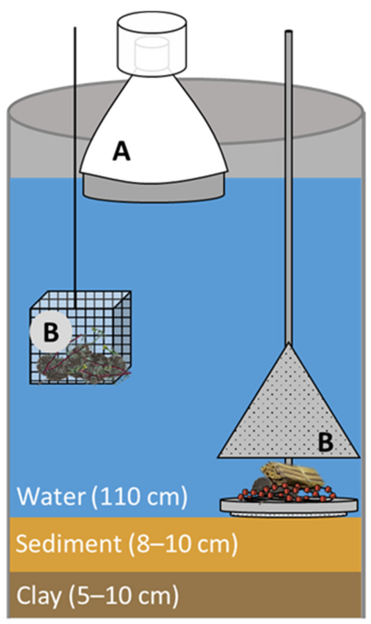
The specimens used in this study derive from a mesocosm experiment, which was conducted in an outdoor enclosure pond system located at the test facility of MESOCOSM GmbH at the Research-Centre Neu-Ulrichstein (FNU) in Homberg (Ohm), Germany (50°45′06.1′′ N 9°02′02.8′′ E). The large pond with a capacity of ~60 m³ was set up in 2018 to allow the establishment of a near-to-nature biocenosis. Both sediment and water originated from a natural pond located at the facility. Chara globularis was planted in the pond to provide oxygen, food, and habitat for the freshly formed organism communities. Five weeks before the start of the actual experiment, stainless steel enclosures were pressed into the clay layer of the pond to form separated but comparable enclosure mesocosm systems. Each replicate system consisted of a 10 cm clay and sediment layer and approximately 1 m3 water (Figure 1). Macrophyte flora (e.g., Chara globularis, Myriophyllum spicatum) in all mesocosms was harmonized thereafter [32].
2.2. Microplastic Application
Spheric polyamide microparticles (Nylon 6, PA 6, Ø 5–50 µm, mean 15–20 µm, Substance No.: PG-29–2020, Batch No.: 300858589, CAS: 25038-54-4) were acquired from Goodfellow Cambridge Limited (Huntingdon PE29 6 WR England). Particle sizes were chosen to be ingestible for planktonic and benthic filter feeders [33,34]. For application, the test substance was suspended in 1 L sieved (63 µm) pond water for 30 min to avoid aggregation and distributed in the mesocosms via a separating funnel. The funnel was rinsed three times with 500 mL sieved pond water to ensure the total transfer of the test substance. This process was performed four times over a span of ten days, applying one-quarter of the total mass of microplastic particles each time. Based on their density (1.13 g cm−3), the particles sunk in the water column. The nominal concentrations of 15 mg L−1 and 150 mg L−1 in each of the three respective replicates were achieved with the fourth application, while five enclosures served as untreated controls. Considering these nominal concentrations in the water and the total area of sediment in each replicate mesocosm, amounts of 16.6 g and 166 g PA per m2 sediment were applied, respectively. The lower concentration was hereby chosen as environmentally relevant [28], while the higher concentration served as the extreme scenario. The application process started in May 2020.
2.3. Macroinvertebrate Sampling
Emerging insects were sampled weekly from May to August 2020, while individuals sampled in late June and early July were used in this study. The emergence traps were stainless steel structures with a conical fabric tent leading the individuals into an eclector head box (Figure 1) filled with tap water and the surfactant Tween (VWR Chemicals) to prevent escaping of the adult insects. All organisms that emerged in a 7-day period were pooled and fixed in ethanol (70%) for further analysis.
To catch the larvae, the mesocosm systems were netted three times in east-west direction (aperture: 27 × 27 cm, mesh size: 450 µm, estimated water volume sampled: 155 L) in September 2020. The organisms were fixed in ethanol (70%) for further analysis. The mesocosms contained larvae of Chaoborus crystallinus and C. obscuripes. Due to their similarity in habitus and behavior, both species were used for further analysis and are henceforth referred to as Chaoborus sp.
2.4. Detection of Microplastics in Chaoborus sp.
For the detection of PA particles, the fluorescent dye Nile Red (Carl Roth) was chosen, which can be used to selectively stain polymers [35,36]. The polymer-specific fluorescence facilitates the differentiation between microplastic particles and organic as well as inorganic substances. It is a faster and more cost-effective method compared to chemical analysis and can be used for the recovery of applied microplastic. It further visualizes the location of ingested particles within the organism, which is a great advantage towards most analytical methods.
In order to identify the PA particles more reliably, the organisms were macerated prior to staining [37]. Maceration of the thorax and abdomen is useful for examining intestinal contents, as it makes it easier to detect microplastic particles. The test organisms were macerated with a 13% KOH solution for 48 h until the thorax and abdomen were transparent. They were then washed with distilled water for ten minutes and fixed with an ethanol series (70–95%) [38].
For staining, Nile Red powder (Carl Roth GmbH) was dissolved in acetone at a concentration of 1 mg L−1 [39] and the fixed animals were stained in it for 2 h.
Chaoborus specimens treated with Nile Red were examined for the presence of PA particles using a Leica DM5500 B fluorescence microscope (excitation filter: BP450/490, dichromatic mirror: DM510, suppression filter: LP515). The number of particles found was documented for each larva and diameters of 120 particles were individually measured using the software ImageJ. A total of 200 larvae were analyzed per concentration, including the control. Additionally, 100 adult specimens were investigated the same way from the highest concentration and the control, respectively. For statistical analysis, the number of particles per larva was compared with the Kruskal−Wallis test, followed by Dunn’s post hoc test with Bonferroni’s correction, using the software “R” with the additional package “PMCMR”.
3. Results
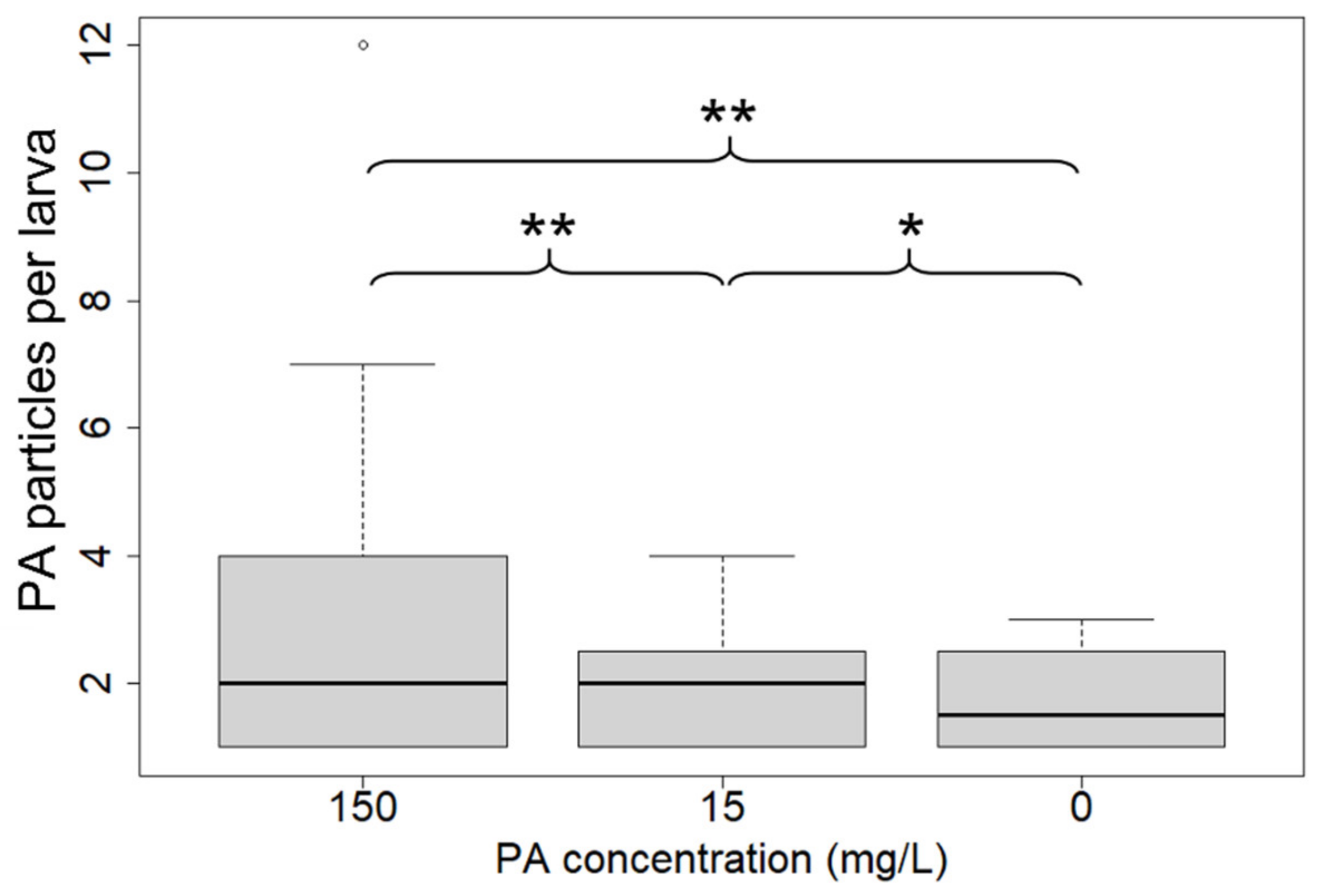
Particles were detected in nearly half of the larvae from mesocosm enclosures with 150 mg L−1 PA and in almost 10% of individuals collected from 15 mg L−1 PA. In the control, four individuals contained plastic particles (Table 1).
Where PA was found, a median of two particles per larva was detected at both 150 mg L−1 and 15 mg L−1 (Figure 2). Significant differences were found among all treatments (p = < 0.05, Kruskal−Wallis test with Dunn’s post hoc test).
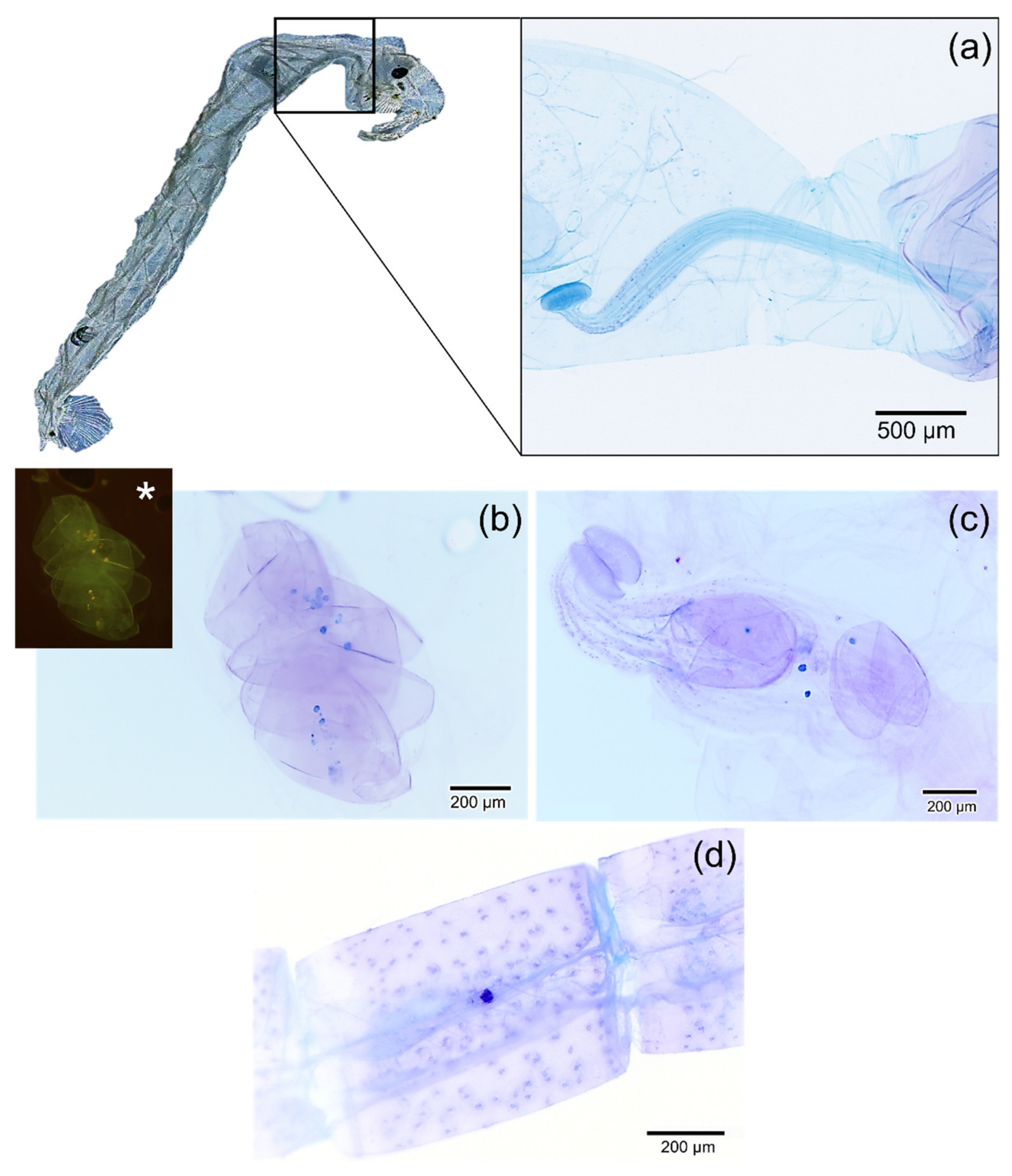
The vast majority of these particles were found in the crop (Figure 3a–c), which was partially distended by the preparation, and only three particles were observed in the intestines of larvae previously exposed to the highest PA concentration. In addition to PA particles, the carapaces of daphnids, ostracods, and possibly other planktonic organisms were visible within the crop. In the control larval samples, in total seven particles in four individuals were identified as plastic within 200 larvae.
Among the imagines, only one individual was found with two PA particles in its intestine and head, respectively (Figure 3d). No PA was found in adult specimens from the control.
The largest diameter of the particles within the larvae had a median of 24.5 µm (min. 8.7 µm; max. 48.9 µm).
4. Discussion
Most studies on microplastic uptake and effects in freshwater organisms take place under small-scale controlled laboratory conditions [16,40,41] which, although accurate through standardization and replicability, cannot reflect complex environmental situations [21,42]. The samples studied in this work are from a large mesocosm facility, where experiments can be conducted under near-natural field conditions. The advantage of mesocosms over laboratory experiments is that a realistic exposure scenario of pollutants can be created and interactions between trophic levels can be enabled and observed [43]. Despite identical preparation of the mesocosms, strong variance can occur within one experiment [44], which leads to the fact that standardized evaluation methods are indispensable. We detected a total of 7 plastic particles in 200 larvae from the control enclosures. Particle contaminations in an outdoor system can never be completely excluded and optical analysis of microplastics in biota can lead to misidentifications of organic particles for microplastics; hence, such methods are mostly recommended for the recovery of applied particles [45,46], as performed in this study. However, our data and additional statistical analyses show significant differences between the three treatments and indicate an uptake of PA particles by Chaoborus sp. via predation, especially in the highest concentration.
It is likely that microplastics that enter a natural environment do not remain stationary but instead are transported between environmental compartments [47]. In mesocosms, there is an opportunity to get a better picture of the actual fate of microplastics within a community and to study its effects on ecosystems and populations [43]. The PA particles used in this study have a density of 1.13 g cm −1 and therefore sink onto the sediment after application [48]. In the environment, this eventually happens with the vast majority of microplastics, since environmental processes such as biofouling or aggregation lead to the deposition of the particles into the sediment [49,50], especially in areas with low flow velocities [51,52]. However, the detection of PA particles in larvae of Chaoborus sp. indicate that even such high-density particles are available to planktonic filter feeders and consequently to pelagic predators. As hypothesized, the uptake of PA particles increased in treatments with higher plastic concentrations.
A comparison to plastic loads in the field is difficult, due to variation in sampling and reported units [53,54]. Although the highest microplastic concentrations used in this study (150 mg L−1) were most likely one or two magnitudes higher than in natural environments [55], the burden of particles was lower than in many laboratory experiments [56]. Using an outdoor model ecosystem allows realistic exposure scenarios and investigation of multiple generations and all life phases, including food consumption, development, and reproduction. Other than in well-mixed beakers in the laboratory, organisms are able to avoid the steadily sinking microplastics but are also exposed to the transmission of particles through the food chain. The planktivorous Chaoborus larvae likely took up filter feeding and grazing organisms (daphnids and ostracods), which are unselective feeders. They filter the suspended particles and graze biofilms, which are in direct contact with sunken particles [57]. Under the microscope, the carapaces of these organisms were clearly visible in the crops of Chaoborus larvae, and a large proportion of the particles appeared to have remained within the prey at the time of sampling (Figure 3b). Our findings offer an important verification of various laboratory studies that observed uptake of microplastics by zooplankton [33,34] and could in conclusion confirm a transferability of its effects into natural scenarios.
Chaoborus sp. actively hunt zooplankton and ingest their prey whole. The initial digestion then takes place in the crop, which is large and muscular (Figure 3a). The entry into the esophagus, however, is blocked by lamellae and only allows entry for liquid components [58]. Solid components, such as the carapaces of the prey, are regurgitated. Here, we could demonstrate that although Chaoborus sp. is able to take up significant amounts of PA particles, these pollutants likely only remain within their bodies for a short time, as almost all particles were found in the crop, rather than the intestines. This might also restrict the toxic potential of the particles for this particular organism and their transfer to even higher trophic levels. Unpublished data on population dynamics in the mesocosms shows no effect of PA exposure on the two Chaoborus species [59]. Thus, these predators do not act as sinks for microplastics, and excreted particles remain in the aquatic ecosystem where they continue to have potentially harmful effects on other organisms. Daphnids have been found to be vulnerable to the effects of microplastic pollution [60] and a constant reintroduction of the pollutant could enhance negative effects. An analogous phenomenon was observed concerning parasitic spores that infected daphnids. When infected individuals were eaten by Chaoborus flavicans, the pathogen was not removed from the ecosystem through ingestion but reintroduced through regurgitation, thus reinfecting new daphnids [61]. However, when microplastic is continuously ingested, Chaoborus sp. might still serve as a vector for microplastics into higher trophic levels, since they serve as prey to e.g., fish and dragonflies themselves [31,62].
In addition to the trophic transfer of microplastics through the food chain, ontogenetic transfer of particles was also examined. After examination of 100 imagines of Chaoborus sp., we detected PA particles in two specimens that were exposed to 150 mg L−1 as larvae and sampled right after emergence. Such ontogenetic transfer was observed in other dipteran species, which has raised concern about affecting terrestrial food chains [19,20]. In both studies, only a fraction of the number of particles detected in the larvae could be found in the adult individuals, as was the case in the present study. Interestingly, the pupae of Culex pipiens still contained more polystyrene than adult mosquitos, indicating a loss of microplastic particles during or after emergence [19]. While a transfer of microplastic pollution from the water into terrestrial ecosystems is evidently possible, it may be more relevant for certain taxa of emerging insects than others. The mechanisms behind the ontogenetic transfer of microplastics are not yet sufficiently understood and further research is necessary to assess the potential impacts of microplastic pollution vectored by aquatic insects. However, the present data indicate towards a reintroduction of ingested microplastic particles into the water before and/or during emergence.
Our findings underline the complexity of the interactions between microplastic pollution and aquatic biota. Additional studies on the fate and effects of microplastics on freshwater ecosystems in mesocosms are needed to better assess the complex effects of these substances. Especially long-term effects of microplastics on population level are scarce and realistic exposure scenarios are needed to verify and support the findings from the laboratory.
Author Contributions
Conceptualization, D.N.M.-K., L.K. and F.G.; methodology, D.N.M.-K., A.H. and L.K.; investigation, D.N.M.-K. and A.H.; formal analysis, D.N.M.-K.; writing—original draft preparation, D.N.M.-K.; writing—review and editing, D.M-K., L.K., K.P.E. and F.G.; visualization, D.N.M.-K.; supervision, K.P.E. and F.G.; project administration, K.P.E. and F.G.; funding acquisition, K.P.E. and F.G. All authors have read and agreed to the published version of the manuscript.
Funding
This research was funded by the German Federal Ministry of Education and Research (BMBF) as part of the MikroPlaTaS (Microplastics in Dams and Reservoirs: Sedimentation, Spread, Effects) project (BMBF grant no. 02WPL1448). We acknowledge support from the Open Access Publication Fund of the University of Muenster.
Data Availability Statement
No applicable.
Acknowledgments
We gratefully acknowledge the working group of Paleobotany of the University of Münster (P. Bomfleur and P. Blomenkemper) for providing access to and assisting with the fluorescence microscope, the laboratory staff of the Institute of Landscape Ecology (University of Muenster) for their assistance in the laboratory, as well as M. Kahlke, M. Hahn and M. Bechstein for their assistance in sampling and taxonomic work.
Conflicts of Interest
The authors declare no conflict of interest.
References
- Haegerbaeumer, A.; Mueller, M.-T.; Fueser, H.; Traunspurger, W. Impacts of Micro- and Nano-Sized Plastic Particles on Benthic Invertebrates: A Literature Review and Gap Analysis. Front. Environ. Sci. 2019, 7, 17. [Google Scholar] [CrossRef] [Green Version]
- Wang, Z.; Zhang, Y.; Kang, S.; Yang, L.; Shi, H.; Tripathee, L.; Gao, T. Research progresses of microplastic pollution in freshwater systems. Sci. Total Environ. 2021, 795, 148888. [Google Scholar] [CrossRef] [PubMed]
- Dris, R.; Imhof, H.K.; Löder, M.G.; Gasperi, J.; Laforsch, C.; Tassin, B. Microplastic Contamination in Freshwater Systems: Methodological Challenges, Occurrence and Sources. In Microplastic Contamination in Aquatic Environments: An Emerging Matter of Environmental Urgency; Zeng, E.Y., Ed.; Elsevier: Amsterdam, The Netherlands, 2018; pp. 51–93. ISBN 9780128137475. [Google Scholar]
- Sarijan, S.; Azman, S.; Said, M.I.M.; Jamal, M.H. Microplastics in freshwater ecosystems: A recent review of occurrence, analysis, potential impacts, and research needs. Environ. Sci. Pollut. Res. Int. 2021, 28, 1341–1356. [Google Scholar] [CrossRef] [PubMed]
- Ivleva, N.P.; Wiesheu, A.C.; Niessner, R. Microplastic in Aquatic Ecosystems. Angew. Chem. Int. Ed. Engl. 2017, 56, 1720–1739. [Google Scholar] [CrossRef]
- Scherer, C.; Weber, A.; Lambert, S.; Wagner, M. Interactions of Microplastics with Freshwater Biota. In Freshwater Microplastics: Emerging Environmental Contaminants? Wagner, M., Lambert, S., Eds.; Springer International Publishing: Cham, Switzerland, 2018; pp. 153–180. ISBN 978-3-319-61615-5. [Google Scholar]
- Lebreton, L.C.M.; van der Zwet, J.; Damsteeg, J.-W.; Slat, B.; Andrady, A.; Reisser, J. River plastic emissions to the world’s oceans. Nat. Commun. 2017, 8, 15611. [Google Scholar] [CrossRef]
- Sá, L.C.; de Oliveira, M.; Ribeiro, F.; Rocha, T.L.; Futter, M.N. Studies of the effects of microplastics on aquatic organisms: What do we know and where should we focus our efforts in the future? Sci. Total Environ. 2018, 645, 1029–1039. [Google Scholar] [CrossRef]
- PlasticsEurope. Plastics–the Facts 2018. An Analysis of European Plastics Production, Demand and Waste Data; Plastics Europe AISBL: Bruessels, Belgium, 2018. [Google Scholar]
- Cole, M.; Lindeque, P.; Fileman, E.; Halsband, C.; Goodhead, R.; Moger, J.; Galloway, T.S. Microplastic ingestion by zooplankton. Environ. Sci. Technol. 2013, 47, 6646–6655. [Google Scholar] [CrossRef]
- Scherer, C.; Brennholt, N.; Reifferscheid, G.; Wagner, M. Feeding type and development drive the ingestion of microplastics by freshwater invertebrates. Sci. Rep. 2017, 7, 17006. [Google Scholar] [CrossRef] [Green Version]
- Browne, M.A.; Galloway, T.; Thompson, R. Microplastic—An emerging contaminant of potential concern? Integr. Environ. Assess. Manag. 2007, 3, 559–561. [Google Scholar] [CrossRef]
- Thompson, R.C.; Moore, C.J.; vom Saal, F.S.; Swan, S.H. Plastics, the environment and human health: Current consensus and future trends. Philos. Trans. R. Soc. Lond. B Biol. Sci. 2009, 364, 2153–2166. [Google Scholar] [CrossRef]
- Silva, C.J.M.; Silva, A.L.P.; Gravato, C.; Pestana, J.L.T. Ingestion of small-sized and irregularly shaped polyethylene microplastics affect Chironomus riparius life-history traits. Sci. Total Environ. 2019, 672, 862–868. [Google Scholar] [CrossRef]
- Ziajahromi, S.; Kumar, A.; Neale, P.A.; Leusch, F.D.L. Environmentally relevant concentrations of polyethylene microplastics negatively impact the survival, growth and emergence of sediment-dwelling invertebrates. Environ. Pollut. 2018, 236, 425–431. [Google Scholar] [CrossRef]
- Cuthbert, R.N.; Al-Jaibachi, R.; Dalu, T.; Dick, J.T.A.; Callaghan, A. The influence of microplastics on trophic interaction strengths and oviposition preferences of dipterans. Sci. Total Environ. 2019, 651, 2420–2423. [Google Scholar] [CrossRef] [Green Version]
- Gouin, T. Toward an Improved Understanding of the Ingestion and Trophic Transfer of Microplastic Particles: Critical Review and Implications for Future Research. Environ. Toxicol. Chem. 2020, 39, 1119–1137. [Google Scholar] [CrossRef]
- Provencher, J.F.; Ammendolia, J.; Rochman, C.M.; Mallory, M.L. Assessing plastic debris in aquatic food webs: What we know and don’t know about uptake and trophic transfer. Environ. Rev. 2019, 27, 304–317. [Google Scholar] [CrossRef]
- Al-Jaibachi, R.; Cuthbert, R.N.; Callaghan, A. Examining effects of ontogenic microplastic transference on Culex mosquito mortality and adult weight. Sci. Total Environ. 2019, 651, 871–876. [Google Scholar] [CrossRef] [Green Version]
- Setyorini, L.; Michler-Kozma, D.; Sures, B.; Gabel, F. Transfer and effects of PET microfibers in Chironomus riparius. Sci. Total Environ. 2021, 757, 143735. [Google Scholar] [CrossRef]
- Weis, J.S.; Palmquist, K.H. Reality Check: Experimental Studies on Microplastics Lack Realism. Appl. Sci. 2021, 11, 8529. [Google Scholar] [CrossRef]
- Ockenden, A.; Tremblay, L.A.; Dikareva, N.; Simon, K.S. Towards more ecologically relevant investigations of the impacts of microplastic pollution in freshwater ecosystems. Sci. Total Environ. 2021, 792, 148507. [Google Scholar] [CrossRef]
- EFSA Panel on Plant Protection Products and Their Residues. Guidance on tiered risk assessment for plant protection products for aquatic organisms in edge-of-field surface waters. EFSA J. 2013, 11, 3290. [Google Scholar] [CrossRef]
- Boyle, T.P.; Fairchild, J.F. The role of mesocosm studies in ecological risk analysis. Ecol. Appl. 1997, 7, 1099–1102. [Google Scholar] [CrossRef]
- Caquet, T.; Lagadic, L.; Jonot, O.; Baturo, W.; Kilanda, M.; Simon, P.; Le Bras, S.; Echaubard, M.; Ramade, F. Outdoor experimental ponds (mesocosms) designed for long-term ecotoxicological studies in aquatic environment. Ecotoxicol. Environ. Saf. 1996, 34, 125–133. [Google Scholar] [CrossRef] [PubMed]
- Caquet, T.; Lagadic, L.; Sheffield, S.R. Mesocosms in ecotoxicology (1): Outdoor aquatic systems. Rev. Environ. Contam. Toxicol. 2000, 165, 1–38. [Google Scholar] [CrossRef] [PubMed]
- Redondo-Hasselerharm, P.E.; Gort, G.; Peeters, E.T.H.M.; Koelmans, A.A. Nano- and microplastics affect the composition of freshwater benthic communities in the long term. Sci. Adv. 2020, 6, eaay4054. [Google Scholar] [CrossRef] [Green Version]
- Stanković, J.; Milošević, D.; Jovanović, B.; Savić-Zdravković, D.; Petrović, A.; Raković, M.; Stanković, N.; Stojković Piperac, M. In Situ Effects of a Microplastic Mixture on the Community Structure of Benthic Macroinvertebrates in a Freshwater Pond. Environ. Toxicol. Chem. 2021. [Google Scholar] [CrossRef]
- Berendonk, T.U.; Bonsall, M.B. The phantom midge and a comparison of metapopulation structures. Ecology 2002, 83, 116–128. [Google Scholar] [CrossRef]
- Yan, N.D.; Keller, W.; MacIsaac, H.J.; McEachern, L.J. Regulation of Zooplankton Community Structure of an Acidified Lake by Chaoborus. Ecol. Appl. 1991, 1, 52–65. [Google Scholar] [CrossRef]
- Parma, S. The life cycle of Chaoborus crystallinus, Diptera, Chaoboridae in a Dutch pond. Int. Ver. Theor. Angew. Limnol. 1969, 17, 888–889. [Google Scholar]
- Pickford, D.B.; Finnegan, M.C.; Baxter, L.R.; Böhmer, W.; Hanson, M.L.; Stegger, P.; Hommen, U.; Hoekstra, P.F.; Hamer, M. Response of the mayfly (Cloeon dipterum) to chronic exposure to thiamethoxam in outdoor mesocosms. Environ. Toxicol. Chem. 2018, 37, 1040–1050. [Google Scholar] [CrossRef] [Green Version]
- Imhof, H.K.; Rusek, J.; Thiel, M.; Wolinska, J.; Laforsch, C. Do microplastic particles affect Daphnia magna at the morphological, life history and molecular level? PLoS ONE 2017, 12, e0187590. [Google Scholar] [CrossRef] [Green Version]
- Elizalde-Velázquez, A.; Carcano, A.M.; Crago, J.; Green, M.J.; Shah, S.A.; Cañas-Carrell, J.E. Translocation, trophic transfer, accumulation and depuration of polystyrene microplastics in Daphnia magna and Pimephales promelas. Environ. Pollut. 2020, 259, 113937. [Google Scholar] [CrossRef]
- Sturm, M.T.; Horn, H.; Schuhen, K. The potential of fluorescent dyes-comparative study of Nile red and three derivatives for the detection of microplastics. Anal. Bioanal. Chem. 2021, 413, 1059–1071. [Google Scholar] [CrossRef]
- Shim, W.J.; Song, Y.K.; Hong, S.H.; Jang, M. Identification and quantification of microplastics using Nile Red staining. Mar. Pollut. Bull. 2016, 113, 469–476. [Google Scholar] [CrossRef]
- Jin, Y.; Li, H.; Mahar, R.B.; Wang, Z.; Nie, Y. Combined alkaline and ultrasonic pretreatment of sludge before aerobic digestion. J. Environ. Sci. 2009, 21, 279–284. [Google Scholar] [CrossRef]
- Erdbeer, L.; Kiel, E. Methodische Entwicklung zur Sichtbarmachung von Mikroplastik in Chironomiden. In Proceedings of the Annual Conference of the German Limnological Society (DGL) and the German and Austrian Section of the International Limnological Society, Kamp-Lintfort, Germany, 10–14 September 2018. [Google Scholar]
- Tamminga, M. Nile Red Staining as a Subsidiary Method for Microplastic Quantifica-tion: A Comparison of Three Solvents and Factors Influencing Application Reliability. SDRP J. Earth Sci. Environ. Stud. 2017, 2, 165–172. [Google Scholar] [CrossRef] [Green Version]
- Rehse, S.; Kloas, W.; Zarfl, C. Short-term exposure with high concentrations of pristine microplastic particles leads to immobilisation of Daphnia magna. Chemosphere 2016, 153, 91–99. [Google Scholar] [CrossRef]
- Chae, Y.; Kim, D.; Kim, S.W.; An, Y.-J. Trophic transfer and individual impact of nano-sized polystyrene in a four-species freshwater food chain. Sci. Rep. 2018, 8, 284. [Google Scholar] [CrossRef]
- Aljaibachi, R.; Laird, W.B.; Stevens, F.; Callaghan, A. Impacts of polystyrene microplastics on Daphnia magna: A laboratory and a mesocosm study. Sci. Total Environ. 2020, 705, 135800. [Google Scholar] [CrossRef]
- Setälä, O.; Norkko, J.; Lehtiniemi, M. Feeding type affects microplastic ingestion in a coastal invertebrate community. Mar. Pollut. Bull. 2016, 102, 95–101. [Google Scholar] [CrossRef]
- Caquet, T.; Lagadic, L.; Monod, G.; Lacaze, J.C.; Couté, A. Variability of physicochemical and biological parameters between replicated outdoor freshwater lentic mesocosms. Ecotoxicology 2001, 10, 51–66. [Google Scholar] [CrossRef]
- Maes, T.; Jessop, R.; Wellner, N.; Haupt, K.; Mayes, A.G. A rapid-screening approach to detect and quantify microplastics based on fluorescent tagging with Nile Red. Sci. Rep. 2017, 7, 44501. [Google Scholar] [CrossRef] [PubMed] [Green Version]
- Baruah, A.; Sharma, A.; Sharma, S.; Nagraik, R. An insight into different microplastic detection methods. Int. J. Environ. Sci. Technol. 2021, 1–10. [Google Scholar] [CrossRef]
- Wagner, M.; Lambert, S. (Eds.) Freshwater Microplastics: Emerging Environmental Contaminants? Springer International Publishing: Cham, Switzerland, 2018; ISBN 978-3-319-61615-5. [Google Scholar]
- Hidalgo-Ruz, V.; Gutow, L.; Thompson, R.C.; Thiel, M. Microplastics in the marine environment: A review of the methods used for identification and quantification. Environ. Sci. Technol. 2012, 46, 3060–3075. [Google Scholar] [CrossRef] [PubMed]
- Kaiser, D.; Kowalski, N.; Waniek, J.J. Effects of biofouling on the sinking behavior of microplastics. Environ. Res. Lett. 2017, 12, 124003. [Google Scholar] [CrossRef] [Green Version]
- Lagarde, F.; Olivier, O.; Zanella, M.; Daniel, P.; Hiard, S.; Caruso, A. Microplastic interactions with freshwater microalgae: Hetero-aggregation and changes in plastic density appear strongly dependent on polymer type. Environ. Pollut. 2016, 215, 331–339. [Google Scholar] [CrossRef]
- Leiser, R.; Wu, G.-M.; Neu, T.R.; Wendt-Potthoff, K. Biofouling, metal sorption and aggregation are related to sinking of microplastics in a stratified reservoir. Water Res. 2020, 176, 115748. [Google Scholar] [CrossRef]
- Hübner, M.K.; Michler-Kozma, D.N.; Gabel, F. Microplastic concentrations at the water surface are reduced by decreasing flow velocities caused by a reservoir. Fundam. Appl. Limnol. 2020, 194, 49–56. [Google Scholar] [CrossRef]
- Lindeque, P.K.; Cole, M.; Coppock, R.L.; Lewis, C.N.; Miller, R.Z.; Watts, A.J.R.; Wilson-McNeal, A.; Wright, S.L.; Galloway, T.S. Are we underestimating microplastic abundance in the marine environment? A comparison of microplastic capture with nets of different mesh-size. Environ. Pollut. 2020, 265, 114721. [Google Scholar] [CrossRef]
- Rios Mendoza, L.M.; Balcer, M. Microplastics in freshwater environments: A review of quantification assessment. TrAC Trends Anal. Chem. 2019, 113, 402–408. [Google Scholar] [CrossRef]
- Horton, A.A.; Walton, A.; Spurgeon, D.J.; Lahive, E.; Svendsen, C. Microplastics in freshwater and terrestrial environments: Evaluating the current understanding to identify the knowledge gaps and future research priorities. Sci. Total Environ. 2017, 586, 127–141. [Google Scholar] [CrossRef] [Green Version]
- Bucci, K.; Tulio, M.; Rochman, C.M. What is known and unknown about the effects of plastic pollution: A meta-analysis and systematic review. Ecol. Appl. 2020, 30, e02044. [Google Scholar] [CrossRef]
- Meisch, C. Crustacea: Ostracoda; Schwoerbel, J., Zwick, P., Eds.; Spektrum Akad. Verl.: Heidelberg, Germany, 2000; pp. 1–522. ISBN 9783827410016. [Google Scholar]
- Gersch, M. Experimentelle Untersuchungen über den Verdauungstraktus der Larve von Chaoborus (Corethra). Z. Vgl. Physiol. 1952, 34, 346–369. [Google Scholar] [CrossRef]
- Kruckenfellner, L.; Ebke, K.P. Institut für Gewässerschutz Mesocosm GmbH, Homberg (Ohm), Germany. 2022; manuscript in preparation. [Google Scholar]
- Bosker, T.; Olthof, G.; Vijver, M.G.; Baas, J.; Barmentlo, S.H. Significant decline of Daphnia magna population biomass due to microplastic exposure. Environ. Pollut. 2019, 250, 669–675. [Google Scholar] [CrossRef] [Green Version]
- Cáceres, C.E.; Knight, C.J.; Hall, S.R. Predator-spreaders: Predation can enhance parasite success in a planktonic host-parasite system. Ecology 2009, 90, 2850–2858. [Google Scholar] [CrossRef] [Green Version]
- Von Ende, C.N. Fish Predation, Interspecific Predation, and the Distribution of Two Chaoborus Species. Ecology 1979, 60, 119–128. [Google Scholar] [CrossRef]
Figure 1.
Schematic cross section through one of 11 mesocosm enclosures with eclectors (A) and macroinvertebrate traps (B) filled with macrophytes, leaves and habitat-forming structures.
Figure 1.
Schematic cross section through one of 11 mesocosm enclosures with eclectors (A) and macroinvertebrate traps (B) filled with macrophytes, leaves and habitat-forming structures.

Figure 2.
Number of PA particles in Chaoborus larvae when PA was detected after exposure to 150 mg L−1 (n = 92), 15 mg L−1 (n = 19) and in the control (n = 4). Brackets indicate significant differences (Kruskal−Wallis test with Dunn’s post hoc test performed with complete datasets; n = 200 per treatment. ** = p < 0.01; * = p < 0.05).

Figure 3.
Habitus of Chaoborus larva with enhanced image of empty crop (a). Fluorescence microscopic images of PA particles (blue) in the crops of Chaoborus sp. larvae with zooplankton shells after exposure to 150 mg L−1 PA (b,c) and PA particles in the intestine of Chaoborus sp. imago after exposure to 150 mg L−1 PA (d). Colors are inverted (asterisk shows original image).


Table 1.
Detection of polyamide (PA) microparticles in larvae and imagines of Chaoborus sp. after exposure in freshwater mesocosms.
Table 1.
Detection of polyamide (PA) microparticles in larvae and imagines of Chaoborus sp. after exposure in freshwater mesocosms.
| Larvae | Imagines | ||||
|---|---|---|---|---|---|
| Treatment (mg L−1 PA) | 150 | 15 | 0 | 150 | 0 |
| Sample size per treatment | 200 | 200 | 200 | 100 | 100 |
| Individuals with PA | 92 | 19 | 4 | 1 | 0 |
| N total particles | 245 | 35 | 7 | 2 | 0 |
Publisher’s Note: MDPI stays neutral with regard to jurisdictional claims in published maps and institutional affiliations. |
© 2022 by the authors. Licensee MDPI, Basel, Switzerland. This article is an open access article distributed under the terms and conditions of the Creative Commons Attribution (CC BY) license (https://creativecommons.org/licenses/by/4.0/).
Share and Cite
MDPI and ACS Style
Michler-Kozma, D.N.; Kruckenfellner, L.; Heitkamp, A.; Ebke, K.P.; Gabel, F. Uptake and Transfer of Polyamide Microplastics in a Freshwater Mesocosm Study. Water 2022, 14, 887. https://doi.org/10.3390/w14060887
AMA Style
Michler-Kozma DN, Kruckenfellner L, Heitkamp A, Ebke KP, Gabel F. Uptake and Transfer of Polyamide Microplastics in a Freshwater Mesocosm Study. Water. 2022; 14(6):887. https://doi.org/10.3390/w14060887
Chicago/Turabian StyleMichler-Kozma, Diana Noemi, Lukas Kruckenfellner, Anna Heitkamp, Klaus Peter Ebke, and Friederike Gabel. 2022. "Uptake and Transfer of Polyamide Microplastics in a Freshwater Mesocosm Study" Water 14, no. 6: 887. https://doi.org/10.3390/w14060887
Note that from the first issue of 2016, this journal uses article numbers instead of page numbers. See further details here.