Article

Application of BiVO4–Microalgae Combined Treatment to Remove High Concentration Mixture of Sulfamethazine and Sulfadiazine

1
Centre for Research on Environmental Ecology and Fish Nutrient, Ministry of Agriculture, Shanghai Ocean University, Shanghai 201306, China
2
Key Laboratory of Exploration and Utilization of Aquatic Genetic Resources, Ministry of Education, Shanghai Ocean University, Shanghai 201306, China
3
Department of Civil and Environmental Engineering, Faculty of Science and Technology, University of Macau, Taipa, Macau 999078, China
4
Shanghai National Engineering Research Center for Urban Water Resources Co., Ltd., Shanghai 200082, China
5
School of Agriculture and Biology, Shanghai Jiao Tong University, Shanghai 200240, China
*
Author to whom correspondence should be addressed.
These authors contributed equally to this work.
Water 2022, 14(5), 718; https://doi.org/10.3390/w14050718
Submission received: 10 January 2022 / Revised: 7 February 2022 / Accepted: 17 February 2022 / Published: 24 February 2022

Abstract

:
Sulfonamides (SAs) are the most common and bio-refractory antibiotics detected in surface water systems, which cause long-term toxic effects on aquatic organisms. This study used the combination of a BiVO4 photocatalyst and freshwater micro-green alga (Dictyosphaerium sp.) to remove sulfadiazine (SD) and sulfamethazine (SM2) at an initial concentration of 5 mg/L (1:1 v/v) for 7 days. We set up three gradient concentrations of BiVO4 (0.5, 1 and 2 g/L) combined with the same concentration (80 mg/L) of Dictyosphaerium sp. and then prepared corresponding concentrations of pure BiVO4 and pure microalgae as controls. We evaluated the ability of BiVO4 and Dictyosphaerium sp. combined technology to remove SAs by observing the removal efficiency of antibiotics and explained the degradation mechanism of antibiotics and the key role of microalgae by studying the changes of reactive oxygen species (ROS) and inorganic ions (nitrogen, sulfur). The results showed that the degradation rate of these two SAs in the 0.5 g/L BiVO4–algae group could reach >96% within 7 d, which was higher than that in the 2 g/L BiVO4 group (93%) and the algae group (28%). The increased degradation efficiency of SAs in BiVO4 and microalgae systems was mainly due to the increased amount of ROS. Meanwhile, more SAs were degraded to inorganic compounds such as NH4+-N, NO3-N and SO42−-S under ROS stress. It was found that microalgae can absorb the degradation products of antibiotics such as NH4+-N for their own growth, thereby reducing the toxicity of antibiotic by-products. In addition, BiVO4 had no damaging effect on the autofluorescence intensity of the microalgae. Our study provides an efficient and eco-economic approach to remove antibiotics using visible-light irradiation in aquatic environments and provides new insights into the biological removal of other antibiotic contaminants in aquatic environments.

1. Introduction

Since they were first discovered in 1929, antibiotics have been frequently detected in surface water, ground water, sediments, and even effluent from wastewater treatment plants [1,2]. Because antibiotics can inhibit the growth of bacteria, fungi, and other microorganisms [3], the widespread use of antibiotics in humans and veterinary medicine has had a major impact on the aquatic environment through domestic excretion, hospital waste, and overuse in agriculture and aquaculture [4]. Sulfonamides (SAs), antibacterial agents that inhibit dihydrofolic acid synthesis, are a major class of antibiotics detected in the aquatic environment [5]. In China, the total concentration of SAs in the Fuzhou section of the Minjiang river basin was 60.9 ng/L [6], and water samples collected from feedlot wastewater pools and irrigation ditches of Shanghai city have shown that the total concentration of SAs was 198–323 μg/L [7]. Although concentrations of contaminated SAs range from ng/L to μg/L, this may increase bacterial resistance and magnify their toxicity via bioaccumulation in the food chain [8]. In light of the non-degradability of SAs and their continuous discharge into surface water, current wastewater treatment plants may not be sufficient to completely remove these compounds [9]. Therefore, we need to develop more effective methods to remove SAs.
Several methods including adsorption, coagulation, and membrane filtration have recently been used to remove antibiotic contaminants [10,11,12]. When compared with conventional removal methods, photocatalytic oxidation has been considered as an environmentally friendly, promising, and efficient alternative to remove antibiotic pollutants [13,14]. Recently, BiVO4 has come into the focus of researchers for its unique properties, such as ferroelasticity [15], ionic conductivity [16], photocatalytic activity for water splitting [17], and ability to degrade harmful pollutants [18]. For instance, pure BiVO4 has been shown to remove 65% of methylene blue (MB) and 90% of ibuprofen under visible-light irradiation [18,19]. BiVO4 is considered an excellent visible-light photocatalyst because it is composed of inexpensive and non-toxic elements [20]. There are three crystal forms of BiVO4: tetragonal zircon, monoclinic scheelite, and tetragonal scheelite. Monoclinic scheelite BiVO4 exhibits a moderate band gap (2.40 eV) and good photocatalytic activity under visible-light irradiation [21]. Despite the advantages of BiVO4, several drawbacks are responsible for the low usage rate of BiVO4 as a photocatalyst. For instance, the photocatalytic activity of BiVO4 depends strongly on its crystalline phase [22] and needs to be improved due to its poor adsorptive performance and high electron-hole recombination rate [23]. Moreover, the preparation method is crucial in terms of the performance of BiVO4. Parameters such as the morphology, crystal plane and surface area of the crystal form are directly dependent on the preparation process [23]. Many methods have been explored, such as doping with metal or non-metal ions, and its combination with metal oxides [24], which may add impurities and increase the overall cost of production [25]. Thus, we need to develop a more cost-effective treatment technique than using pure BiVO4.
Microalgae-mediated bioremediation is attracting a great deal of attention in the scientific community due to its dual role of nutrient uptake and biodegradation/detoxification of hazardous organic pollutants from water bodies, such as phenolics, polyaromatic hydrocarbons and pharmaceuticals [26]. Previous studies have shown that the removal of antibiotics directly caused by microalgae mainly includes bioadsorption, bioaccumulation, and biodegradation [8,27,28]. Microalgae remove antibiotics mainly by breaking down complex toxic compounds into simpler and less toxic products [29]. According to the highest reported degradation rate by other researchers, 32% of sulfamethoxazole can be removed using Scenedesmus obliquus [30] and 29.3% of sulfamethazine (SM2) can be removed using Chlorella pyrenoidosa [31]. Our previous study found that Dictyosphaerium sp. (FACHB-1902) can effectively improve the degradation efficiency of sulfadiazine (SD) and SM2 to 45% and 51%, respectively [32]. Bioremediation has the advantage of low cost; thus, the combination of a photocatalyst and microalgae may be a potential technology for degrading antibiotics.
Our previous studies [32,33] have mainly focused on the removal rate of a single antibiotic using microalgae alone or a combination of BiVO4 and microalgae. However, in this study, the monoclinic scheelite BiVO4 and microalgae Dictyosphaerium sp. were selected, and SD and SM2 mixed SAs were explored for the first time. In our previous study, different antibiotic classes had significantly different removal efficiencies even at the same concentration level [32]. Microalgae have different sensitivities to different antibiotics, and generally many antibiotics exist in the water environment at the same time. Therefore, we chose the mixture of SD and SM2 as the experimental object to observe whether the combined technology of catalyst and microalgae can improve the degradation efficiency of different antibiotics. The main objectives are to (1) evaluate the removal ability of the photocatalyst and microalgae combination technology toward antibiotics based on the removal kinetics of a SD and SM2 mixture and the change in the amount of reactive oxygen species (ROS), and (2) illustrate the degradation mechanism of antibiotics and the key role of microalgae by investigating the changes in the inorganic ion (nitrogen and sulfur) contents in the environment. This study provides a cost-effective way to remove antibiotic mixtures using visible light in an aquatic environment, thereby providing theoretical and technical support for the treatment of environmental pollution caused by multi-antibiotics. Moreover, it may provide new insights in terms of the bio-removal of other antibiotic contaminants in aquatic systems.

2. Materials and Methods

2.1. Chemicals

All the chemicals used in this study were of analytical grade, and special grade chemicals are stated otherwise in the text. SD and SM2 were purchased from Sangon Biotech Co., Ltd. (Shanghai, China) with >98% purity (Table S1). Bi(NO3)3·5H2O and NH4VO3 were purchased from Aladdin (Shanghai, China).

2.2. Algal Culture

The freshwater micro-green algae Dictyosphaerium sp. (FACHB-1902) were purchased from the Institute of the Freshwater Algae Culture Collection of the Institute of Hydrobiology (FACHB-Collection), Wuhan City, China, and was cultured in BG-11 medium [34] within a homeothermic incubator at 25 ± 1 °C under 4000 lux illuminations with a light–dark period of 12:12 h. To retain the exponential growth phase, the algae were aseptically transferred to fresh media every 3–4 d and shaken three times per day [35].

2.3. Experimental Design

All experiments were performed in a homeothermic incubator at 25 ± 1 °C under 4000 lux illuminations with a light–dark period of 12:12 h. The BiVO4 photocatalyst was prepared as per the method described in our previous study [33] (Figure S2). The stock solution of SAs was obtained by dissolving 25 mg of SD and 25 mg of SM2 into 500 mL of BG-11 medium. We used NaOH solution to dissolve SAs, and after the SAs were completely dissolved, we used HCl to adjust the pH of the test medium to 7. The BiVO4 stock suspension was prepared using SAs stock solution. Prior to illumination, the suspension was magnetically stirred in the dark for 1 h to ensure the adsorption/desorption equilibrium of the SAs with the photocatalyst [18]. The final concentrations of BiVO4 prepared in 100 mL of 5 mg/L SAs were 0.5, 1, and 2 g/L. It is necessary to dilute a certain volume of the algae culture with BG-11 medium to achieve an initial algae cell biomass of 80 mg/L. In short, the experiment was conducted in 250 mL Erlenmeyer flasks containing 100 mL of the test solution comprised of SAs, BiVO4, and algae or no algae for 7 d under the same conditions used for the inoculum culture. Three treatment groups (0.5 g/L BiVO4–algae, 1 g/L BiVO4–algae, and 2 g/L BiVO4–algae) and four control groups (algae, 0.5 g/L BiVO4, 1 g/L BiVO4, and 2 g/L BiVO4) were tested in triplicate (n = 3). The flasks were gently shaken, and their positions were randomized and changed every 24 h to ensure the uniform distribution of light [36]. Before the start of the experiment, we carried out algae removal experiments of the two antibiotics under the same experimental conditions, taking the natural degradation of the antibiotics as a control, and found that Dictyosphaerium sp. could improve the removal rates of the two antibiotics (Figure S1).

2.4. Determination of the Antibiotic Concentration

The concentration of antibiotics in the environmental solution was measured every 24 h. The concentrations of SD and SM2 were measured using ultra performance liquid chromatography mass spectrometry (UPLC-MS/MS). Before being used for UPLC-MS/MS analysis, the supernatants of the samples were decanted and filtered through a 0.22 μm polytetrafluoroethylene filter within 7 d. A CORTES UPLC C18 column (100 × 2.1 mm, 1.6 μm) was used for the separation at a constant flow rate of 0.3 mL/min. The auto-sampler draws 2 μL of each sample. The mobile phase was composed of methanol (LCMS-grade) and an aqueous solution containing 0.1% formic acid (LCMS-grade). All experiments used Waters Xevo TQ-S triple-quadrupole mass spectrometry (MS) in electrospray-positive (ESI+) mode and multiple reactive ion monitoring mode for qualitative analysis.
The removal rate (η) of antibiotics in different treatment groups was calculated using the following equation:
η % = C 0 C t C 0 × 100 %
where C0 (mg/L) is the initial concentration of antibiotics at time 0, and Ct (mg/L) is the concentration of antibiotics at time t.

2.5. Determination of the Algal Autofluorescence Intensity

On days 3 and 7 of the experiment, a drop of the algae suspension was taken from the sample with algae and placed on a glass slide, after which the slide was immediately observed and photographed under a confocal laser scanning microscope (CLSM, Leica SP8, Shanghai, China), and the confocal system was controlled using Lasersharp software [37]. The samples were placed on an CLSM platform to focus on the most luminous scanning plane of the chloroplast autofluorescence. The excitation wavelength was 588 nm, the receiving range of the emitted light was set at 411–695 nm, and the pinhole size was 1 AiryUnit. The scanning resolution (frame size) was 1024 × 1024, the scanning speed was 3.15 µs/pixel and the averaging number was 2. The algal autofluorescence intensity was analyzed digitally using Lasersharp software (Bio-Rad Laboratories, Hercules, CA, USA).

2.6. Biochemical Indicators Measurements

2.6.1. Determination of the Superoxide (•O2) and Hydroxyl (•OH)

The concentrations of the •O2 and •OH radicals in the environmental solution were measured every 24 h using an ultraviolet–visible spectrophotometer (Unico UV-2800, Shanghai, China). The contents of •O2 were determined by measuring the nitrite formed from hydroxylamine in the presence of •O2 using the method described in a previous study by our group [38] with some modification. A total of 1.5 mL filtered sample was mixed with 0.5 mL of hydroxylamine hydrochloride (10 mmol/L) and incubated at 25 °C for 20 min. The supernatant (2 mL) was harvested by centrifugation at 3500 rpm for 10 min (Eppendorf, Centrifuge 5804R). Then, p-aminobenzene sulfonic acid (17 mmol/L) and α-naphthylamine (7 mmol/L) were added to the supernatant, which was incubated at 25 °C for 20 min. The absorbance of the aqueous solution at 530 nm was measured. A total of 1.5 mL of the filtered sample was mixed with 1.5 mL of MB solution (40 mol/L) in a centrifuge tube to react for 5 min in order to test the •OH content [39]. The optical density was measured at 660 nm. The concentration of MB consumed in the reaction was the •OH concentration captured by MB.

2.6.2. Determination of the Nitrogen and Sulfur Contents

In order to explore changes in the inorganic ions in the environmental solution, the contents of dissolved inorganic nitrogen and sulfur in the culture medium were measured on days 3 and 7 of the experiment according to the Chinese Water Analysis Methods Standard [40]. Briefly, ammoniacal–nitrogen (NH4+-N) was determined using Nessler’s reagent spectrophotometry, nitrate–nitrogen (NO3-N) was determined using the zinc-cadmium reduction method, and sulfate (SO42−-S) was determined using the barium chromate method.

2.7. Statistical Analysis

The differences in the SAs removal rate, algal autofluorescence intensity, ROS (•O2 and •OH) contents, and inorganic ion (NH4+-N, NO3-N and SO42−-S) contents between the different groups were analyzed using one-way analysis of variance (ANOVA) with IBM SPSS Statistics for Windows (version 18, Chicago, IL, USA). Prior to the one-way ANOVA test, normal distribution of data and homogeneity of variance were checked using Shapiro–Wilk tests and Levene’s test, respectively. Pairwise comparisons were made using the post hoc analysis of Fisher’s least significant difference (LSD) test. The difference was considered significant at p < 0.05. All figures were drawn using Origin 2017 software (OriginLab, Northampton, MA, USA), and the data shown in the figures are mean ± standard deviation.

3. Results

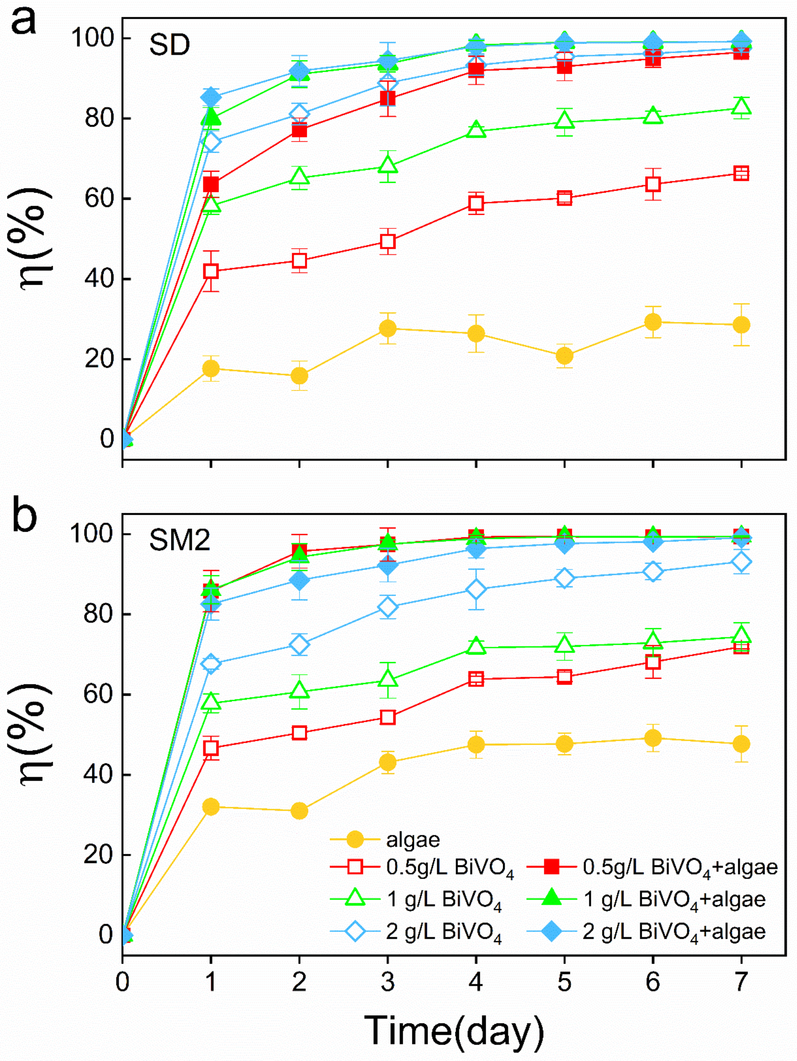
3.1. Synergistic Effect of BiVO4 and Microalgae on the Degradation of SAs

The removal rate of SD and SM2 by the BiVO4 and microalgae systems was investigated for 7 d (Figure 1). For SD, all treatment groups exhibited a relatively high removal rate on day 1, and then slowly increased (Figure 1a). The removal rates of the BiVO4–algae group were significantly higher than those of the algae group by >70% (p < 0.05); when the BiVO4–algae and BiVO4 groups were compared, at the same BiVO4 concentration, the addition of algae can significantly increase the removal rate (p < 0.05); namely, the maximum SD removal rates were from 66% (0.5 g/L BiVO4) to 96.5% (0.5 g/L BiVO4–algae), from 82% (1 g/L BiVO4) to 99.2% (1 g/L BiVO4–algae), and from 97% (2 g/L BiVO4) to 99.2% (2 g/L BiVO4–algae). Evidently, the combined effect of low concentrations (0.5 g/L and 1 g/L) of BiVO4 and microalgae can reach or exceed the effect of a high concentration (2 g/L) of pure BiVO4.
For SM2, the overall removal trend within 7 d was similar to SD (Figure 1b). Unlike SD, the 0.5 g/L BiVO4–algae group exhibited the best SM2 removal effect, whose maximum η value was 99.4%. Moreover, the removal rates observed in the BiVO4–algae group were all higher than those in the BiVO4 group (p < 0.05). In addition, the combined removal effect of SM2 by BiVO4 and microalgae was better than that of SD (Figure 1).

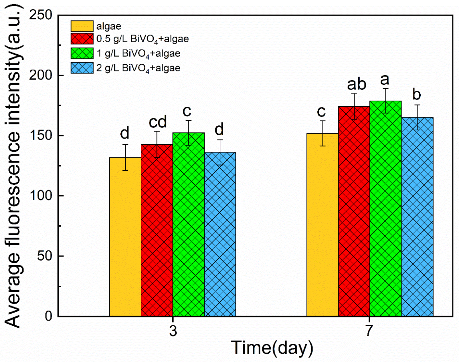
3.2. Effects of BiVO4 on the Algal Photosynthetic System

When compared with the control group (algae), the autofluorescence intensity of chloroplast in Dictyosphaerium sp. increased when exposed to 0.5, 1, and 2 g/L BiVO4 on days 3 and 7 (Figure 2). With the extension of the culture time, the chloroplast autofluorescence intensity of the microalgae was observed to be higher on day 7 when compared to day 3. In the three treatment groups, the autofluorescence intensity of chloroplasts was the lowest at 2 g/L BiVO4.

3.3. Changes in the •O2 and •OH Radical Contents

The •O2 contents in all of the treatment groups (BiVO4–algae) were lower when compared with the control group (algae) and significantly higher than those in the other control group (BiVO4) (p < 0.05) (Figure 3a). In the BiVO4–algae group, the •O2 concentration first decreased and then increased, and as the concentration of BiVO4 increased, the •O2 concentration basically does not change. Similarly, in the BiVO4 group, while increasing the concentration of BiVO4, the concentration of •O2 remained essentially unchanged over 7 d.
When compared to the algae group, the •OH contents in all of the BiVO4–algae groups and BiVO4 group increased (Figure 3b). There was no significant difference in the •OH concentration between these two groups with different concentrations of BiVO4. In addition, two peaks were observed during the experiment, one peak appeared from day 2 to 3, and the other appeared from day 5 to 7. In other words, the •OH concentrations at day 4 were relatively low.

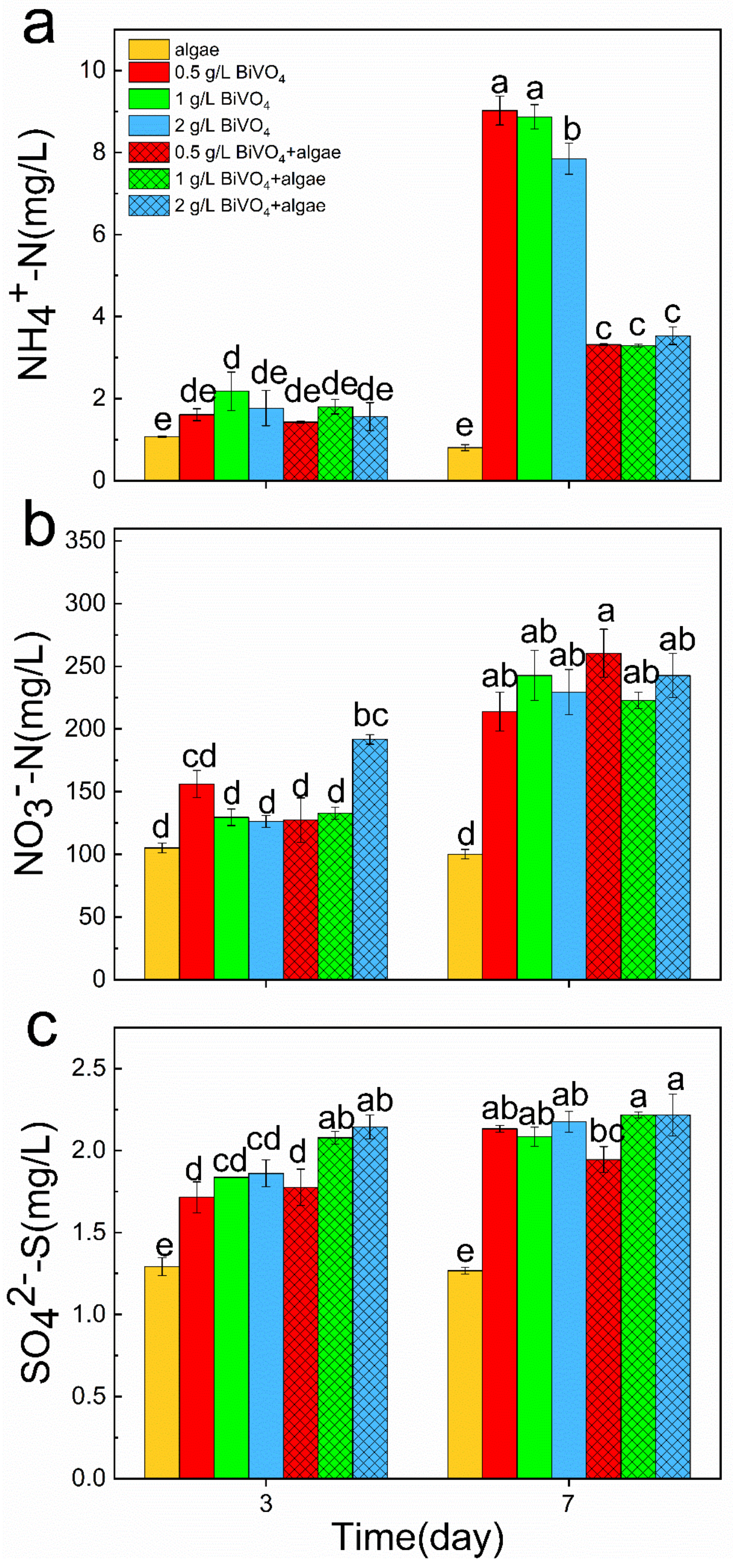
3.4. Changes in the Nitrogen and Sulfur Contents

On days 3 and 7, when compared with the algae group, the NH4+-N concentrations of the BiVO4–algae group increased; when compared with the BiVO4 group, the NH4+-N concentrations of the BiVO4–algae group decreased (Figure 4a). The NH4+-N concentrations of the BiVO4–algae group were significantly lower than those of the BiVO4 group and significantly higher than those of the algae group on day 7. The NH4+-N concentrations of the algae group decreased slightly on day 7 when compared to day 3. However, the concentrations of NH4+-N in the BiVO4 group and BiVO4–algae group were significantly higher on day 7 than on day 3.
The NO3-N contents in all of the BiVO4–algae group increased significantly on days 3 and 7 when compared with the algae group, and the contents on day 7 were significantly higher than those on day 3 (Figure 4b). In addition, on day 7, the highest NO3-N content was observed in the 0.5 g/L BiVO4–algae group. On day 3 or day 7, there was no significant difference in NO3-N contents between the BiVO4–algae group and BiVO4 group. However, the NO3-N contents in the same group were significantly different between these two days.
Figure 4c shows that the SO42−-S contents in solution hardly change in the algae and BiVO4–algae groups on days 3 and 7. However, the SO42−-S contents of the BiVO4 group on day 7 were significantly higher than those on day 3. In addition, from day 3 to 7, the SO42−-S contents changed from a difference to no difference between the BiVO4–algae and BiVO4 groups.

4. Discussion

Generally, the degradation of organic pollutants catalyzed by a semiconductor is an oxidative process using either the direct oxidizing power of photogenerated holes or indirectly via oxidation by ROS [41]. ROS can convert chemically stable organic pollutants into simpler non-toxic products [42]. A photocatalyst can use light as energy to generate strong oxidizing ROS to degrade organic pollutants. For example, BiVO4 mainly triggers the photocatalytic degradation of ibuprofen by generating •O2 and •OH [18]. In this study, the BiVO4 groups exhibit high concentrations of •OH; thus, SAs degradation was achieved through their reaction with •OH, which has also been confirmed by Yan et al. [43] and Wang et al. [44]. However, excessive use of BiVO4 will cause catalyst accumulation [18]. It may be that aggregation reduces the contact area between BiVO4 and light; thus, the ROS contents of the high-concentration (2 g/L) BiVO4 group were lower than those of the low-concentration (0.5 g/L) BiVO4 group (Figure 3).
The BiVO4–algae group can efficiently remove antibiotics not only because BiVO4 can produce ROS, but because the algae can also produce ROS [45]. There are studies showing that microalgae can produce a large amount of ROS under stress, which can oxidatively degrade antibiotics and other pollutants [46,47]. For example, Xue et al. [48] reported that the main oxidation species observed in the degradation of rhodamine B is •O2. In addition, Zhang et al. [49] showed that the main oxidative species observed during the degradation of phenol are holes and •O2. The addition of algae increased the •O2 content and the total amount of ROS in the water column, thereby greatly improving the SAs removal rate. In addition, the main components of the algae extracellular polymer are protein and polysaccharide [50]. Microalgae can absorb pollutants via glycoproteins formed from a combination of protein and polysaccharide in the cell wall to produce the effect of removing antibiotics in water [51]. The removal of antibiotics using algae is an indirect photodegradation process that relies on active oxidants produced by photosensitizers, such as extracellular organic matter (EOM) [52]. Therefore, we speculate that the exposure of antibiotics can stimulate algal cells to continuously secrete ROS, extracellular polymer, and EOM in order to degrade pollutants.
With the addition of BiVO4 and Dictyosphaerium sp., more SAs are degraded into inorganic compounds such as NH4+-N, NO3-N, and SO42−-S under the stress of more ROS. However, NH4+-N is also one of the most important pollutants found in water bodies, which has a significant impact on the community structure of zooplankton [53]; concentrations of >1.2 mg/L will cause Daphnia magna and Daphnia similoides to delay sexual maturation, reduce reproduction rate, and even die [54,55,56]. In this study, the NH4+-N content of the BiVO4 group on day 7 could reach >8 mg/L (Figure 4). It can be seen that the use of photocatalytic materials alone to remove antibiotics may bring secondary hazards. However, when algae were added, the NH4+-N contents in the BiVO4–algae group were significantly lower than those in the BiVO4 group. This may be because algae use NH4+-N as their own nutrient source [57], which can reduce the secondary harm caused by the degradation of SAs. Simultaneously, the NO3-N contents in the treatment group on day 7 were significantly higher than those on day 3, indicating that more antibiotics were degraded into inorganic ions. In addition, the SO42−-S contents in the treatment group showed no significant difference between days 3 and 7, indicating that the microalgae also slowly utilize the SO42−-S degraded by SAs after using the SO42−-S in the BG-11 medium.
In order to explore whether BiVO4 has an adverse effect on algae cells, we measured the changes in the autofluorescence intensity of algae. The autofluorescence intensities of the BiVO4–algae group were higher than those of the algae group (Figure 2), indicating that the growth state of microalgae was not inhibited but improved after adding BiVO4. This may be because when BiVO4 was added, more SAs were degraded into algal nutrients, which accelerated the growth of algae and increased the autofluorescence intensity. Moreover, in our previous studies, it was also demonstrated that glucose metabolism in microalgae can efficiently biodegrade antibiotics in the BiVO4–microalgae system [30]. In addition, we found that the degradation efficiencies of SAs by 0.5 g/L BiVO4–algae were better than those using 2 g/L BiVO4. The preparation cost of BiVO4 is higher than the cultivation cost of microalgae. In this situation, using microalgae to replace parts of the BiVO4 used to remove antibiotics cannot only surpass the effect achieved by high-concentration BiVO4, but it is also economical and environmentally friendly.

5. Conclusions

This study showed that a combined 0.5 g/L BiVO4 and Dictyosphaerium sp. treatment can remove 96% of SD and 99% of SM2, and the removal effect was higher than that of 2 g/L pure BiVO4 and significantly higher than that of pure algae. The increase in the photocatalytic degradation rate of SAs was due to the increase in the contents of •O2 and •OH radicals after the combination of the photocatalyst and microalgae. Moreover, the degradation products of antibiotics (i.e., inorganic ions) can also be absorbed by microalgae to promote their own growth, thereby reducing the toxicity of antibiotic by-products. The economic cost of the low-concentration photocatalyst and microalgae was lower than that of the high-concentration photocatalyst, and the detoxification ability was stronger than that of the algae or high-concentration photocatalyst alone. Therefore, this study has illustrated that under visible light irradiation, the use of a low-concentration photocatalyst and freshwater microalgae for the photocatalytic degradation of SD and SM2 mixtures has application prospects. However, the subsequent recovery process of the photocatalyst and the reuse efficiency of the combination of the photocatalyst and microalgae still require further investigation.

Supplementary Materials

The following supporting information can be downloaded at: https://www.mdpi.com/article/10.3390/w14050718/s1, Figure S1: Effect of algae on the removal of SAs under vis-light irradiation; Figure S2: TEM image of BiVO4; Table S1: Physicochemical characteristics of the test pharmaceuticals.

Author Contributions

Conceptualization, W.L. and S.C.; Formal analysis, W.L., S.C. and H.Z.; Experiment, W.L. and S.C.; Visualization, W.L., S.C. and H.Z.; Writing—original draft, W.L.; Writing—review and editing, W.L., S.C. and W.Z.; Resources, X.W.; Project administration, H.X., L.W. and W.Z.; Supervision, L.W., W.Z. and L.C. All authors have read and agreed to the published version of the manuscript.

Funding

This work was supported by the Chinese Major Science and Technology Program for Water Pollution Control and Treatment (no. 2017ZX07207002-03) and the Science and Technology Commission of Shanghai Municipality (nos. 19050501900 and 19DZ1204504).

Institutional Review Board Statement

Not applicable.

Informed Consent Statement

Not applicable.

Data Availability Statement

Experimental data that support the findings of this study are available from the corresponding author upon request.

Conflicts of Interest

The authors declare no conflict of interest.

References

  1. Cheng, D.; Ngo, H.H.; Guo, W.; Lee, D.; Nghiem, D.L.; Zhang, J.; Liang, S.; Varjani, S.; Wang, J. Performance of microbial fuel cell for treating swine wastewater containing sulfonamide antibiotics. Bioresour. Technol. 2020, 311, 123588. [Google Scholar] [CrossRef] [PubMed]
  2. Zheng, J.; Su, C.; Zhou, J.; Xu, L.; Qian, Y.; Chen, H. Effects and mechanisms of ultraviolet, chlorination, and ozone disinfection on antibiotic resistance genes in secondary effluents of municipal wastewater treatment plants. Chem. Eng. J. 2017, 317, 309–316. [Google Scholar] [CrossRef]
  3. Pariyarath, R.V.; Inagaki, Y.; Sakakibara, Y. Phycoremediation of tetracycline via bio-Fenton process using diatoms. J. Water Process Eng. 2021, 40, 101851. [Google Scholar] [CrossRef]
  4. Kiki, C.; Rashid, A.; Wang, Y.; Li, Y.; Zeng, Q.; Yu, C.-P.; Sun, Q. Dissipation of antibiotics by microalgae: Kinetics, identification of transformation products and pathways. J. Hazard. Mater. 2020, 387, 121985. [Google Scholar] [CrossRef]
  5. Xiong, J.-Q.; Kim, S.-J.; Kurade, M.B.; Govindwar, S.; Abou-Shanab, R.A.I.; Kim, J.-R.; Roh, H.-S.; Khan, M.A.; Jeon, B.-H. Combined effects of sulfamethazine and sulfamethoxazole on a freshwater microalga, Scenedesmus obliquus: Toxicity, biodegradation, and metabolic fate. J. Hazard. Mater. 2019, 370, 138–146. [Google Scholar] [CrossRef]
  6. Li, S.; Shi, W.; Liu, W.; Li, H.; Zhang, W.; Hu, J.; Ke, Y.; Sun, W.; Ni, J. A duodecennial national synthesis of antibiotics in China’s major rivers and seas (2005–2016). Sci. Total Environ. 2018, 615, 906–917. [Google Scholar] [CrossRef]
  7. Ji, X.; Liu, F.; Shen, Q.; Liu, Y. Quantitative detection of sulfonamides and tetracycline antibiotics and resistance genes in sewage farms. Ecol. Environ. Sci. 2011, 20, 927–933. [Google Scholar]
  8. Xiong, J.-Q.; Kurade, M.B.; Jeon, B.-H. Can Microalgae Remove Pharmaceutical Contaminants from Water? Trends Biotechnol. 2018, 36, 30–44. [Google Scholar] [CrossRef]
  9. Al-Ahmad, A.; Daschner, F.; Kummerer, K. Biodegradability of cefotiam, ciprofloxacin, meropenem, penicillin G, and sulfa-methoxazole and inhibition of waste water bacteria. Arch. Environ. Contam. Toxicol. 1999, 37, 158–163. [Google Scholar] [CrossRef]
  10. Cao, Y.; Lei, X.; Chen, Q.; Kang, C.; Li, W.; Liu, B. Enhanced photocatalytic degradation of tetracycline hydrochloride by novel porous hollow cube ZnFe2O4. J. Photochem. Photobiol. A Chem. 2018, 364, 794–800. [Google Scholar] [CrossRef]
  11. Chen, C.; Zeng, H.; Yi, M.; Xiao, G.; Xu, S.; Shen, S.; Feng, B. In-situ growth of Ag3PO4 on calcined Zn-Al layered double hy-droxides for enhanced photocatalytic degradation of tetracycline under simulated solar light irradiation and toxicity assess-ment. Appl. Catal. B. 2019, 252, 47–54. [Google Scholar] [CrossRef]
  12. Solehudin, M.; Sirimahachai, U.; Ali, G.A.; Chong, K.F.; Wongnawa, S. One-pot synthesis of isotype heterojunction g-C3N4-MU photocatalyst for effective tetracycline hydrochloride antibiotic and reactive orange 16 dye removal. Adv. Powder Technol. 2020, 31, 1891–1902. [Google Scholar] [CrossRef]
  13. Raj, C.; Han, L. Advanced oxidation processes for wastewater treatment: Optimization of UV/H2O2 process through a statis-tical technique. Chem. Eng. Sci. 2005, 60, 5305–5311. [Google Scholar]
  14. Gou, J.; Dai, Y.; Liu, L.; Chen, Y.; Zhou, Q. Research Progress on Photocatalytic Oxidation Technology in Environmental Management. Guangdong Chem. Ind. 2019, 46, 85–86. (In Chinese) [Google Scholar]
  15. Lim, A.R.; Choh, S.H.; Jang, M.S. Prominent ferroelastic domain walls in BiVO4 crystal. J. Physics: Condens. Matter 1995, 7, 7309–7323. [Google Scholar] [CrossRef]
  16. Rullens, F.; Laschewsky, A.; Devillers, M. Bulk and Thin Films of Bismuth Vanadates Prepared from Hybrid Materials Made from an Organic Polymer and Inorganic Salts. Chem. Mater. 2006, 18, 771–777. [Google Scholar] [CrossRef]
  17. Khan, I.; Ali, S.; Mansha, M.; Qurashi, A. Sonochemical assisted hydrothermal synthesis of pseudo-flower shaped Bismuth vanadate (BiVO4) and their solar-driven water splitting application. Ultrason. Sonochem. 2017, 36, 386–392. [Google Scholar] [CrossRef]
  18. Li, F.; Kang, Y.; Chen, M.; Liu, G.; Lv, W.; Yao, K.; Chen, P.; Huang, H. Photocatalytic degradation and removal mechanism of ibuprofen via monoclinic BiVO4 under simulated solar light. Chemosphere 2016, 150, 139–144. [Google Scholar] [CrossRef]
  19. Lopes, O.F.; Carvalho, K.T.G.; Macedo, G.K.; de Mendonça, V.R.; Avansi, W.; Ribeiro, C. Synthesis of BiVO4 via oxidant peroxo-method: Insights into the photocatalytic performance and degradation mechanism of pollutants. New J. Chem. 2015, 39, 6231–6237. [Google Scholar] [CrossRef]
  20. Wang, Z.; Huang, X.; Wang, X. Recent progresses in the design of BiVO4-based photocatalysts for efficient solar water splitting. Catal. Today 2019, 335, 31–38. [Google Scholar] [CrossRef]
  21. Tokunaga, S.; Kato, H.; Kudo, A. ChemInform Abstract: Selective Preparation of Monoclinic and Tetragonal BiVO4 with Scheelite Structure and Their Photocatalytic Properties. ChemInform 2002, 33, 46. [Google Scholar] [CrossRef]
  22. Li, H.; Liu, G.; Duan, X. Monoclinic BiVO4 with regular morphologies: Hydrothermal synthesis, characterization and photocatalytic properties. Mater. Chem. Phys. 2009, 115, 9–13. [Google Scholar] [CrossRef]
  23. Gan, J.; Lu, X.; Tong, Y. Towards highly efficient photoanodes: Boosting sunlight-driven semiconductor nanomaterials for water oxidation. Nanoscale 2014, 6, 7142–7164. [Google Scholar] [CrossRef] [PubMed]
  24. Park, Y.; McDonald, K.J.; Choi, K.-S. Progress in bismuth vanadate photoanodes for use in solar water oxidation. Chem. Soc. Rev. 2013, 42, 2321–2337. [Google Scholar] [CrossRef] [PubMed]
  25. Puttaswamy, M.; Malahalli, K.; Tadashi, I.; Kenji, T.; Kazuyoshi, U.; Mineo, S. Hydrothermal synthesis of meso/macroporous BiVO4 hierarchical particles and their photocatalytic degradation properties under visible light irradiation. Environ. Sci. Pollut. Res. Int. 2013, 20, 6638–6645. [Google Scholar]
  26. Xiong, J.-Q.; Kurade, M.; Jeon, B.-H. Biodegradation of levofloxacin by an acclimated freshwater microalga, Chlorella vulgaris. Chem. Eng. J. 2017, 313, 1251–1257. [Google Scholar] [CrossRef]
  27. Leng, L.; Wei, L.; Xiong, Q.; Xu, S.; Li, W.; Lv, S.; Lu, Q.; Wan, L.; Wen, Z.; Zhou, W. Use of microalgae based technology for the removal of antibiotics from wastewater: A review. Chemosphere 2020, 238, 124680. [Google Scholar] [CrossRef]
  28. Mustafa, S.; Bhatti, H.N.; Maqbool, M.; Iqbal, M. Microalgae biosorption, bioaccumulation and biodegradation efficiency for the remediation of wastewater and carbon dioxide mitigation: Prospects, challenges and opportunities. J. Water Process Eng. 2021, 41, 102009. [Google Scholar] [CrossRef]
  29. Tiwari, B.; Sellamuthu, B.; Ouarda, Y.; Drogui, P.; Tyagi, R.D.; Buelna, G. Review on fate and mechanism of removal of pharmaceutical pollutants from wastewater using biological approach. Bioresour. Technol. 2017, 224, 1–12. [Google Scholar] [CrossRef] [Green Version]
  30. Xiong, J.-Q.; Govindwar, S.; Kurade, M.B.; Paeng, K.-J.; Roh, H.-S.; Khan, M.; Jeon, B.-H. Toxicity of sulfamethazine and sulfa-methoxazole and their removal by a green microalga, Scenedesmus obliquus. Chemosphere 2019, 218, 551–558. [Google Scholar] [CrossRef]
  31. Sun, M.; Lin, H.; Guo, W.; Zhao, F.; Li, J. Bioaccumulation and biodegradation of sulfamethazine in Chlorella pyrenoidosa. J. Ocean Univ. China 2017, 16, 1167–1174. [Google Scholar] [CrossRef]
  32. Chen, S.; Li, J.; Feng, W.; Yuan, M.; Zhang, W.; Xu, H.; Zheng, X.; Wang, L. Biochemical responses of the freshwater microalga Dictyosphaerium sp. upon exposure to three sulfonamides. J. Environ. Sci. 2020, 97, 141–148. [Google Scholar] [CrossRef] [PubMed]
  33. Chen, S.; Yuan, M.; Feng, W.; Liu, W.; Zhang, W.; Xu, H.; Zheng, X.; Shen, G.; Guo, C.; Wang, L. Catalytic degradation mechanism of sulfamethazine via photosynergy of monoclinic BiVO4 and microalgae under visible-light irradiation. Water Res. 2020, 185, 116220. [Google Scholar] [CrossRef] [PubMed]
  34. Aderemi, A.O.; Novais, S.C.; Lemos, M.F.; Alves, L.M.; Hunter, C.; Pahl, O. Oxidative stress responses and cellular energy allocation changes in microalgae following exposure to widely used human antibiotics. Aquat. Toxicol. 2018, 203, 130–139. [Google Scholar] [CrossRef] [PubMed] [Green Version]
  35. Rippka, R.; Deruelles, J.; Waterbury, J.B.; Herdman, M.; Stanier, R.Y. Generic Assignments, Strain Histories and Properties of Pure Cultures of Cyanobacteria. Microbiology 1979, 111, 1–61. [Google Scholar] [CrossRef] [Green Version]
  36. Lewis, P.A.; Klemm, D.J.; Lazorchak, J.M.; Norberg-King, T.J.; Peltier, W.H. Short-Term Methods for Estimating the Chronic Toxicity of Effluents and Receiving Waters to Freshwater Organisms, 3rd ed.; EPA: Santa Monica, CA, USA, 1994. [Google Scholar]
  37. Paddock, S.W. Confocal Microscopy: Methods and Protocols; Methods in Molecular Biology; Humana Press: New York, NY, USA, 2014; Volume 122, pp. 1–443. [Google Scholar]
  38. Zhu, L.; Xu, H.; Xiao, W.; Lu, J.; Lu, D.; Chen, X.; Zheng, X.; Jeppesen, E.; Zhang, W.; Wang, L. Ecotoxicological effects of sulfonamide on and its removal by the submerged plant Vallisneria natans (Lour.) Hara. Water Res. 2020, 170, 115354. [Google Scholar] [CrossRef]
  39. Wang, J.; Wang, X.; Guo, W.; Guo, P.; Gu, Z. Photometric Determination of Hydroxyl Free Radical by Its Reaction with Methylene Blue. Phys. Test. Chem. Anal. Part B Chem. Anal. 2007, 43, 495–497. (In Chinese) [Google Scholar]
  40. Editorial Board of the State Environmental Protection Administration. Determination Methods for Examination of Water and Wastewater; China Environmental Science Press: Beijing, China, 2002. [Google Scholar]
  41. Monfort, O.; Plesch, G. Bismuth vanadate-based semiconductor photocatalysts: A short critical review on the efficiency and the mechanism of photodegradation of organic pollutants. Environ. Sci. Pollut. Res. 2018, 25, 19362–19379. [Google Scholar] [CrossRef]
  42. Chueca, J.J.R.; Ormad, M.P.; Mosteo, R.; Canalis, S.; Ovelleiro, J.L. Escherichia coli Inactivation in Fresh Water Through Photocatalysis with TiO2—Effect of H2O2on Disinfection Kinetics. CLEAN Soil Air Water 2016, 44, 515–524. [Google Scholar] [CrossRef]
  43. Yan, J.; Lei, M.; Zhu, L.; Anjum, M.N.; Zou, J.; Tang, H. Degradation of sulfamonomethoxine with Fe3O4 magnetic nanoparticles as heterogeneous activator of persulfate. J. Hazard. Mater. 2011, 186, 1398–1404. [Google Scholar] [CrossRef]
  44. Wang, H.; Li, Y.; Si, Y.; Wu, K.; Zhou, C.; Yousaf, A. Electrocataytic oxidative degradation of sulfonamide in water. Chin. J. Environ. Eng. 2018, 12, 779–787. (In Chinese) [Google Scholar]
  45. Xiong, J.-Q.; Kurade, M.B.; Kim, J.R.; Roh, H.-S.; Jeon, B.-H. Ciprofloxacin toxicity and its co-metabolic removal by a freshwater microalga Chlamydomonas mexicana. J. Hazard. Mater. 2017, 323, 212–219. [Google Scholar] [CrossRef]
  46. Seth, V.; Banerjee, B.D.; Chakravorty, A.K. Lipid Peroxidation, Free Radical Scavenging Enzymes, and Glutathione Redox System in Blood of Rats Exposed to Propoxur. Pestic. Biochem. Physiol. 2001, 71, 133–139. [Google Scholar] [CrossRef]
  47. Tian, Y.; Wei, L.; Yin, Z.; Feng, L.; Zhang, L.; Liu, Y.; Zhang, L. Photosensitization mechanism of algogenic extracellular organic matters (EOMs) in the photo-transformation of chlortetracycline: Role of chemical constituents and structure. Water Res. 2019, 164, 114940. [Google Scholar] [CrossRef]
  48. Xue, S.; He, H.; Wu, Z.; Yu, C.; Fan, Q.; Peng, G.; Yang, K. An interesting Eu, F-codoped BiVO4 microsphere with enhanced photocatalytic performance. J. Alloy. Compd. 2016, 694, 989–997. [Google Scholar] [CrossRef]
  49. Zhang, K.; Liu, Y.; Deng, J.; Xie, S.; Zhao, X. Co-Pd/BiVO4: High-performance photocatalysts for the degradation of phenol under visible light irradiation. Appl. Catal. B. 2018, 224, 350–359. [Google Scholar] [CrossRef]
  50. Zhi, T.; Cheng, L.; Xu, X.; Zhang, L.; Chen, H. Advances on Heavy Metals Removal from Aqueous Solution by Algae. Prog. Chem. 2011, 23, 1782–1794. (In Chinese) [Google Scholar]
  51. Zhang, J.; Fu, D.; Wu, J. Photodegradation of Norfloxacin in aqueous solution containing algae. J. Environ. Sci. 2012, 24, 743–749. [Google Scholar] [CrossRef]
  52. Wei, L.; Li, H.; Lu, J. Algae-induced photodegradation of antibiotics: A review. Environ. Pollut. 2021, 272, 115589. [Google Scholar] [CrossRef]
  53. Yang, J.; Zhang, X.; Xie, Y.; Song, C.; Sun, J.; Zhang, Y.; Giesy, J.P.; Yu, H. Ecogenomics of Zooplankton Community Reveals Ecological Threshold of Ammonia Nitrogen. Environ. Sci. Technol. 2017, 51, 3057–3064. [Google Scholar] [CrossRef]
  54. Arauzo, M. Harmful effects of un-ionised ammonia on the zooplankton community in a deep waste treatment pond. Water Res. 2003, 37, 1048–1054. [Google Scholar] [CrossRef] [Green Version]
  55. Martins, J.; Teles, L.O.; Vasconcelos, V. Assays with Daphnia magna and Danio rerio as alert systems in aquatic toxicology. Environ. Int. 2007, 33, 414–425. [Google Scholar] [CrossRef]
  56. Xiang, F.; Yang, W.; Chen, Y.; Yang, Z. Acute Toxicity of Nitrite and Ammonia to Daphnia similoides of Different Develop-mental Stages: Using the Modified Gaussian Model to Describe. Bull. Environ. Contam. Toxicol. 2010, 84, 708. [Google Scholar] [CrossRef]
  57. Mycielski, M.R. Intensive culture of Chlorella vulgaris/AA as the second stage of biological purification of nitrogen industry wastewaters. Water Res. 1984, 18, 1–7. [Google Scholar]
Figure 1. Effect of the treatment group (BiVO4–algae) and control groups (algae and BiVO4) on the removal of SD (a) and SM2 (b) during the experiment.
Water 14 00718 g001
Figure 2. Effect of the treatment group (BiVO4–algae) and control group (algae) on average autofluorescence intensity of Dictyosphaerium sp. on days 3 and 7 of the experiment. Identical letters denote no significant difference (p < 0.05).
Water 14 00718 g002
Figure 3. Changes of •O2 (a) and •OH (b) contents in the treatment group (BiVO4-algae) and control groups (algae and BiVO4) during the experiment.
Water 14 00718 g003
Figure 4. Changes of NH4+-N (a), NO3-N (b), and SO42−-S (c) contents in the treatment group (BiVO4-algae) and control groups (algae and BiVO4) during the experiment. Identical letters denote no significant difference (p < 0.05).
Water 14 00718 g004
Publisher’s Note: MDPI stays neutral with regard to jurisdictional claims in published maps and institutional affiliations.

