Article

Combined Effects of Polystyrene Nanoplastics and Enrofloxacin on the Life Histories and Gut Microbiota of Daphnia magna

by
Piotr Maszczyk
1,*,
Bartosz Kiersztyn
1,
Sebastiano Gozzo
2,
Grzegorz Kowalczyk
1,
Javier Jimenez-Lamana
2,
Joanna Szpunar
2,
Joanna Pijanowska
1,
Cristina Jines-Muñoz
1,
Marcin Lukasz Zebrowski
1 and
Ewa Babkiewicz
1
1
Department of Hydrobiology, Institute of Functional Biology and Ecology, Faculty of Biology, University of Warsaw, 00-927 Warsaw, Poland
2
IPREM, UMR 5254, E2S UPPA, CNRS, Universite de Pau et des Pays de l’Adour, 64053 Pau, France
*
Author to whom correspondence should be addressed.
Water 2022, 14(21), 3403; https://doi.org/10.3390/w14213403
Submission received: 22 August 2022 / Revised: 22 October 2022 / Accepted: 24 October 2022 / Published: 27 October 2022

Abstract

:
The effect of nanoplastics (NPs) has been shown to interact with the effect of pollutants, including antibiotics. However, little is known about studies performed on freshwater organisms. In this study, we aimed to test the hypothesis that both NPs and antibiotics affect the life history traits of freshwater planktonic Daphnia magna, a model organism in ecotoxicological research, as well as the metabolic and taxonomic fingerprint of their gut microbiota, and whether there is an interaction in the effect of both stressors. To assess this, we experimented with the effect of different spherical polystyrene nanoplastic concentrations and antibiotic enrofloxacin measured through (i) the Daphnia body size and their selected reproductive parameters (the clutch size, egg volume, and total reproductive investment), (ii) the metabolomic diversity of gut microbiota (the respiration rate and the relative use of different carbon sources), and (iii) the microbial taxonomic diversity in the Daphnia intestine. Our results supported the hypothesis as each of the stressors on its own significantly influenced most of the measured parameters, and because there was a significant interaction in the effect of both stressors on all of the measured parameters. Therefore, the results suggest an interactive negative effect of the stressors and a possible link between the observed effects at the different levels of a biological organisation.

1. Introduction

Plastics comprise a wide range of synthetic or semi-synthetic organic compounds, usually polymers with a high molecular mass [1]. For many decades, their production and use has continued to increase worldwide, and many studies reported their increasing presence in natural environments [2,3,4]. Discarded plastic wastes can directly or indirectly enter the environment, and degrade due to solar radiation, mechanical forces, and the biological activity of organisms to smaller-sized pieces, including micro- (MPs, particles smaller than 5 mm) and nanoplastics (NPs, particles smaller than 1 µm, e.g., [5]; or smaller than 0.1 µm, [6]). In addition, primary nanoparticles originating from engineered materials (e.g., personal health care products) can be found. Among the MPs and NPs found in marine and freshwater environments, polystyrene (PS) is one of the most common types [7]. NPs pollution is an issue of special concern because of its unique features that include: (i) their colloidal properties, (ii) their tendency to aggregate, (iii) their high surface area to the volume ratio, resulting in a high ability to absorb and release chemicals, and (iv) the ability to penetrate cell membranes [8]. Because of their small size, adequate quantitative analytical techniques are not currently available to assess the NP concentrations in the environment [9]. While MPs’ presence in marine and freshwater systems has been studied for several years, NPs have been detected in the North Atlantic Gyre only recently [10]. It is assumed that NP concentrations are even 1014 times higher than those currently measured for MPs [6].
Several studies have indicated that many organisms ingest NPs or absorb them on their surfaces, e.g., in [11,12,13], and that because of their small sizes and colloidal properties, they may cross biological barriers [14,15], negatively affecting the organisms. The negative effects depend on the particle type, size, density, charge, and origin (primary or secondary), and this may be related to mechanical (e.g., adhering to external surfaces hindering the mobility) and chemical effects [8]. In addition, chemical harmfulness results from the presence of additives that have the potential to leach into the environment, causing damage to organisms. Among the most common additives are plasticisers, which may affect life history and morphology in Daphnia magna [16], flame retardants that may cause induced significant sublethal chronic toxicity to D. magna [17], antioxidants that may reduce the hatching rates, increase the malformation rates and decrease the length of calcified vertebrae [18], and UV stabilisers that recently have been revealed to cause potential immune dysfunction [19]. Moreover, chemical harmfulness may be due to the particles that can act as a carrier for other co-occurring pollutants, resulting in organisms’ accumulation of harmful hydrophobic substances from the surrounding water [13,15].
The ability of NPs, due to their high surface area to volume ratio, to adsorb, concentrate, and act as a vector of toxic pollutants can modify the environmental impact of the latter. In fact, it has been revealed that most of the combined toxic effects are not simply additive, but rather synergistic or antagonistic [20]. On the other hand, NPs may decrease the toxicity of other pollutants by absorbing and then agglomerating them to form larger particles, reducing the ease of uptake by organisms [21,22,23]. Additionally, their presence may cause an enhancement of toxicity occurring from the on-surface pre-concentration (“The Trojan horse effect”) [22,24]. Although many ecotoxicological studies suggest that realistic environmental concentrations of micro- and nanoplastics may not induce significant detrimental effects on marine organisms nor threaten their survival [13,25], the co-exposure to NPs and other associated contaminants/stressors could exacerbate their effects [26,27,28].
A significant group of such pollutants are antibiotics, which are detectable in surface waters, including rivers, lakes, and seas [13], in the ng L−1 up to µg L−1 range, exceeding sometimes the predicted no-effect environmental concentration [29]. Their extensive and irregular use has induced multifaceted adverse impacts in recent years, such as the propagation of multi-drug-resistant bacteria, antibiotic-resistant bacteria (ARB), and antibiotic-resistant genes (ARGs) in the aquatic environment [30,31]. Their antibacterial impacts are not strain-specific; thus, while the pathogenic bacteria are killed, some bacteria which are beneficial for organisms’ health are also targeted, which may cause several adverse effects, such as an intestinal flora imbalance [32,33]. In addition, different classes of antibiotics have been shown to be toxic to organisms at different trophic levels, such as algae, bacteria, crustaceans, and fish [34,35].
Among the most widely used antibiotics are fluoroquinolones (FQs), which are broad-spectrum synthetic antibiotics commonly used in human and veterinary medicine [36,37] and in agriculture and aquaculture [38]. Among FQs, enrofloxacin is used to prevent and treat a broad spectrum of gram-positive and -negative bacterial infections in livestock. Due to the spread of antibiotic resistance [39], it is listed among the compounds that can be considered to be of a high ecotoxicological concern [40]. It is usually detected in the effluents of municipal sewage plants and the related aquatic environments in the range of ng and μg L−1 [41,42] or even in extreme cases in mg L−1 [43]. Other examples concern their concentrations in surface waters (up to 248 ng L−1 [44]), in groundwater [45], and up to 7.7 mg kg−1 in sediments [46].
Some published studies have investigated the combined effect of NPs and antibiotics on cyanobacteria [47], algae [48], bivalvia [49], and fish [23,24,50,51]. However, there are no reviews yet on other aquatic organisms, including planktonic animals such as D. magna, a keystone species in the food webs of fishless ponds. The combined effect of the stressors may be different for different organisms. It is important to build experimental datasets using a range of different organisms to quantify and predict the factors and mechanisms responsible for the pattern under different contexts. The endpoint of many published studies has focused mainly on the effect at the molecular level: the integrated biomarkers response, antioxidant indexes, gene expression, and histological symptoms [24,52] rather than at the organismal level, e.g., as the combined effect on the life history traits and gut microbiota.
The present work aimed to test several hypotheses concerning the single and combined effects of polystyrene NPs and enrofloxacin on the selected life history traits of D. magna as well as the metabolic and taxonomic diversity of the bacterial community in their intestinal tracts. First, the presence of each of the stressors results in decreasing Daphnia’s body size and reproductive parameters. The effect of enrofloxacin differs in the presence and absence of NPs. Second, as the NPs presence increases, the enrofloxacin presence decreases the metabolic rate of the gut microbiota of Daphnia. Third, the metabolic fingerprint measured as the relative use of various carbon sources is different in the presence of each of the stressors on its own and combined. Finally, those stressors affect Daphnia’s taxonomic diversity in the gut microbiota. On the whole, there is an interaction in the effect of both stressors.

2. Materials and Methods

2.1. Experimental Animals

Three replicates of the experiments were performed. In order to assess the species—rather than the clone-specific effects—each replicate was performed using a different clone of D. magna (MB, MN, and MD of body size at first reproduction 1.86 ± 0.22, 1.82 ± 0.13, and 1.83 ± 0.31 mm, respectively) [53]. Clone MB was sampled from Lake Binnen (54°19′29″ N; 10°37′39″ E, Germany), clone MN from the Nový Rybnik pond (50°13′27.8″ N; 14°4′3.1″ E, Czech Republic), and clone MD from the Domin pond (49°00′21.3″ N; 14°26′29.1″ E, Czech Republic). Daphnia was cultured in 5 L containers, with 25 individuals per container, at room temperature and with a natural photoperiod. The daily food supply was added ad libitum in the amount of 1.6 mg C × L−1 of unicellular green algae, Chlamydomonas klinobasis (strain SAG 56) from a stationary phase, a chemostat culture grown in a WC medium [54]. The algal concentration was assessed using a portable fluorometer (AquaFluor handheld fluorometer, Turner Designs®, San Jose, CA, USA).

2.2. Chemicals

Polystyrene (PS) NPs were synthesised in IPREM, Institut des Sciences Analytiques et de Physico-Chimie pour l’Environnement et les Matériaux, Pau, France, avoiding any additives, especially surfactants, bactericides (e.g., sodium azide), and the trace metals usually present in commercial standards [55,56]. The synthesis and characteristics of soap-free polystyrene models have been detailed elsewhere, e.g., in [55]. Spherical PS NPs were used (diameter of 420 ± 20 nm determined by scanning electron microscopy) with a surface functionalized by carboxylic groups, a low polydispersity (PDI of 0.009), and the zeta potential of −46 mV at a pH value of 7.
Enrofloxacin powder (purity ≥98%) was acquired from Sigma-Aldrich (St. Louis, MO, USA). The stock solution was prepared daily to get 100 µg ml−1 by dissolving the weighted amount in milliQ water in an ultrasonic bath. The stock solution of enrofloxacin was prepared without using organic solvents or buffer solutions to avoid changing the water parameters.

2.3. Experimental System

The system was installed in a room with a constant photoperiod (16 light:8 dark) and comprised of 12 glass containers (L = 25 cm, W = 25 cm, H = 40 cm, large enough to minimise the scale-effects) filled with a 9 L media placed in a water bath (L = 150 cm, W = 50 cm, H = 50 cm, and V = 200 L) with a submersible water-heater (Aquael Neoheather 150 W, Warsaw, Poland) and water pumps (Aquael Circulator 500, Warsaw, Poland) to maintain a stable temperature. The water bath had opaque walls with mounted warm white (3000 K) LED lamps (5.76 Watts, manufacturer ID: FSLEDWW1200-EF, Green Lighting®, Worcester, UK) inside.

2.4. Experimental Design

2.4.1. Experimental Protocol

We performed the experiments at the Hydrobiological field station of the University of Warsaw in Pilchy (https://pilchy.biol.uw.edu.pl/, accessed on 1 June 2020). The samples obtained during the experiments were analysed at the station during and after the end of the experiments to assess the community-level metabolic fingerprinting and the life history parameters of Daphnia. Some analyses (e.g., the assessment of the taxonomic diversity of bacterial communities) were performed in the laboratories of the Department of Hydrobiology, Faculty of Biology, University of Warsaw. We completed the experimental part between May and July 2021 in 12 variants that represent the combination of 3 enrofloxacin concentrations (0, El = 10 and Eh = 100 ng × L−1) and 4 densities of PS-NPs (0, Nl = 1 × 10³, Nm = 1 × 10⁶, and Nh = 1 × 10⁹ particles × L−1). The concentrations used in our study were within the environmental concentration range [41,42,44,45,46]. The concentration 1 × 10⁹ × L−1 of NPs corresponds to the range of bacteria abundance in the lake samples. We fixed the temperature at 23 ± 0.3 °C, which is close to the thermal optimum of D. magna [57], and we supplied the media daily with the same amount of algal food (Ch. klinobasis) set close to the limiting concentration of 0.6 mg Corg × L−1. We calculated the organic carbon content from the calibration curve relating the organic carbon concentration to the absorbance level at 800 nm. We chose a high temperature and meagre food for the experiment to increase the Daphnia filtration rate. The LED lamps inside the water bath provided homogeneity throughout the water column and a low light intensity (1.0 ± 0.4 µmol × m−2 × s−1) measured by a Li-Cor 189 quantum sensor that measures the radiance (LiCor Biosciences), and it was used at a low light intensity. According to the literature, photodegradation is the main cause of the deactivation of fluoroquinolones, including enrofloxacin, in the environment [58].
At the beginning of each experiment, we added tap water to each of the 12 containers (one container per variant), filtered through 0.45 µm pore membrane filters and aerated for 24 h to reach an oxygen concentration up to 8.00 ± 0.08 mg × L−1 (pH = 7.4, µS cm−1 = 373 ± 0.7). The physicochemical parameters (the temperature, conductivity, and oxygen concentration) were determined using a multiparametric YSI 6000 probe (Yellow Spring, YSI Inc., Yellow Springs, OH, USA/Xylem Inc., Washington, DC, USA). We added NPs, enrofloxacin, and algal food in the following order. After, 90 newborn (0–24 h) Daphnia were collected and randomly distributed into all containers to a final density of 10 ind. × L−1 in each variant. Every six hours, the media was gently mixed. The new media was prepared and replaced every 24 h. During the media preparation, we removed the Daphnia from each container using a strainer with a plankton net and placed them temporarily in 250 mL glass containers with the respective media. We prepared the new media in the same order as the initial one. The experiments lasted five days when at least 50% of individuals produced eggs in each variant. At the end of each of the replicates of the experiments, we placed Daphnia temporarily in 250 mL glass containers with the respective media. Then, we photographed all of the individuals in order to determine the life history traits. The photographed individuals were transferred to sterile 100 mL plastic containers with milliQ water to remove non-symbiotic bacteria from their guts. Then, we used randomly selected individuals from each variant to determine the diversity of the bacterial community in Daphnia guts, both metabolic (25 individuals) and taxonomic (10 individuals).

2.4.2. Life History Parameters

We photographed all the individuals collected in each variant from the lateral side under a dissecting microscope connected with the camera and computer. The length and height of each Daphnia were measured in the photographs using the NIS program (Nikon Nis Elements). The length was measured from the top of the eye to the base of the tail spine and the height was measured along the body, starting from its greatest dimension. Based on these measurements, the body volume of each individual was calculated, assuming an ellipsoidal shape for each individual according to the formula 4/3π × 1/2a × 1/2b × 1/2c, where a stands for the length, b the height, and c the width (assuming that the body width is equal to the body height [59]. For each ovigerous female, we counted the eggs, and the volume of an egg (upon the mean dimensions for at least two eggs in the clutch) was calculated using the same formula as that which was employed for body volume evaluation. Finally, the clutch volume as the common currency of the reproductive investment was calculated as the number of eggs multiplied by the mean egg volume for each ovigerous female.

2.4.3. Metabolomic Diversity of Gut Microbiota

The extracted Daphnia guts were homogenised in 1 mL of milliQ water and transferred into 15 mL of their respective media, which were earlier filtered through 0.45 µm Nylon filters and autoclaved. The standard method of a Biolog EcoPlate [60,61] was used to analyse the metabolic diversity of the gut microbiota in these samples. An EcoPlate (Biolog, Hayward, CA, USA) was employed to measure the ability of the bacterial community to utilise the different carbon substrates. An EcoPlate is a 96-well microplate composed of the triplicates of control wells (containing no additional carbon source) and 31 wells containing various carbon sources (Table 1).
Twelve plates were used, one for each variant. Each well of a single plate, except the control one (filled with milliQ water), was filled with 150 μL aliquots of homogenised and the diluted content of Daphnia guts from one variant. We incubated the plates in darkness at a temperature of 22 °C for 72 h. Because of the reduction in the tetrazolium chloride by the electrons which derived mainly from the oxidation chains, the colour in the wells increased the proportionally to the respiration rate. We measured the absorbance every four hours at 590 nm wavelength using a Biotek Synergy H1 plate reader (Biotek Corporation, Broadview, IL, USA). For the analysis, we used the maximal colour development rates (Vmax). In calculating the Vmax values, we used Gen 5 software (Biotec Corporation, Broadview, IL, USA). Additionally, we identified the slope for every four consecutive reads. The Vmax was calculated using a linear regression model by determining the maximum slope during the 72 h incubation time. For the maximal rate of colour development, the Vmax (mOD × min−1) was treated as an indicator of the intensity of the respiration of a single carbon source by the Daphnia gut microbial communities. The difference in the Vmax between the carbon-containing wells and the control wells was calculated to determine the influence of each additional carbon source on the respiration rate (delta). When the delta was lower than zero or equal to zero, we assumed the microorganism community did not use the source, and we set this value of Vmax to zero. For the analyses of the community-level physiological fingerprinting, we used the relative values of the respiration rate for each carbon source, calculated as the percentage share of each carbon source in the sum of the Vmax for the whole plate. The overall microbial activity in each microplate was expressed as the mean Vmax for the plate, and was calculated as the average of all the wells, including the control.

2.4.4. Taxonomic Diversity of Gut Microbiota

The DNA was isolated from the microorganisms, inhabiting the intestinal tract of Daphnia from all replications of the experiment. The isolated DNA was then mixed v/v to obtain a representative sample and was then sequenced. In order to analyse the microbiota, we used a standard Miseq illumine sequencing method used in numerous previous studies, e.g., in [62,63,64,65]. The D. magna, the digestive tracts were collected in 1.5 mL Eppendorf and stored at −20 °C. Afterwards, the DNA extraction was performed in the spin-column-based method using the GeneMATRIX DNA Purification kit (EurX, Gdańsk, Poland), according to the manufacturer’s procedure. The total DNA was assessed for its quality and quantity by the absorbance measurement in a Synergy H1 microplate reader (Gen5 software, BioTek, Broadview, IL, USA) equipped with a Take3 microvolume plate. We then stored the samples at −20 °C for further analysis. We performed the phylogenetic analysis of the bacterial community using Illumina sequencing method [66]. The 16S rRNA genes, V3–V4 hypervariable regions (amplicons of approximately 459 bp), were selected. The PCR amplification was carried out using a Q5 Hot Start High-Fidelity 2X Master Mix, using reaction conditions as recommended by the manufacturer (95 °C for 3 min, 25 cycles of 95 °C for 30 s, 55 °C for 30 s, 72 °C for 30 s, and, after the last cycle, 72 °C for 5 min) with region-specific (341F and 785R) [67] primers that include the Illumina flow cell adapter sequences. The primer sequence was as follows: the forward primer was 5′ CGGGNGGCWGCAG 3′ and the reverse primer was 5′ GACTACHVGGGTATCTAATCC 3′. The Illumina overhang adapter sequences added to the locus-specific sequences were as follows: the forward overhang was 5′ TCGTCGGCAGCGTCAGATGTGTATAAGAGACAG (locus-specific sequence) and the reverse overhang was 5′ GTCTCGTGGGCTCGGAGATGTGTATAAGAGACAG (locus-specific sequence).
The amplicons were sequenced using a MiSeq (Illumina, San Diego, CA, USA) platform on a single run using the MiSeq Reagent Kit v2 (Illumina, San Diego, CA, USA) and the paired-end method (2 × 300 bp), according to the standard protocols by Genomed (Warsaw, Poland). A demultiplexing and trimming of the Illumina adapter sequences (cutadapt software [68]) were performed. FastQC (https://www.bioinformatics.babraham.ac.uk/projects/fastqc; acecssed on 4 October 2018) and MultiQC [69] were used to achieve the quality inspection, visualisation, and assessment of the raw FASTQ files. The sequences were processed using the DADA2 plugin within QIIME 2 [70]. We trimmed the sequence at 270 nt, while the first 8 nt were truncated. The alpha rarefaction plots confirmed that the number of the remaining sequences is sufficient for detecting the current microbial diversity. Taxonomies were assigned to the resulting amplicon sequence variants (ASV) with a q2-feature-classifier plug-in using a pre-trained Naive Bayes classifier based on a 16S rRNA silva 138 SILVA SSU gene database at 99% similarity. The core diversity metrics pipeline was the tool for calculating the phylogenetic and non-phylogenetic core diversity metrics. Data for this purpose were rarefied to a sampling-depth equal to the lowest frequency among the samples (23,500 reads).

2.5. Statistical Analysis

To assess the effect of the NPs and enrofloxacin on the life history parameters the (body length, body volume, clutch size, egg volume, and clutch volume) and on the respiration rate of the gut microbiota of Daphnia, we used an Aligned Rank Transform for a nonparametric factorial two-way ANOVA (ART ANOVA; ARTool package v.0.11.1 [71], with the “art” function (ARTool package 0.11.1 [72] Washington, USA), which allowed us to fit the model despite the non-normality and heteroscedasticity of the initial data distribution. To verify that the ART procedure was correctly applied and is appropriate for this dataset, the “summary()” function was used. To test the differences in the pairwise combinations of levels between the factors in the interactions, the ART-C method (multifactor contrast test) was conducted by using the “art.con” function (ARTool, [73]). The statistical analysis was performed using the R platform (v.4.2.0, R Team, Vienna, Austria) by setting the level of significance at α  =  0.05 for all of the statistics.
A Bray–Curtis-based NMDS (non-metric multidimensional scaling) was applied (PAST3 software, [74]), aiming to group the experimental variants according to the microorganism metabolic and phylogenetic differences. The Bray–Curtis dissimilarity was used because, unlike many other common statistical tools (e.g., Jaccard and unweighted UniFrac), it takes into account not only the number of observed ASVs, but also their relative abundance. For the analyses of community-level physiological fingerprinting, we used the relative values of the respiration rate for each carbon source, calculated as the percentage share of each carbon source in the sum of the Vmax for the whole plate. For taxonomic NMDS, we managed the relative abundance of ASV at the family level. The NMDS analysis and data visualisation were performed in Statistica 13 software (StatSoft, TIBCO Software Inc., Palo Alto, CA, USA).
Additionally, a Mantel correlation between the taxonomic and metabolic profiles was performed (PAST3 software) to reveal the correlation between two Bray–Curtis similarity-based matrices of the relative metabolic and relative phylogenetic data.

3. Results

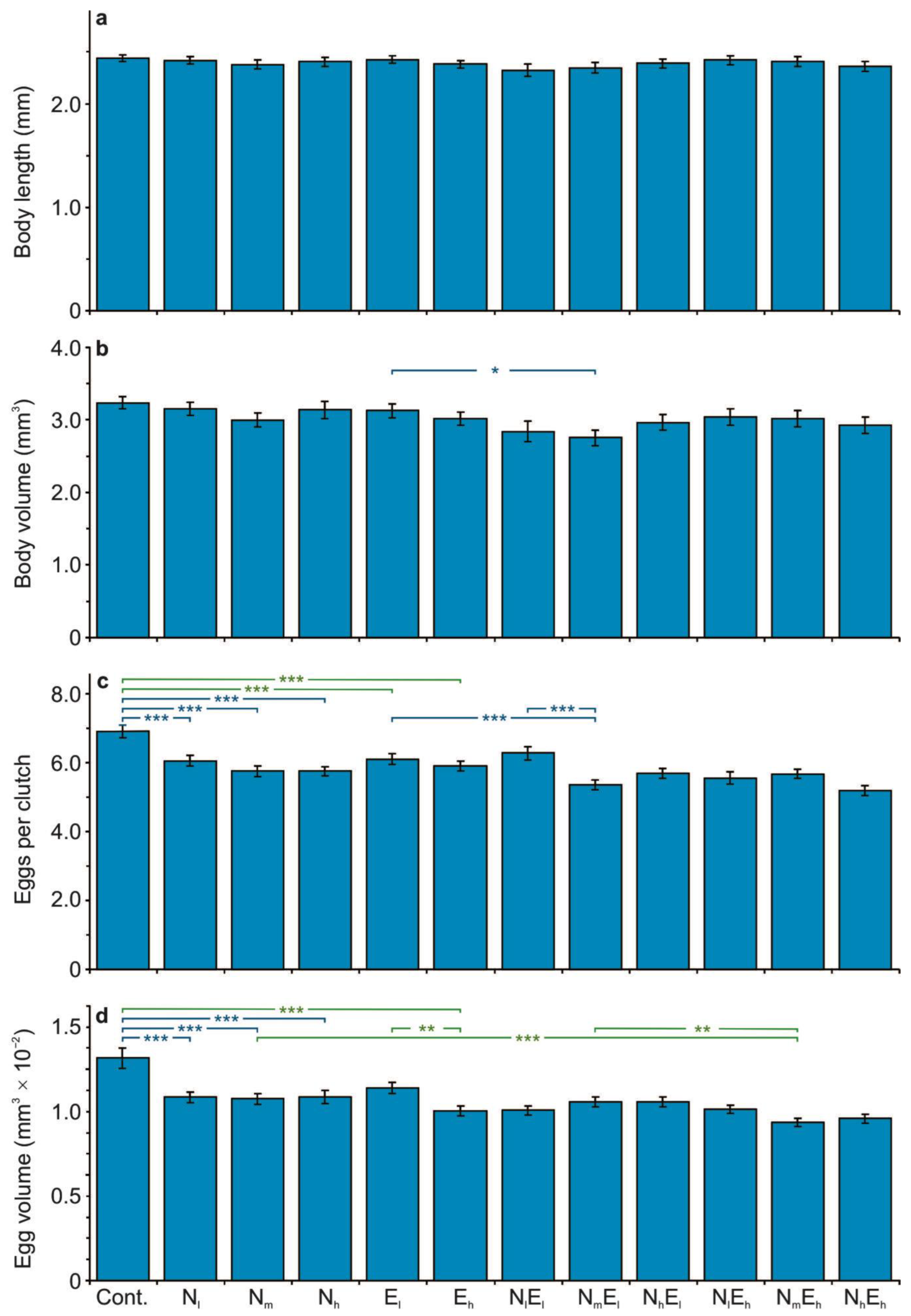
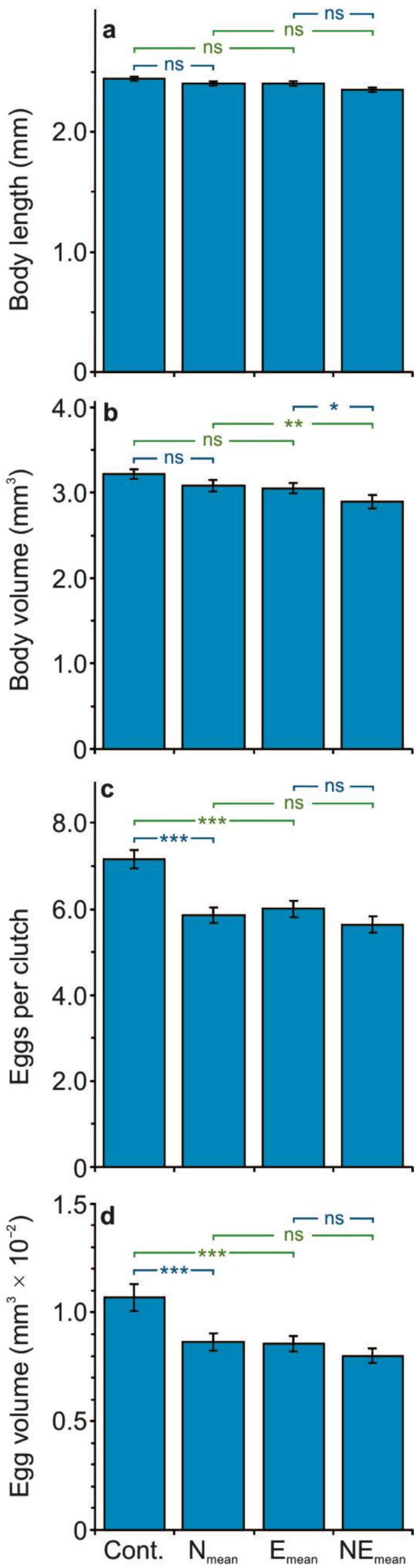
3.1. The Effect of the Stressors on the Life History Parameters

None of the stressors (enrofloxacin and NPs) had a significant effect on the Daphnia body length (Table S1, Supplementary Materials). The interaction between the stressors was also not significant (at p < 0.001, Table S1), that is, the effect of one stressor has not been modified by the effect of another stressor. Neither the effect of a single stressor nor the effect of combined stressors was significant (Figure 1a and Figure 2a), neither for the combined data from all the concentrations of NPs and enrofloxacin (Table S2, Figure 2a), nor for the data from each of the concentrations assessed, separately (Table S3, Figure 1a).
Both stressors significantly affected the Daphnia body volume (at p ≤ 0.001, Table S1). The effect of the NPs was negative, which was apparent in the significant difference between the NEmean and Emean (Table S2, Figure 2b) and between NmEl and El treatments (Table S4, Figure 1b). The effect of enrofloxacin was also negative, which was apparent in the significant difference between the NEmean and Nmean treatments (Table S2, Figure 2b). The interaction between the stressors was also significant (at p = 0.008, Table S1); more specifically, the presence of one stressor resulted in increasing the negative effect of another one (Tables S2 and S4, Figure 1b and Figure 2b). This was apparent: (1) in the significant difference between the NEmean and Emean treatments (at p = 0.031, Table S2, Figure 2b) in comparison to the non-significant difference between the Nmean and control treatments (Table S2, Figure 2b), (2) in the significant difference between the NEmean and Nmean (at p = 0.001, Table S2, Figure 2b) in comparison to the non-significant difference between the Emean and control treatments (Table S2, Figure 2b), and (3) in the significant difference between the NmEl and El treatments (at p ≤ 0.017, Table S4, Figure 1b) in comparison to the non-significant difference between the Nm and control treatments (Table S4, Figure 1b).
The NPs and enrofloxacin significantly affected the clutch size (at p < 0.001, Table S1). The effect of the NPs was negative, which was apparent in the significant difference between: (1) the Nmean and control treatments (at p < 0.001, Table S2, Figure 2c), and (2) each of the concentrations of the NPs and the control, NmEl and El, as well as the NmEl and NlEl treatments (at p ≤ 0.005, Table S5, Figure 1c). The impact of enrofloxacin was also negative, which was apparent in the significant difference between: (1) the Emean and control treatments (at p < 0.001, Table S2, Figure 2c), and (2) each of the two concentrations in relation to the control (at p ≤ 0.005, Table S5, Figure 1c). The interaction between the stressors was also significant (at p < 0.001, Table S1); more specifically, the presence of one stressor resulted in decreasing the negative effect of another one (Tables S2 and S5, Figure 1c and Figure 2c). This was apparent: (1) in the non-significant difference between the NEmean and Emean treatments in comparison to the significant difference between the Nmean and control treatments (at p < 0.001, Table S2, Figure 2c), (2) in the non-significant difference between the NEmean and Nmean treatments in comparison to the significant difference between the Emean and control treatments (at p < 0.001, Table S2, Figure 2c), and (3) in the non-significant differences in the majority of comparisons between the single and combined stressors in relation to the significant difference between the single stressors and control for the data from each of the concentrations of NPs and enrofloxacin, separately (Table S5, Figure 1c). The only exception was the significant differences between the NmEl and El, and between the NmEl and NlEl treatments (Table S5, Figure 1c). The number of ovigerous females in relation to the females without eggs was the greatest in the control (84%), moderate in the presence of NPs on their own (81–83%), and in the presence of enrofloxacin on its own (79–80%), and was the lowest in the NmEl (70%) and NhEh (75%) treatments.
The NPs and enrofloxacin also significantly affected the egg volume (at p < 0.001, Table S1). The effect of the NPs was negative, which was apparent in the significant difference between: (1) the Nmean and control treatments (at p < 0.001, Table S2, Figure 2d), and (2) each of the concentrations of NPs and the control (at p ≤ 0.001, Table S6, Figure 1d). The impact of enrofloxacin was also negative, which was apparent in the significant difference between: (1) the Emean and control treatments (at p < 0.001, Table S2, Figure 2d), and (2) the Eh and the control, NmEh and Nm, Eh and El, as well as the NmEh and NmEl treatments (at p ≤ 0.018, Table S6, Figure 1d). The interaction between the stressors was also significant (at p < 0.001, Table S1); more specifically, the presence of one stressor resulted in decreasing the negative effect of another one (Tables S2 and S6, Figure 1d and Figure 2d). This was apparent: (1) in the non-significant difference between the NEmean and Emean treatments in comparison to the significant difference between the Nmean and control treatments (at p < 0.001, Table S2, Figure 2d), (2) in the non-significant difference between the NEmean and Nmean treatments in comparison to the significant difference between the Emean and control treatments (at p < 0.001, Table S2, Figure 2d), and (3) in the non-significant differences in the majority of comparisons between the single and combined stressors in relation to the significant difference between the single stressors and the control for the data from each of the concentrations of NPs and enrofloxacin, separately (Table S6, Figure 1d).
Additionally, the NPs and enrofloxacin significantly affected the clutch volume (at p ≤ 0.001, Table S1). The effect of the NPs was negative, which was apparent in the significant difference between: (1) the Nmean and control treatments (at p < 0.001, Table S2), and (2) each of the concentrations of the NPs and the control, NmEl and El, NhEl and El, as well as the NmEh and Eh treatments (at p ≤ 0.040, Table S7). The impact of enrofloxacin was also negative, which was apparent in the significant difference between: (1) the Emean and control treatments (at p < 0.001, Table S2), and (2) each of the two concentrations in relation to the control, the NlEh and Nl, NmEh and Nm, and the NhEh and Nh treatments (at p ≤ 0.031, Table S7). The interaction between the stressors was also significant (at p < 0.001, Table S1); more specifically, the presence of one stressor resulted in decreasing the negative effect of another one (Tables S2 and S7). This was apparent: (1) in the non-significant difference between the NEmean and Emean treatments in comparison to the significant difference between the Nmean and control treatments (at p < 0.001, Table S2), (2) in the non-significant difference between the NEmean and Nmean treatments in comparison to the significant difference between the Emean and control treatments (at p < 0.001, Table S2), and (3) in the non-significant differences in the majority of comparisons between the single and combined stressors in relation to the significant difference between the single stressors and the control for the data from each of the concentrations of NPs and enrofloxacin, separately (Table S7).

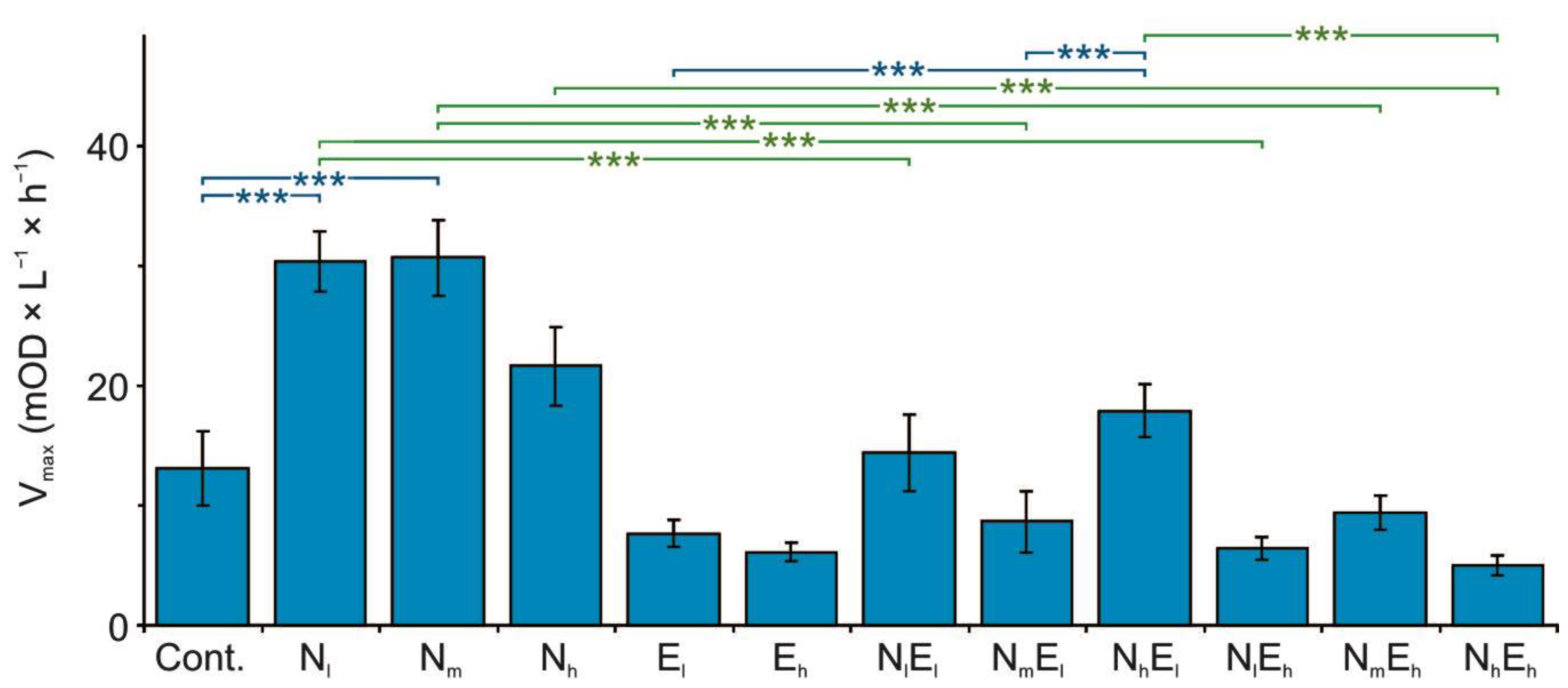
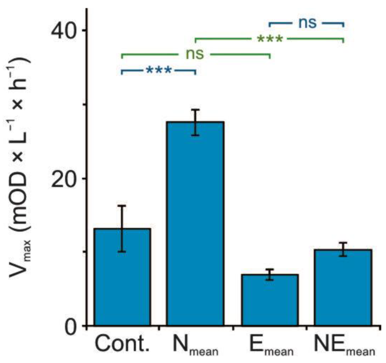
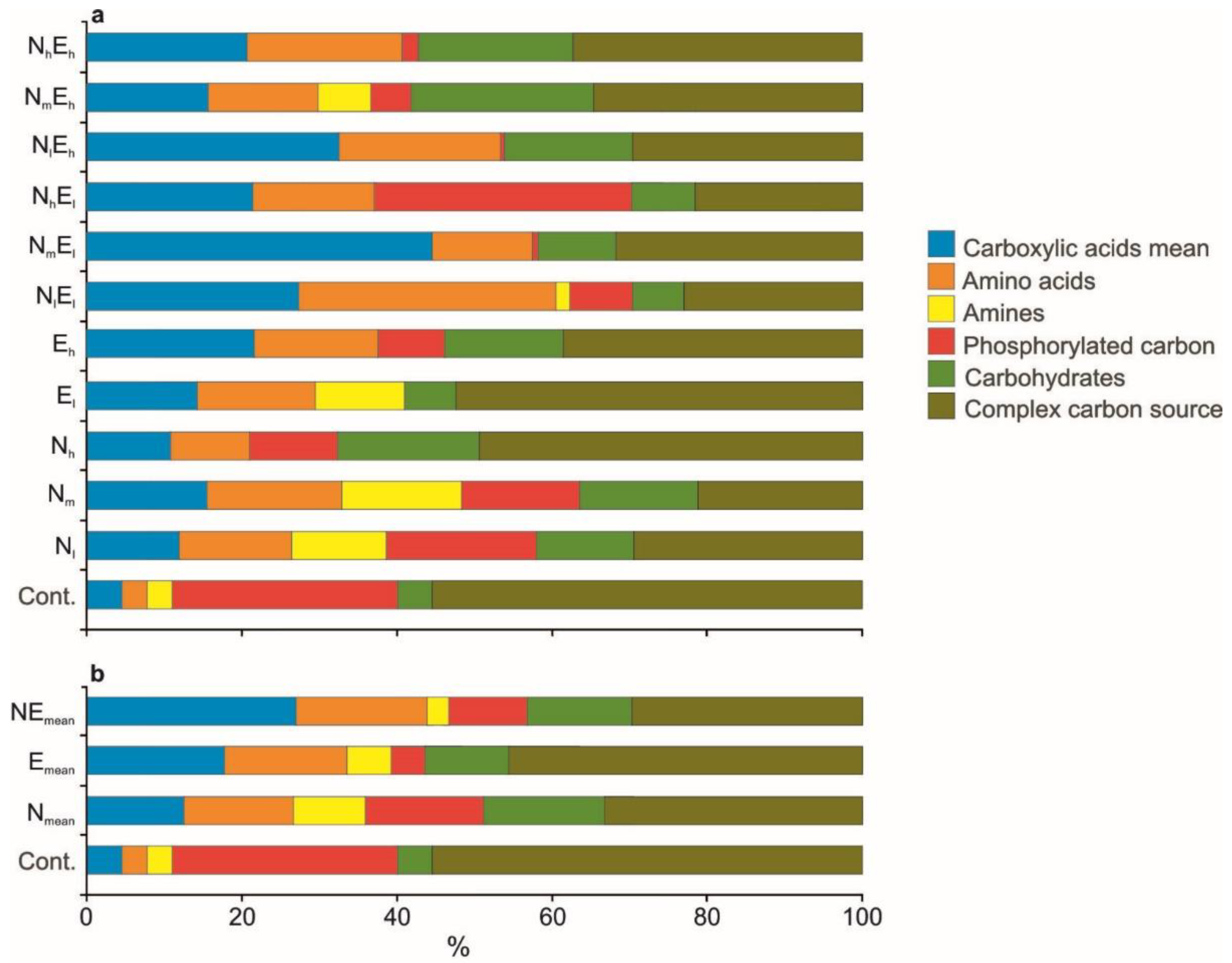
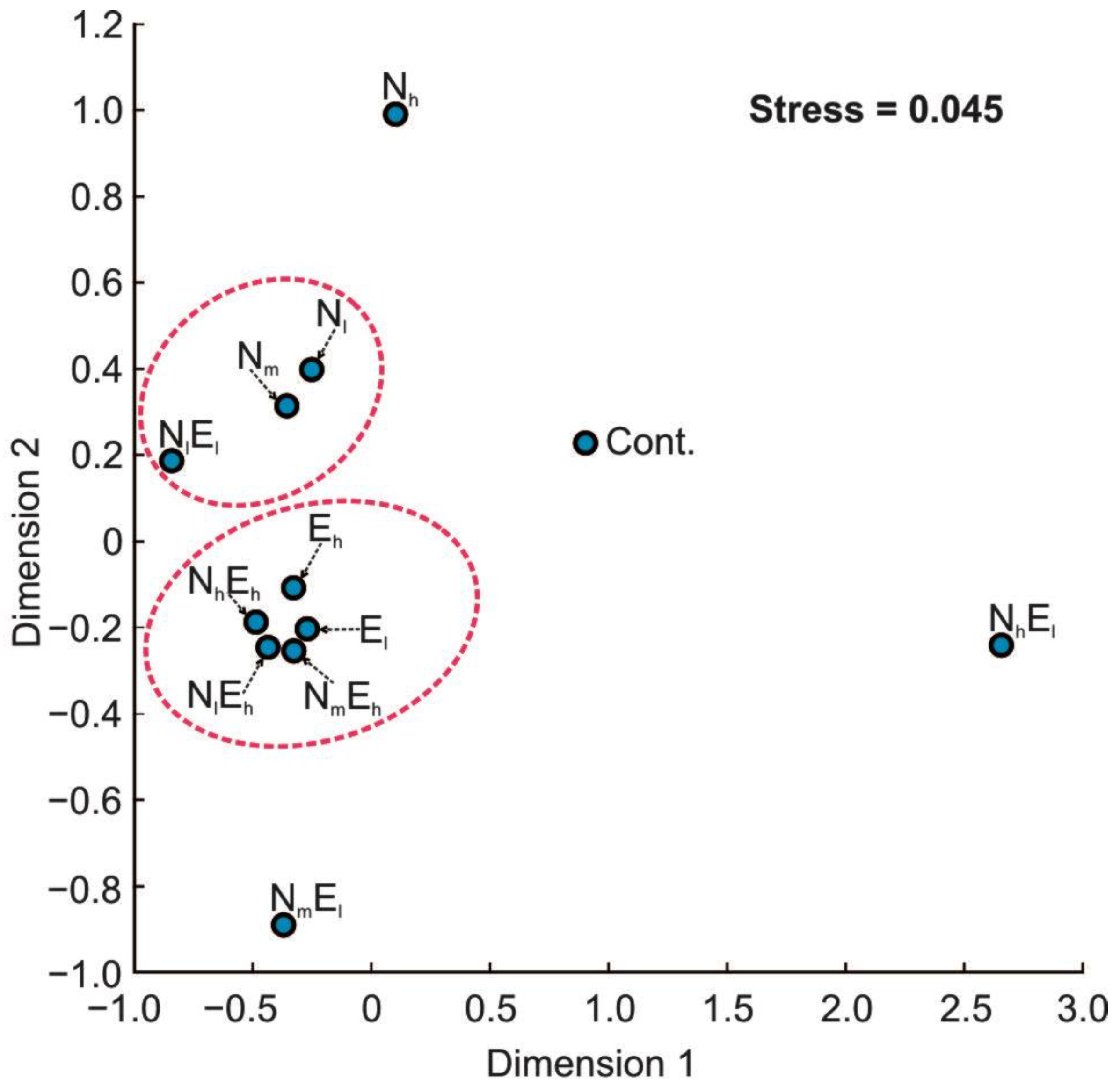
3.2. Metabolomic Diversity of Gut Microbiota

The NPs and enrofloxacin significantly affected the mean respiration rate of the gut microbiota expressed as the Vmax values (at p < 0.001, Table S1). The effect of the NPs was positive (Tables S2 and S8, Figure 3 and Figure 4), which was apparent in the significant difference between: (1) the Nmean and control treatments (at p < 0.001, Table S2, Figure 4), and (2) the Nl and control, Nm and control, NhEl and El, as well as the NhEl and NmEl treatments (at p ≤ 0.002, Table S8, Figure 3). The impact of enrofloxacin was negative, which was apparent in the significant difference between: (1) the NEmean and Nmean treatments (at p < 0.001, Table S2, Figure 4), and (2) the NlEl and Nl, NlEh and Nl, NmEl and Nm, NmEh and Nm, NhEh and Nh, as well as the NhEh and NhEl treatments (at p ≤ 0.001, Table S8, Figure 3). The interaction between the stressors was significant (at p < 0.001, Table S1), more specifically in the majority of comparisons, the presence of NPs increased the inhibitory effect of enrofloxacin, and the presence of enrofloxacin reduced the positive effect of the NPs (Tables S2 and S8, Figure 3 and Figure 4). The former was apparent: (1) in the non-significant difference between the Emean and control in comparison to the significant difference between the NEmean and Nmean treatments (at p < 0.001, Table S2, Figure 4), and (2) in the non-significant difference between the El and control and the Eh and control in relation to the significant difference in the majority of comparisons (in six among nine) between the treatments in which enrofloxacin was combined with the NPs (at p < 0.001, Table S8, Figure 3). The latter was apparent (1) in the significant difference between the Nmean and the control (at p < 0.001, Table S2, Figure 4) in comparison to the non-significant difference between the NEmean and Emean treatments (Table S2, Figure 4), and (2) in the significant difference between the Nl and the control and the Nm and the control (at p < 0.001, Table S8, Figure 3) in relation to the non-significant difference in the majority of comparisons (in 10 among 12) between the treatments in which NPs were combined with enrofloxacin (Table S8, Figure 3).
The analysis of the percentage share of the respiration rate of different carbon sources by the gut microbiota of Daphnia revealed that the presence of each of the stressors resulted in a relative increase in the usage of carboxylic acids, amino acids, and carbohydrates, as well as a relative decrease in the usage of phosphorylated carbons and complex carbon sources with the control (Figure 5a,b). The pattern was similar in the presence of single and combined stressors, which suggests a negative interaction between their effects.
A Bray–Curtis-based NMDS analysis revealed two distinct groups of variants: variants with a high concentration of enrofloxacin (in the presence and absence of NPs) and variants with a low and medium density of NPs, which suggests a different effect of each of the stressors on the metabolic profile of the gut microbial community (Figure 6). In the first group, a relatively low usage of phosphorylated carbon and amines compared to the control and the majority of the remaining variants was observed (Figure 5a). In the second group, there was relatively even usage of the different carbon sources with a relatively low usage of the complex carbon sources, as well as a relatively high usage of amines concerning the majority of the remaining variants (Figure 5a).

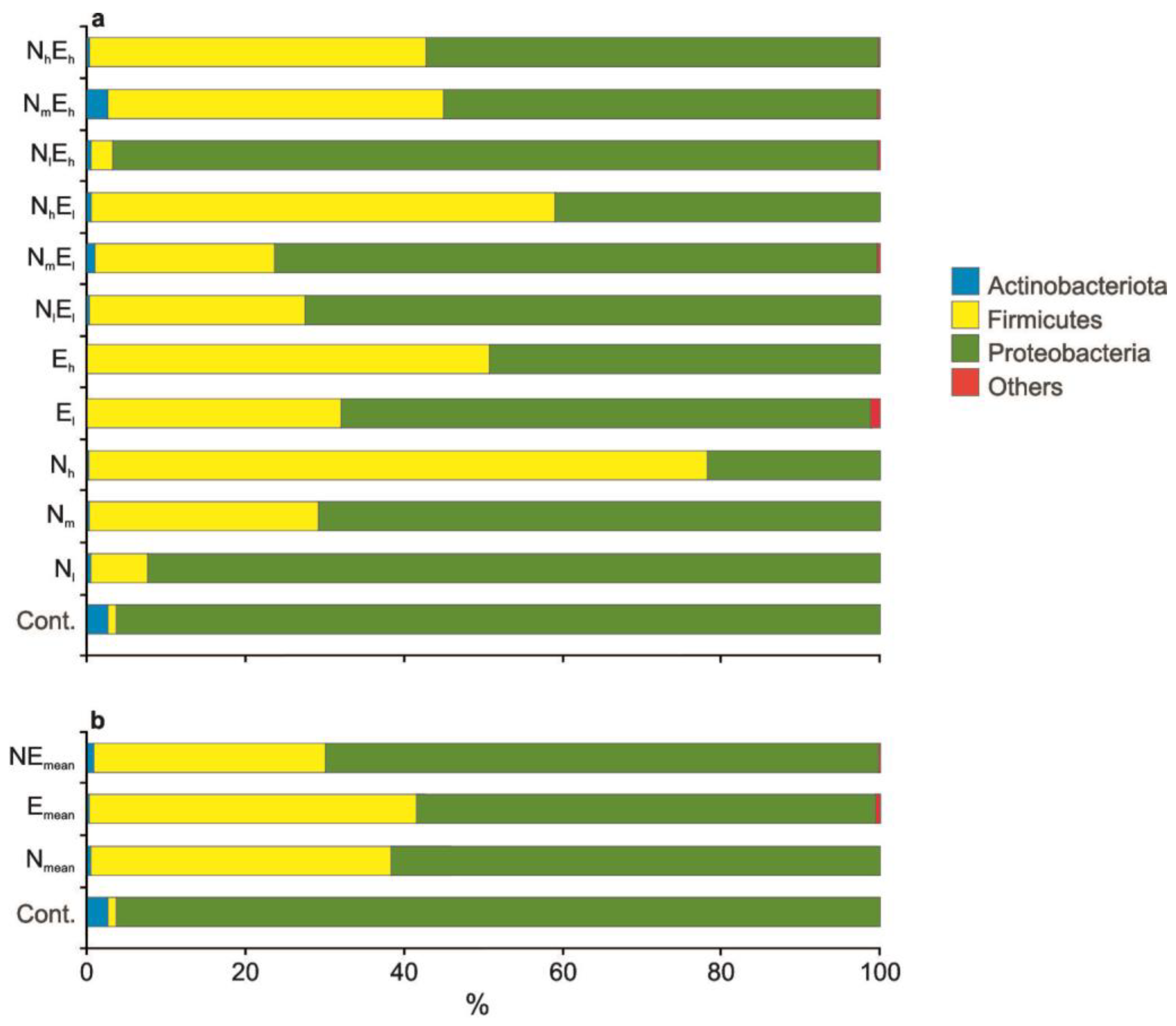
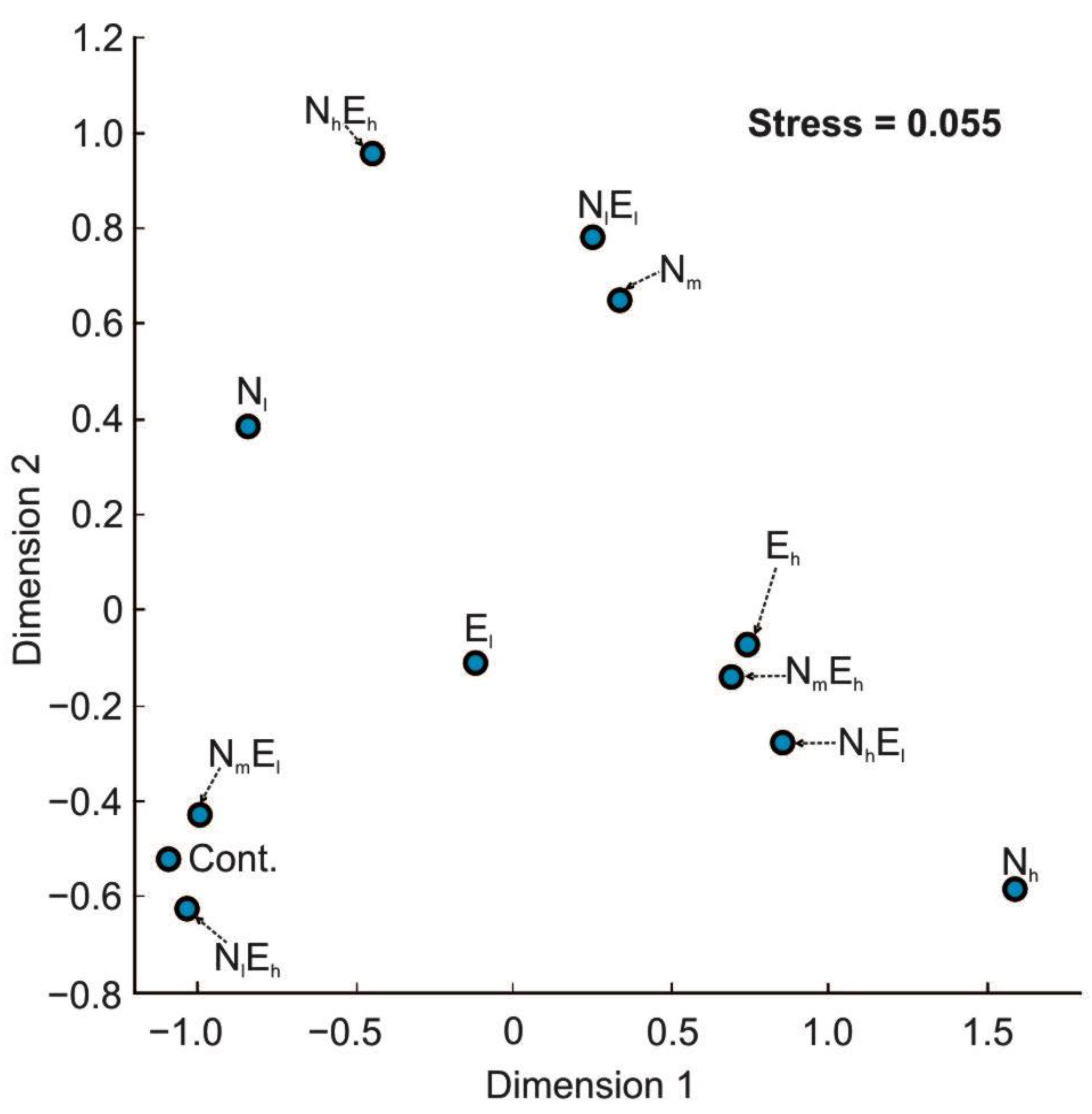
3.3. Taxonomic Diversity of Gut Microbiota

We observed a low taxonomic diversity of the bacteria living in the digestive tract of Daphnia compared to typical diversity of the bacteria living in the lake waters and bacteria from the Daphnia digestive tracts described in other studies, e.g., in [61,75]. The gut microbiota was mainly represented by the bacteria belonging to three phyla—Actinobacteriota, Firmicutes, and Proteobacteria—with the predominance of Proteobacteria and Firmicutes (Figure 7a,b). The presence of each of the stressors, especially the high concentration of NPs, increased the Firmicutes’ participation (Figure 7a,b). This effect was also apparent with the increasing concentrations of enrofloxacin (Figure 5). Despite the clear effect of both stressors on their own—the Firmicutes share the increase—the pattern was similar in the presence of single and combined stressors, which suggest a negative interaction between their effects (Figure 7a,b). The Bray–Curtis-based NMDS analyses at the family level did not show any apparent group of similar variants (Figure 8), which suggests the different taxonomic composition within phylum Firmicutes, whose relative abundance increased after adding both stressors. However, it revealed that the variants with the combined effect of both stressors and with high concentrations of each of the stressors on its own are further away from the control than the other variants (i.e., with low concentrations of each of the stressors on its own and combined).
No significant correlation between the taxonomic profile (at the family level) and the Eco Plate-based metabolic profile was observed (Mantel Correlation, R = 0.153, p = 0.2052, permutation n = 9999).

4. Discussion

4.1. General Effect

The results of our study revealed that each of the stressors (the NPs and enrofloxacin) on their own influenced most of the measured life history parameters of D. magna (except the body length of 5-day-old individuals) and the metabolic and taxonomic diversity of the Daphnia gastrointestinal microbiota, and there was an interaction in the effect of both stressors on all of the measured parameters. On the one hand, since we used in our study temperature and food conditions very close to the optimal ones (23 °C and 0.6 mg Corg × L−1), it is rather unlikely that the stress caused by the suboptimal experimental conditions had any impact on the observed effects of NPs and enrofloxacin. On the other hand, it can be expected that the use of a lower temperature and a higher food concentration could reduce the Daphnia filtration rate and, consequently, could reduce the observed effects of the NPs and enrofloxacin, although they would not change the direction of the effects.

4.2. The Effect of Single and Combined Stressors on the Life History Parameters

The results confirmed our first hypothesis, as both stressors resulted in a decrease in the Daphnia body size and their reproductive parameters, including an average egg volume in the brood cavity and the number of eggs in the clutch of individuals during the first reproduction, which also resulted in decreasing the clutch volume being a common currency of the reproductive investment, which suggests that each of the stressors has a negative effect on the fitness of an individual. Although we did not assess the age of the first reproduction of Daphnia in the experiments, the greatest number of ovigerous females (in relation to the females without eggs) during the fifth day of the experiment in the control, moderate in the presence of each of the stressors on its own, and the lowest in the treatments with the combined stressors, suggests that each of the stressors delayed the start of reproduction and that there was an interaction in the effect of both stressors.
In the case of the NPs, these results may have been caused by the clogging of the filtration appendages and the gut which, in turn, results in decreasing the filtration and assimilation rate, as demonstrated in several earlier studies, e.g., in [76]. These findings are consistent with a great number of earlier studies in which the presence of NPs suspended in water alters the life history traits of various animals, and that these alterations are manifold [13], including a reduction in the body size of adult and juvenile individuals, reproduction (i.e., decreased numbers and body size of neonates), the individual growth rate and survival of freshwater, e.g., in [77], and saline lakes, e.g., in [78], planktonic animals, cnidarians [79], and fishes, e.g., in [23,80], although other studies did not find any effect of an acute exposure on the life history parameters, including negligible effects on the survival rate and development of Danio rerio, e.g., in [81], on the survival and individual growth rate of Gammarus pulex [82].
Several studies revealed the significance of antimicrobial drugs on aquatic organisms [83,84]. For example, the parental exposure of marine fish (Oryzias melastigma) to sulfamethazine (4.62 mg × g−1) may negatively affect the growth performance in adults [14,85]. In the case of enrofloxacin, the decrease in all the measured life history parameters is consistent with the numerous earlier studies performed on fish [23,85] and Daphnia [75]. Another study revealed the negative effect of tetracycline (1 µg × L−1) on the reproduction and survival of D. magna [75]. However, it should be pointed out that not all studies reported the effect of antibiotics on the life history parameters. For example, Nunes et al. [86] showed that ecologically relevant ciprofloxacin concentrations (0.005–0.195 mg × L−1), did not cause significant impacts on the growth rate and reproductive parameters of D. magna, and Ma et al. [87] did not find the effect of tetracycline (10 mg × L−1) on the growth rate of the soil annelid Enchytraeus crypticus. Moreover, in a single study, it was revealed that at very low ciprofloxacin concentrations (10 ng L−1), that is, at a level of a lower concentration that the enrofloxacin used in our study, the growth and fecundity of D. magna were even higher than that in the control of animals [88].
We are aware of only two earlier studies in which the combined effect of the NPs and antibiotics on the life history parameters was determined [23,87]. While the first study revealed that the effect of tetracycline on the dry weight of E. crypticus after a seven-day exposure was stronger in the presence of polystyrene [87], the second study revealed that the adverse impact of the mixture of polystyrene NPs and sulfamethazine on the dry weight in the O. melastigma was weaker than the sole effect of the NPs [23]. In our study, the negative effect of each of the stressors (the NPs and enrofloxacin) on the body size was stronger, and on the reproductive parameters was lower in the presence of another factor, which may suggest that in the presence of cumulative stress, Daphnia redirects more resources to reproduction at the expense of somatic growth.

4.3. The Effect of Single and Combined Stressors on the Metabolomic Diversity of Gut Microbiota

The results also confirmed our second hypothesis, since NPs resulted in an increase and enrofloxacin resulted in the decrease in the overall carbon respiration rate of the Daphnia gut microbiota. This may be due to a changing bacteria abundance, their metabolic condition, and their ability of utilising different carbon sources. The decrease in the metabolic rate in the presence of enrofloxacin is consistent with the existing data on the negative effect of antibiotics on the bacteria metabolism, since there is a link between antibiotic-induced cellular respiration and bactericidal lethality. Antibiotics disturb the metabolic state of bacteria, which impacts the antibiotic efficacy [89,90]. The positive effect of NPs on the respiration of the bacteria in the Daphnia gastrointestinal tract may be due to the provision of an additional sorption surface, which not only facilitates the formation of bacterial biofilms, but may also affect the ability to degrade organic compounds that absorb on such surfaces. For example, the sorption of proteins on surfaces can change their conformation, making them more accessible to proteolytic enzymes and subject to faster hydrolysis [91]. Our results are the first to demonstrate the effect of NPs on the metabolic profile of the gut microbiota. Moreover, the results are also the first to demonstrate the interaction between the stressors on the metabolic profile of the gut microbiota, which was apparent in the increased inhibitory effect of enrofloxacin in the presence of NPs, and in the reduced positive effect of NPs in the presence of enrofloxacin.
The results also confirmed our third hypothesis, as the presence of NPs and enrofloxacin affected the metabolic fingerprints measured as the relative use of different carbon sources as compared to the control. The functional structure of the bacteria community had changed. The presence of each of the stressors resulted in a relative increase in the usage of carboxylic acids, amino acids, and carbohydrates and in the relative decrease in the usage of phosphorylated carbons and complex carbon sources with the control, which suggests that both the enrofloxacin and NPs forced the bacteria to exploit the easily digestible carbon sources [92]. In general, these results are in accordance with the existing literature. For instance, it has been shown that the presence of polystyrene NPs altered the carbohydrate metabolism of marine medaka fish gut microbiota [23]. However, Zhang et al. [51] did not show any significant effect of NPs on the predicted metabolic pathways of the gut microbiota in the same fish species. A picture of the negative impact of NPs on the metabolic fingerprint emerges from the research on the freshwater biofilm done with the Biolog EcoPlate method, in which the NPs’ presence (1, 5 and 10 mg × L−1) reduced the microbial metabolic functional diversity. The total carbon metabolism remained constant with growing NP concentrations, but the utilisation of some specific carbon sources (e.g., esters) had changed [93]. It was revealed that the presence of sulfamethazine affected the function of the gut microbiota of medaka fish, so the carbohydrate metabolism was significantly decreased [23]. Zhang et al. [51] presented similar results, showing a decrease in the carbohydrate metabolism in marine medaka females who had been exposed to low concentrations of sulfamethazine (0.5 mg × g−1 supplied in fish food); thus, also the lipid and amino acid metabolism was enhanced under these conditions. In the murine model, other antibiotics, like amoxicillin, have been shown to elevate the expression of genes responsible for starch utilisation by Bacteroides thetaiotaomicron [94].
Moreover, the NPs and enrofloxacin which were acting together had a different effect on the metabolic fingerprints than the NPs and enrofloxacin which were acting separately, suggesting the existence of the interaction between the two stressors. The simultaneous presence of sulfamethazine and NPs also significantly altered the carbohydrate metabolism, as He et al. [23] discovered. However, it should be pointed out that the results from He et al. [23] and Zhang et al. [51] concerning the effects of antibiotics and NPs on changes in the gut microbial metabolism fingerprint were obtained by the bioinformatics prediction. Our results stem from the direct measurement of the microbial metabolic fingerprint as the function of different respiration intensities of various carbon sources by the Daphnia gut microbial community.

4.4. The Effect of Single and Combined Stressors on the Taxonomic Diversity of Gut Microbiota

Finally, the results also confirmed the fourth hypothesis, as the taxonomic diversity of bacteria was affected by the presence of each of the two stressors on its own, which was apparent in the relative increase in the Firmicutes (mainly the Bacillaceae family, representatives of which occur in the digestive tracts of many aquatic animals, e.g., in [95]), in relation to Actinobacteria and Proteobacteria at the phylum level. Despite this, in most of the experimental variants, the dominant taxa at the phylum level were Proteobacteria, which is consistent with the previous studies on the microbiome of other animals, including the soil fauna [88,96,97]. On the one hand, the relative increase in Firmicutes may have a positive effect on Daphnia, as it increases the diversity of the microbial composition, which is often equated with the improvement of the host’s health [98]. It has been disclosed that Firmicutes produce short-chain fatty acids, which could be used for the de novo synthesis of lipids or glucose and an additional energy source for the host [23,99,100]. Therefore, from the perspective of the host energy input, Firmicutes in the gut may play a positive role. On the other hand, it has been shown that an increased proportion of Firmicutes to other bacteria phyla is an indicator of metabolic disorders in animals [101,102], which may explain the reduction in the body size and reproductive potential of Daphnia by each of the stressors on its own in our study.
In the case of NPs, the relative increase in Firmicutes in the gut microbiota may be due to the fact that NPs can provide them with a better matrix for biofilm growth compared to other bacteria taxa. The results are consistent with several recent studies, which found that NPs may affect a community of free-living bacteria [103,104] and the microbiome of E. crypticus [67]. This also includes a marine mollusk Mytilus galloprovincialis [105] and various fish species, including O. melastigma [106] and D. rerio, e.g., [107,108], which can induce dysbiosis and inflammation in their intestine. For example, it has been discovered that the dietary NPs (1 mg × g−1) affected the relative proportions of Microbacteriaceae, Streptococcaceae, Enterobacteriaceae, and Rhodocyclaceae in the whole body microbial community of E. crypticus [87]. It has been recognised that all of these taxa potentially negatively (with Enterobacteriaceae [109]) or positively (with the remaining taxa [110,111]) affect the host. However, our study contradicts the results of He et al. [23], who found a decrease in the relative abundance of Firmicutes in males exposed to (3.45 mg × g−1) dietary polystyrene NPs. It is worth noting that, in our study, the taxonomic diversity of the bacteria increased in the presence of NPs, as the relative abundance of different taxa was more even compared to the controls, which is consistent with the earlier studies for the diversity of the microbial communities in the gut of D. rerio, e.g., [107].
The increase in the relative abundance of Firmicutes in the presence of enrofloxacin is consistent with several previous studies, which revealed that antibiotics might change the taxonomic diversity of the gut microbiota of animals, including mice [112] soil organisms [87,102], fish [23], and Daphnia [76]. For example, it has been revealed that an exposure to tetracycline (1 µg × L−1) resulted in an increase in the relative abundance of Pseudomonaceae in the intestinal microbial community of D. magna [75]. Other studies have shown that dietary tetracycline (0.01 mg × g−1) affected the relative proportions of Microbacteriaceae, Streptococcaceae, Enterobacteriaceae, and Rhodocyclaceae in the whole body microbial community of E. crypticus [87]. In another study, Motiei et al. [88] showed that G + bacteria, mostly Actinobacteria and Firmicutes, was better equipped to withstand an exposure to ciprofloxacin (of a similar mechanism of action as enrofloxacin) as their relative abundance increased with the antibiotic concentration.
Moreover, the results also confirmed the fourth hypothesis as we observed an interaction between the two stressors on the bacteria community structure, which was apparent in the weaker effect of each of the stressors on its own than combined. To our knowledge, only two earlier studies investigated the combined effect of NPs and antibiotics [23,88]. The first study showed that the combined exposure of tetracycline and polystyrene NPs negatively affected the abundance of bacteria belonging to several families, including Microbacteriaceae, Streptococcaceae, and Enterobacteriaceae in the whole body microbiome of E. crypticus, and additionally, significantly higher ratios of Planococcaceae/Chitinophagaceae and Bacillaceae/Chitinophagaceae were observed after a tetracycline and polystyrene exposure, especially when the two pollutants were combined [87]. The second study revealed that a parental exposure to polystyrene NPs and sulfamethazine had a weaker effect on the gut microbial communities in the offspring of marine fish (O. melastigma) than each of the stressors on its own [23]. Moreover, it has been released that after terminating the exposure, the microbiome was not permanently changed but impacted reversibly [87].
Summing up, the NPs and enrofloxacin altered the taxonomic (and also metabolic) structure of the bacterial communities living in the gut of Daphnia. The interaction of the enrofloxacin and NPs resulted in changes in the taxonomic composition of the bacterial communities slightly different from those induced by each of the stressors acting separately. This fact may result from the system’s complexity, where NPs constitute the sorption surface for the dissolved organic compounds and antibiotics, causing both their local concentration to increase and a change in the conformation of sorbed molecules that can change their physical properties. The formation of biofilms on aggregated plastic particles may also change the dominant bacteria’s taxonomic structure. No clear relationship has been found between the bacterial taxonomic composition and metabolic fingerprints. Thus, this may indicate the occurrence of the phenomenon of redundancy and the high plasticity of microorganism communities which, when changing the taxonomic composition, can replace their environmental functions.

5. Conclusions

In conclusion, despite the growing interest in the effects of NPs and antibiotics on the biology of organisms, knowledge on the subject is still fragmentary and is often based on contradictory results. Our study seems to be the first one to investigate the combined effect of NPs and antibiotics on the life history parameters of freshwater organisms and the metabolomic and taxonomic diversity of their intestinal microbial community. Among the most important results that we obtained is that both stressors: (i) negatively affected most of the Daphnia life history parameters, (ii) modified the use of different carbon sources by the intestinal microbiota, (iii) increased the Firmicute phyla participation in the microbiota taxonomic composition, and (iv) interacted with each other, affecting all the measured parameters. Moreover, we also observed that the NPs increased and enrofloxacin decreased the metabolic rate of the gut microbiota. The results of our study suggest a possible link between the observed effects. Future studies concerning the issue should primarily focus on integrating the results from different levels of the biological organisation on the combined effect of both stressors on different taxa.

Supplementary Materials

The following are available online at https://www.mdpi.com/article/10.3390/w14213403/s1, Tables S1–S8: The results of the analysis of ART two-way ANOVA.

Author Contributions

Conceptualization: P.M. and B.K.; methodology: P.M. and B.K.; investigation: E.B., S.G., P.M., C.J.-M. and B.K.; resources: P.M.; data curation: S.G., B.K., P.M., G.K. and M.L.Z.; writing—original draft preparation: P.M.; writing—review and editing: J.P., J.S., J.J.-L., B.K., P.M., E.B. and C.J.-M.; visualization: B.K. and P.M.; supervision: B.K. and P.M.; project administration: P.M.; funding acquisition: P.M. and E.B. All authors have read and agreed to the published version of the manuscript.

Funding

The research described here was supported by the grants no. 2018/31/N/NZ8/03269 and 2019/35/B/NZ8/04523 from the National Science Center, Poland.

Institutional Review Board Statement

Not applicable.

Informed Consent Statement

Not applicable.

Data Availability Statement

Not applicable.

Conflicts of Interest

The authors declare no conflict of interest.

References

  1. MacLeod, M.; Arp, H.P.H.; Tekman, M.B.; Jahnke, A. The global threat from plastic pollution. Science 2021, 373, 61–65. [Google Scholar] [CrossRef] [PubMed]
  2. Barnes, D.K.; Galgani, F.; Thompson, R.C.; Barlaz, M. Accumulation and fragmentation of plastic debris in global environments. Philos. Trans. R. Soc. B Biol. Sci. 2009, 364, 1985–1998. [Google Scholar] [CrossRef] [PubMed] [Green Version]
  3. Kedzierski, M.; Frčre, D.; Le Maguer, G.; Bruzaud, S. Why is there plastic packaging in the natural environment? Understanding the roots of our individual plastic waste management behaviours. Sci. Total Environ. 2020, 740, 139985. [Google Scholar] [CrossRef] [PubMed]
  4. Geyer, R.; Jambeck, J.R.; Law, K.L. Production, use, and fate of all plastics ever made. Sci. Adv. 2017, 3, e1700782. [Google Scholar] [CrossRef] [PubMed] [Green Version]
  5. Gigault, J.; Ter Halle, A.; Baudrimont, M.; Pascal, P.Y.; Gauffre, F.; Phi, T.L.; Reynaud, S. Current opinion: What is a nanoplastic? Environ. Pollut. 2018, 235, 1030–1034. [Google Scholar] [CrossRef]
  6. Besseling, E.; Redondo-Hasselerharm, P.; Foekema, E.M.; Koelmans, A.A. Quantifying ecological risks of aquatic micro-and nanoplastic. Crit. Rev. Environ. Sci. Technol. 2019, 49, 32–80. [Google Scholar] [CrossRef] [Green Version]
  7. Egessa, R.; Nankabirwa, A.; Ocaya, H.; Pabire, W.G. Microplastic pollution in surface water of Lake Victoria. Sci. Total Environ. 2020, 741, 140201. [Google Scholar] [CrossRef]
  8. Huang, D.; Chen, H.; Shen, M.; Tao, J.; Chen, S.; Yin, L.; Li, R. Recent advances on the transport of microplastics/nanoplastics in abiotic and biotic compartments. J. Hazard. Mater. 2022, 438, 129515. [Google Scholar] [CrossRef]
  9. Koelmans, A.A.; Besseling, E.; Shim, W.J. Nanoplastics in the aquatic environment. Critical review. Mar. Anthropog. Litter 2015, 325–340. [Google Scholar]
  10. Ter Halle, A.; Jeanneau, L.; Martignac, M.; Jardé, E.; Pedrono, B.; Brach, L.; Gigault, J. Nanoplastic in the North Atlantic subtropical gyre. Environ. Sci. Technol. 2017, 51, 13689–13697. [Google Scholar] [CrossRef]
  11. Bhattacharya, P.; Lin, S.; Turner, J.P.; Ke, P.C. Physical adsorption of charged plastic nanoparticles affects algal photosynthesis. J. Phys. Chem. C 2010, 114, 16556–16561. [Google Scholar] [CrossRef]
  12. Booth, A.M.; Hansen, B.H.; Frenzel, M.; Johnsen, H.; Altin, D. Uptake and toxicity of methylmethacrylate-based nanoplastic particles in aquatic organisms. Environ. Toxicol. Chem. 2016, 35, 1641–1649. [Google Scholar] [CrossRef] [PubMed] [Green Version]
  13. Maszczyk, P.; Pijanowska, J.; Mrówka, P.; Babkiewicz, E. Effects of nanoplastics on aquatic organisms. In Environmental Nanopollutants: Sources, Occurrence, Analysis and Fate; CRC Press: Boca Raton, FL, USA, 2022. [Google Scholar]
  14. El Hadri, H.; Gigault, J.; Maxit, B.; Grassl, B.; Reynaud, S. Nanoplastic from mechanically degraded primary and secondary microplastics for environmental assessments. NanoImpact 2020, 17, 100206. [Google Scholar] [CrossRef]
  15. Gaylarde, C.C.; Neto, J.A.B.; da Fonseca, E.M. Nanoplastics in aquatic systems-are they more hazardous than microplastics? Environ. Pollut. 2021, 272, 115950. [Google Scholar] [CrossRef] [PubMed]
  16. Schrank, I.; Trotter, B.; Dummert, J.; Scholz-Böttcher, B.M.; Löder, M.G.; Laforsch, C. Effects of microplastic particles and leaching additive on the life history and morphology of Daphnia magna. Environ. Pollut. 2019, 255, 113233. [Google Scholar] [CrossRef] [PubMed]
  17. Song, J.; Na, J.; An, D.; Jung, J. Role of benzophenone-3 additive in chronic toxicity of polyethylene microplastic fragments to Daphnia magna. Sci. Total Environ. 2021, 800, 149638. [Google Scholar] [CrossRef]
  18. Zhao, H.J.; Xu, J.K.; Yan, Z.H.; Ren, H.Q.; Zhang, Y. Microplastics enhance the developmental toxicity of synthetic phenolic antioxidants by disturbing the thyroid function and metabolism in developing zebrafish. Environ. Int. 2020, 140, 105750. [Google Scholar] [CrossRef]
  19. Li, Z.; Li, W.; Zha, J.; Chen, H.; Martyniuk, C.J.; Liang, X. Transcriptome analysis reveals benzotriazole ultraviolet stabilizers regulate networks related to inflammation in juvenile zebrafish (Danio rerio) brain. Environ. Toxicol. 2019, 34, 112–122. [Google Scholar] [CrossRef]
  20. Gauthier, P.T.; Norwood, W.P.; Prepas, E.E.; Pyle, G.G. Metal–PAH mixtures in the aquatic environment: A review of co-toxic mechanisms leading to more-than-additive outcomes. Aquat. Toxicol. 2014, 154, 253–269. [Google Scholar] [CrossRef]
  21. Trevisan, R.; Voy, C.; Chen, S.; Di Giulio, R.T. Nanoplastics decrease the toxicity of a complex PAH mixture but impair mitochondrial energy production in developing zebrafish. Environ. Sci. Technol. 2019, 53, 8405–8415. [Google Scholar] [CrossRef]
  22. Menéndez-Pedriza, A.; Jaumot, J. Interaction of environmental pollutants with microplastics: A critical review of sorption factors, bioaccumulation and ecotoxicological effects. Toxics 2020, 8, 40. [Google Scholar] [CrossRef] [PubMed]
  23. He, S.; Li, D.; Wang, F.; Zhang, C.; Yue, C.; HuangF, Y.; Mu, J. Parental exposure to sulfamethazine and nanoplastics alters the gut microbial communities in the offspring of marine madaka (Oryzias melastigma). J. Hazard. Mater. 2022, 423, 127003. [Google Scholar] [CrossRef]
  24. Liu, S.; Yan, L.; Zhang, Y.; Junaid, M.; Wang, J. Polystyrene nanoplastics exacerbated the ecotoxicological and potential carcinogenic effects of tetracycline in juvenile grass carp (Ctenopharyngodon idella). Sci. Total Environ. 2022, 803, 150027. [Google Scholar] [CrossRef] [PubMed]
  25. Canniff, P.M.; Hoang, T.C. Microplastic ingestion by Daphnia magna and its enhancement on algal growth. Sci. Total Environ. 2018, 633, 500–507. [Google Scholar] [CrossRef] [PubMed] [Green Version]
  26. Trevisan, R.; Ranasinghe, P.; Jayasundara, N.; Di Giulio, R.T. Nanoplastics in Aquatic Environments: Impacts on Aquatic Species and Interactions with Environmental Factors and Pollutants. Toxics 2022, 10, 326. [Google Scholar] [CrossRef] [PubMed]
  27. Chen, Q.; Yin, D.; Jia, Y.; Schiwy, S.; Legradi, J.; Yang, S.; Hollert, H. Enhanced uptake of BPA in the presence of nanoplastics can lead to neurotoxic effects in adult zebrafish. Sci. Total Environ. 2017, 609, 1312–1321. [Google Scholar] [CrossRef]
  28. Mendes, L.A.; Barreto, A.; Santos, J.; Amorim, M.J.; Maria, V.L. Co-Exposure of Nanopolystyrene and Other Environmental Contaminants—Their Toxic Effects on the Survival and Reproduction of Enchytraeus crypticus. Toxics 2022, 10, 193. [Google Scholar] [CrossRef]
  29. Booth, A.; Aga, D.S.; Wester, A.L. Retrospective analysis of the global antibiotic residues that exceed the predicted no effect concentration for antimicrobial resistance in various environmental matrices. Environ. Int. 2020, 141, 105796. [Google Scholar] [CrossRef]
  30. Fair, R.J.; Tor, Y. Antibiotics and bacterial resistance in the 21st century. Perspect. Med. Chem. 2014, 6, S14459. [Google Scholar] [CrossRef] [Green Version]
  31. Zainab, S.M.; Junaid, M.; Xu, N.; Malik, R.N. Antibiotics and antibiotic resistant genes (ARGs) in groundwater: A global review on dissemination, sources, interactions, environmental and human health risks. Water Res. 2020, 187, 116455. [Google Scholar] [CrossRef]
  32. Hill, D.A.; Hoffmann, C.; Abt, M.C.; Du, Y.; Kobuley, D.; Kirn, T.J.; Artis, D. Metagenomic analyses reveal antibiotic-induced temporal and spatial changes in intestinal microbiota with associated alterations in immune cell homeostasis. Mucosal Immunol. 2010, 3, 148–158. [Google Scholar] [CrossRef] [PubMed]
  33. Zhou, L.; Limbu, S.M.; Shen, M.; Zhai, W.; Qiao, F.; He, A.; Zhang, M. Environmental concentrations of antibiotics impair zebrafish gut health. Environ. Pollut. 2018, 235, 245–254. [Google Scholar] [CrossRef] [PubMed]
  34. Kovalakova, P.; Cizmas, L.; McDonald, T.J.; Marsalek, B.; Feng, M.; Sharma, V.K. Occurrence and toxicity of antibiotics in the aquatic environment: A review. Chemosphere 2020, 251, 126351. [Google Scholar] [CrossRef] [PubMed]
  35. Duan, W.; Cui, H.; Jia, X.; Huang, X. Occurrence and ecotoxicity of sulfonamides in the aquatic environment: A review. Sci. Total Environ. 2022, 820, 153178. [Google Scholar] [CrossRef]
  36. Baggio, D.; Ananda-Rajah, M.R. Fluoroquinolone antibiotics and adverse events. Aust. Prescr. 2021, 44, 161. [Google Scholar] [CrossRef]
  37. Brown, S.A. Fluoroquinolones in animal health. J. Vet. Pharmacol. Ther. 1996, 19, 1–14. [Google Scholar] [CrossRef]
  38. Migliore, L.; Cozzolino, S.; Fiori, M. Phytotoxicity to and uptake of flumequine used in intensive aquaculture on the aquatic weed, Lythrum salicaria L. Chemosphere 2000, 40, 741–750. [Google Scholar] [CrossRef] [Green Version]
  39. Jacoby, G.A. Mechanisms of resistance to quinolones. Clin. Infect. Dis. 2005, 41 (Suppl. 2), S120–S126. [Google Scholar] [CrossRef] [Green Version]
  40. Välitalo, P.; Kruglova, A.; Mikola, A.; Vahala, R. Toxicological impacts of antibiotics on aquatic micro-organisms: A mini-review. Int. J. Hyg. Environ. Health 2017, 220, 558–569. [Google Scholar] [CrossRef] [Green Version]
  41. Kim, Y.; Jung, J.; Kim, M.; Park, J.; Boxall, A.B.; Choi, K. Prioritizing veterinary pharmaceuticals for aquatic environment in Korea. Environ. Toxicol. Pharmacol. 2008, 26, 167–176. [Google Scholar] [CrossRef]
  42. Kern, W.V. New plasmid-borne quinolone-resistance determinant in Escherichia coli. Future Microbiol. 2007, 2, 473–475. [Google Scholar] [CrossRef]
  43. Ding, G.; Chen, G.; Liu, Y.; Li, M.; Liu, X. Occurrence and risk assessment of fluoroquinolone antibiotics in reclaimed water and receiving groundwater with different replenishment pathways. Sci. Total Environ. 2020, 738, 139802. [Google Scholar] [CrossRef] [PubMed]
  44. Wagil, M.; Kumirska, J.; Stolte, S.; Puckowski, A.; Maszkowska, J.; Stepnowski, P.; Białk-Bielińska, A. Development of sensitive and reliable LC-MS/MS methods for the determination of three fluoroquinolones in water and fish tissue samples and preliminary environmental risk assessment of their presence in two rivers in northern Poland. Sci. Total Environ. 2014, 493, 1006–1013. [Google Scholar] [CrossRef]
  45. Ma, Y.; Li, M.; Wu, M.; Li, Z.; Liu, X. Occurrences and regional distributions of 20 antibiotics in water bodies during groundwater recharge. Sci. Total Environ. 2015, 518, 498–506. [Google Scholar] [CrossRef] [PubMed]
  46. Hu, Y.; Yan, X.; Shen, Y.; Di, M.; Wang, J. Antibiotics in surface water and sediments from Hanjiang River, Central China: Occurrence, behavior and risk assessment. Ecotoxicol. Environ. Saf. 2018, 157, 150–158. [Google Scholar] [CrossRef] [PubMed]
  47. You, X.; Cao, X.; Zhang, X.; Guo, J.; Sun, W. Unraveling individual and combined toxicity of nano/microplastics and ciprofloxacin to Synechocystis sp. at the cellular and molecular levels. Environ. Int. 2021, 157, 106842. [Google Scholar] [CrossRef]
  48. Feng, L.J.; Shi, Y.; Li, X.Y.; Sun, X.D.; Xiao, F.; Sun, J.W.; Yuan, X.Z. Behavior of tetracycline and polystyrene nanoparticles in estuaries and their joint toxicity on marine microalgae Skeletonema costatum. Environ. Pollut. 2020, 263, 114453. [Google Scholar] [CrossRef] [PubMed]
  49. Guo, X.; Cai, Y.; Ma, C.; Han, L.; Yang, Z. Combined toxicity of micro/nano scale polystyrene plastics and ciprofloxacin to Corbicula fluminea in freshwater sediments. Sci. Total Environ. 2020, 789, 147887. [Google Scholar] [CrossRef]
  50. Almeida, M.; Martins, M.A.; Soares, A.M.; Cuesta, A.; Oliveira, M. Polystyrene nanoplastics alter the cytotoxicity of human pharmaceuticals on marine fish cell lines. Environ. Toxicol. Pharmacol. 2019, 69, 57–65. [Google Scholar] [CrossRef]
  51. Zhang, Y.T.; Chen, H.; He, S.; Wang, F.; Liu, Y.; Chen, M.; Mu, J. Subchronic toxicity of dietary sulfamethazine and nanoplastics in marine medaka (Oryzias melastigma): Insights from the gut microbiota and intestinal oxidative status. Ecotoxicol. Environ. Saf. 2021, 226, 112820. [Google Scholar] [CrossRef]
  52. Baysal, A.; Saygin, H. Co-occurence of antibiotics and micro (nano) plastics: A systematic review between 2016–2021. J. Environ. Sci. Health Part A 2022, 57, 519–539. [Google Scholar] [CrossRef] [PubMed]
  53. Zebrowski, M.; Babkiewicz, E.; Błażejewska, A.; Pukos, S.; Wawrzeńczak, J.; Wilczynski, W.; Zebrowski, J.; Slusarczyk, M.; Maszczyk, P. The effect of microplastics on the interspecific competition of Daphnia. Environ. Pollut. 2022, 313, 120121. [Google Scholar] [CrossRef] [PubMed]
  54. Guillard, R.R.L. Culture of phytoplankton for feeding marine invertebrates. In Culture of Marine Invertebrate Animals; Smith, W.L., Chanley, M.H., Eds.; Plenum Press: New York, NY, USA, 1975; pp. 29–60. [Google Scholar]
  55. Pessoni, L.; Veclin, C.; El Hadri, H.; Cugnet, C.; Davranche, M.; Pierson-Wickmann, A.C.; Reynaud, S. Soap-and metal-free polystyrene latex particles as a nanoplastic model. Environ. Sci. Nano 2019, 6, 2253–2258. [Google Scholar] [CrossRef]
  56. Jiménez-Lamana, J.; Marigliano, L.; Allouche, J.; Grassl, B.; Szpunar, J.; Reynaud, S. A novel strategy for the detection and quantification of nanoplastics by single particle inductively coupled plasma mass spectrometry (ICP-MS). Anal. Chem. 2020, 92, 11664–11672. [Google Scholar] [CrossRef] [PubMed]
  57. Bruijning, M.; ten Berge, A.C.; Jongejans, E. Population-level responses to temperature, density and clonal differences in Daphnia magna as revealed by integral projection modelling. Funct. Ecol. 2018, 32, 2407–2422. [Google Scholar] [CrossRef] [Green Version]
  58. Ge, L.; Na, G.; Zhang, S.; Li, K.; Zhang, P.; Ren, H.; Yao, Z. New insights into the aquatic photochemistry of fluoroquinolone antibiotics: Direct photodegradation, hydroxyl-radical oxidation, and antibacterial activity changes. Sci. Total Environ. 2015, 527, 12–17. [Google Scholar] [CrossRef] [PubMed]
  59. Bartosiewicz, M.; Jabłoński, J.; Kozłowski, J.; Maszczyk, P. Brood space limitation of reproduction may explain growth after maturity in differently sized Daphnia species. J. Plankton Res. 2015, 37, 417–428. [Google Scholar] [CrossRef] [Green Version]
  60. Dickerson, T.L.; Williams, H.N. Functional diversity of bacterioplankton in three North Florida freshwater lakes over an Annual Cycle. Microb. Ecol. 2011, 67, 34–44. [Google Scholar] [CrossRef]
  61. Kiersztyn, B.; Chróst, R.; Kaliński, T.; Siuda, W.; Bukowska, A.; Kowalczyk, G.; Grabowska, K. Structural and functional microbial diversity along a eutrophication gradient of interconnected lakes undergoing anthropopressure. Sci. Rep. 2019, 9, 11144. [Google Scholar] [CrossRef] [Green Version]
  62. Macke, E.; Callens, M.; De Meester, L.; Decaestecker, E. Host-genotype dependent gut microbiota drives zooplankton tolerance to toxic cyanobacteria. Nat. Commun. 2017, 8, 1608. [Google Scholar] [CrossRef] [Green Version]
  63. Callens, M.; Watanabe, H.; Kato, Y.; Miura, J.; Decaestecker, E. Microbiota inoculum composition affects holobiont assembly and host growth in Daphnia. Microbiome 2018, 6, 56. [Google Scholar] [CrossRef] [PubMed]
  64. Hegg, A.; Radersma, R.; Uller, T. A field experiment reveals seasonal variation in the Daphnia gut microbiome. Oikos 2021, 130, 2191–2201. [Google Scholar] [CrossRef]
  65. Bulteel, L.; Houwenhuyse, S.; Declerck, S.A.; Decaestecker, E. The role of microbiome and genotype in Daphnia magna upon parasite re-exposure. Genes 2021, 12, 70. [Google Scholar] [CrossRef] [PubMed]
  66. Caporaso, J.G.; Kuczynski, J.; Stombaugh, J.; Bittinger, K.; Bushman, F.D.; Costello, E.K.; Knight, R. QIIME allows analysis of high-throughput community sequencing data. Nat. Methods 2010, 7, 335–336. [Google Scholar] [CrossRef] [PubMed] [Green Version]
  67. Beckers, B.; Op De Beeck, M.; Thijs, S.; Truyens, S.; Weyens, N.; Boerjan, W.; Vangronsveld, J. Performance of 16s rDNA primer pairs in the study of rhizosphere and endosphere bacterial microbiomes in metabarcoding studies. Front. Microbiol. 2016, 7, 650. [Google Scholar] [CrossRef] [Green Version]
  68. Martin, M. Cutadapt removes adapter sequences from high-throughput sequencing reads. EMBnet. J. 2011, 17, 10–12. [Google Scholar] [CrossRef]
  69. Ewels, P.; Magnusson, M.; Lundin, S.; Käller, M. MultiQC: Summarize analysis results for multiple tools and samples in a single report. Bioinformatics 2016, 32, 3047–3048. [Google Scholar] [CrossRef] [Green Version]
  70. Bolyen, E.; Rideout, J.R.; Dillon, M.R.; Bokulich, N.A.; Abnet, C.C.; Al-Ghalith, G.A.; Caporaso, J.G. Reproducible, interactive, scalable and extensible microbiome data science using QIIME 2. Nat. Biotechnol. 2019, 37, 852–857. [Google Scholar] [CrossRef]
  71. Kay, M.; Elkin, L.A.; Higgins, J.J.; Wobbrock, J.O. ARTool: Aligned Rank Transform for Nonparametric Factorial ANOVAs, R package version 0.11.1; Association for Computing Machinery: New York, NY, USA, 2021; Available online: https://github.com/mjskay/ARTool (accessed on 27 April 2021). [CrossRef]
  72. Wobbrock, J.O.; Findlater, L.; Gergle, D.; Higgins, J.J. The aligned rank transform for nonparametric factorial analyses using only ANOVA procedures. In Proceedings of the ACM Conference on Human Factors in Computing Systems (CHI 2011), Vancouver, BC, Canada, 7–12 May 2011; ACM Press: New York, NY, USA, 2011; pp. 143–146. Available online: https://depts.washington.edu/acelab/proj/art/ (accessed on 7 May 2011). [CrossRef] [Green Version]
  73. Elkin, L.A.; Kay, M.; Higgins, J.J.; Wobbrock, J.O. An aligned rank transform procedure for multifactor contrast tests. In Proceedings of the ACM Symposium on User Interface Software and Technology (UIST 2021), Virtual Event, 10–14 October 2021; ACM Press: New York, NY, USA, 2021; pp. 754–768. [Google Scholar] [CrossRef]
  74. Hammer, Ř.; Harper, D.A.; Ryan, D.D. Past: Paleontological statistics software package for education and data analysis. Palaeontol. Electron. 2001, 4, 9. [Google Scholar]
  75. Akbar, S.; Gu, L.; Sun, Y.; Zhou, Q.; Zhang, L.; Lyu, K.; Yang, Z. Changes in the life history traits of Daphnia magna are associated with the gut microbiota composition shaped by diet and antibiotics. Sci. Total Environ. 2020, 705, 135827. [Google Scholar] [CrossRef]
  76. Rist, S.; Baun, A.; Hartmann, N.B. Ingestion of micro- and nanoplastics in Daphnia magna—Quantification of body burdens and assessment of feeding rates and reproduction. Environ. Pollut. 2017, 228, 398–407. [Google Scholar] [CrossRef] [PubMed] [Green Version]
  77. Besseling, E.; Wang, B.; Lurling, M.; Koelmans, A.A. Nanoplastic affects growth of S. obliquus and reproduction of D. magna. Environ. Sci. Technol. 2014, 48, 12336–12343. [Google Scholar] [CrossRef] [PubMed]
  78. Bergami, E.; Pugnalini, S.; Vannuccini, M.L.; Manfra, L.; Faleri, C.; Savorelli, F.; Corsi, I. Long-term toxicity of surface-charged polystyrene nanoplastics to marine planktonic species Dunaliella tertiolecta and Artemia franciscana. Aquat. Toxicol. 2017, 189, 159–169. [Google Scholar] [CrossRef] [PubMed]
  79. Venâncio, C.; Ferreira, I.; Martins, M.A.; Soares, A.M.; Lopes, I.; Oliveira, M. The effects of nanoplastics on marine plankton: A case study with polymethylmethacrylate. Ecotoxicol. Environ. Saf. 2019, 184, 109632. [Google Scholar] [CrossRef] [PubMed]
  80. Sökmen, T.Ö.; Sulukan, E.; Türkoğlu, M.; Baran, A.; Özkaraca, M.; Ceyhun, S.B. Polystyrene nanoplastics (20 nm) are able to bioaccumulate and cause oxidative DNA damages in the brain tissue of zebrafish embryo (Danio rerio). Neurotoxicology 2020, 77, 51–59. [Google Scholar] [CrossRef]
  81. Brun, N.R.; Koch, B.E.; Varela, M.; Peijnenburg, W.J.; Spaink, H.P.; Vijver, M.G. Nanoparticles induce dermal and intestinal innate immune system responses in zebrafish embryos. Environ. Sci. Nano 2018, 5, 904–916. [Google Scholar] [CrossRef]
  82. Redondo-Hasselerharm, P.E.; Vink, G.; Mitrano, D.M.; Koelmans, A.A. Metal-doping of nanoplastics enables accurate assessment of uptake and effects on Gammarus pulex. Environ. Sci. Nano 2021, 8, 1761–1770. [Google Scholar] [CrossRef]
  83. Fernandez, R.; Colás-Ruiz, N.R.; Bolívar-Anillo, H.J.; Anfuso, G.; Hampel, M. Occurrence and Effects of Antimicrobials Drugs in Aquatic Ecosystems. Sustainability 2021, 13, 13428. [Google Scholar] [CrossRef]
  84. Kostich, M.S.; Lazorchak, J.M. Risks to aquatic organisms posed by human pharmaceutical use. Sci. Total Environm. 2008, 389, 329–339. [Google Scholar] [CrossRef]
  85. Qiu, W.; Fang, M.; Magnuson, J.T.; Greer, J.B.; Chen, Q.; Zheng, Y.; Xiong, Y.; Luo, S.; Zheng, C.; Schlenk, D. Maternal exposure to environmental antibiotic mixture during gravid period predicts gastrointestinal effects in zebrafish offspring. J. Hazard. Mater. 2020, 399, 123009. [Google Scholar] [CrossRef]
  86. Nunes, B.; Leal, C.; Rodrigues, S.; Antunes, S.C. Assessment of ecotoxicological effects of ciprofloxacin in Daphnia magna: Life-history traits, biochemical and genotoxic effects. Water Sci. Technol. 2018, 2017, 835–844. [Google Scholar] [CrossRef] [PubMed]
  87. Ma, J.; Sheng, G.D.; Chen, Q.L.; O’Connor, P. Do combined nanoscale polystyrene and tetracycline impact on the incidence of resistance genes and microbial community disturbance in Enchytraeus crypticus? J. Hazard. Mater. 2020, 387, 122012. [Google Scholar] [CrossRef] [PubMed]
  88. Motiei, A.; Brindefalk, B.; Ogonowski, M.; El-Shehawy, R.; Pastuszek, P.; Ek, K.; Gorokhova, E. Disparate effects of antibiotic-induced microbiome change and enhanced fitness in Daphnia magna. PLoS ONE 2020, 15, e0214833. [Google Scholar] [CrossRef] [PubMed] [Green Version]
  89. Lobritz, M.A.; Belenky, P.; Porter, C.B.; Gutierrez, A.; Yang, J.H.; Schwarz, E.G.; Collins, J.J. Antibiotic efficacy is linked to bacterial cellular respiration. Proc. Natl. Acad. Sci. USA 2015, 112, 8173–8180. [Google Scholar] [CrossRef] [Green Version]
  90. Stokes, J.M.; Lopatkin, A.J.; Lobritz, M.A.; Collins, J.J. Bacterial metabolism and antibiotic efficacy. Cell Metab. 2019, 30, 251–259. [Google Scholar] [CrossRef]
  91. Siuda, W.; Kiersztyn, B.; Chrost, R.J. The dynamics of protein decomposition in lakes of different trophic status-reflections on the assessment of the real proteolytic activity in situ. J. Microbiol. Biotechnol. 2007, 17, 897–904. [Google Scholar]
  92. Kataria, R.; Ruhal, R. Microbiological metabolism under chemical stress. In Microbial Biodegradation and Bioremediation; Elsevier: Amsterdam, The Netherlands, 2014; pp. 497–509. [Google Scholar]
  93. Miao, L.; Guo, S.; Liu, Z.; Liu, S.; You, G.; Qu, H.; Hou, J. Effects of nanoplastics on freshwater biofilm microbial metabolic functions as determined by BIOLOG ECO microplates. Int. J. Environ. Res. Public Health 2019, 16, 4639. [Google Scholar] [CrossRef] [Green Version]
  94. Cabral, D.J.; Penumutchu, S.; Reinhart, E.M.; Zhang, C.; Korry, B.J.; Wurster, J.I.; Belenky, P. Microbial metabolism modulates antibiotic susceptibility within the murine gut microbiome. Cell Metab. 2019, 30, 800–823. [Google Scholar] [CrossRef]
  95. Zuo, Y.; Xie, W.; Pang, Y.; Li, T.; Li, Q.; Li, Y. Bacterial community composition in the gut content of Lampetra japonica revealed by 16S rRNA gene pyrosequencing. PLoS ONE 2017, 12, e0188919. [Google Scholar] [CrossRef] [Green Version]
  96. Zheng, F.; Zhu, D.; Giles, M.; Daniell, T.; Neilson, R.; Zhu, Y.G.; Yang, X.R. Mineral and organic fertilization alters the microbiome of a soil nematode Dorylaimus stagnalis and its resistome. Sci. Total Environ. 2019, 680, 70–78. [Google Scholar] [CrossRef]
  97. Berg, M.; Stenuit, B.; Ho, J.; Wang, A.; Parke, C.; Knight, M.; Alvarez-Cohen, L.; Shapira, M. Assembly of the Caenorhabditis elegans gut microbiota from diverse soil microbial environments. ISME J. 2016, 10, 1998–2009. [Google Scholar] [CrossRef] [PubMed]
  98. Xiong, J.B.; Nie, L.; Chen, J. Current understanding on the roles of gut microbiota in fish disease and immunity. Zool. Res. 2019, 40, 70–76. [Google Scholar] [PubMed] [Green Version]
  99. Wolever, T.; Brighenti, F.; Royall, D.; Jenkins, A.L.; Jenkins, D.J. Effect of rectal infusion of short chain fatty acids in human subjects. Am. J. Gastroenterol. 1989, 84, 1027–1033. [Google Scholar] [PubMed]
  100. Schwiertz, A.; Taras, D.; Schäfer, K.; Beijer, S.; Bos, N.A.; Donus, C.; Hardt, P.D. Microbiota and SCFA in lean and overweight healthy subjects. Obesity 2010, 18, 190–195. [Google Scholar] [CrossRef]
  101. Shin, N.R.; Whon, T.W.; Bae, J.W. Proteobacteria: Microbial signature of dysbiosis in gut microbiota. Trends Biotechnol. 2015, 33, 496–503. [Google Scholar] [CrossRef]
  102. Zhu, D.; An, X.L.; Chen, Q.L.; Yang, X.R.; Christie, P.; Ke, X.; Wu, L.H.; Zhu, Y.G. Antibiotics disturb the microbiome and increase the incidence of resistance genes in the gut of a common soil collembolan. Environ. Sci. Technol. 2018, 52, 3081–3090. [Google Scholar] [CrossRef]
  103. Saygin, H.; Baysal, A. Similarities and discrepancies between bio-based and conventional submicron-sized plastics: In relation to clinically important bacteria. Bull. Environ. Contam. Toxicol. 2020, 105, 26–35. [Google Scholar] [CrossRef]
  104. Zhu, D.; Li, G.; Wang, H.T.; Duan, G.L. Effects of nano-or microplastic exposure combined with arsenic on soil bacterial, fungal, and protistan communities. Chemosphere 2021, 281, 130998. [Google Scholar] [CrossRef]
  105. Auguste, M.; Lasa, A.; Balbi, T.; Pallavicini, A.; Vezzulli, L.; Canesi, L. Impact of nanoplastics on hemolymph immune parameters and microbiota composition in Mytilus galloprovincialis. Mar. Environ. Res. 2020, 159, 105017. [Google Scholar] [CrossRef]
  106. Kang, H.M.; Byeon, E.; Jeong, H.; Kim, M.S.; Chen, Q.; Lee, J.S. Different effects of nano-and microplastics on oxidative status and gut microbiota in the marine medaka Oryzias melastigma. J. Hazard. Mater. 2021, 405, 124207. [Google Scholar] [CrossRef]
  107. Gu, W.; Liu, S.; Chen, L.; Liu, Y.; Gu, C.; Ren, H.Q.; Wu, B. Single-cell RNA sequencing reveals size-dependent effects of polystyrene microplastics on immune and secretory cell populations from zebrafish intestines. Environ. Sci. Technol. 2020, 54, 3417–3427. [Google Scholar] [CrossRef] [PubMed]
  108. Xie, S.; Zhou, A.; Wei, T.; Li, S.; Yang, B.; Xu, G.; Zou, J. Nanoplastics induce more serious microbiota dysbiosis and inflammation in the gut of adult zebrafish than microplastics. Bull. Environ. Contam. Toxicol. 2021, 107, 640–650. [Google Scholar] [CrossRef]
  109. Yoon, J.H.; Kang, S.J.; Schumann, P.; Oh, T.K. Yonghaparkia alkaliphila gen. nov., sp nov., a novel member of the family Microbacteriaceae isolated from an alkaline soil. Int. J. Syst. Evol. Microbiol. 2006, 56, 2415–2420. [Google Scholar] [CrossRef] [PubMed]
  110. Pishgar, R.; Dominic, J.A.; Sheng, Z.; Tay, J.H. Denitrification performance and microbial versatility in response to different selection pressures. Bioresour. Technol. 2019, 281, 72–83. [Google Scholar] [CrossRef]
  111. Oh, S.; Choi, D. Microbial community enhances biodegradation of bisphenol A through selection of Sphingomonadaceae. Microb. Ecol. 2019, 77, 631–639. [Google Scholar] [CrossRef] [PubMed]
  112. Yin, J.B.; Zhang, X.X.; Wu, B.; Xian, Q.M. Metagenomic insights into tetracycline effects on microbial community and antibiotic resistance of mouse gut. Ecotoxicology 2015, 24, 2125–2132. [Google Scholar] [CrossRef]
Figure 1. Mean values (± 1 SE) of: (a) body length, (b) body volume, (c) clutch size (the number of eggs per ovigerous female), and (d) egg volume of 5-day-old D. magna from the control variant (Cont.) and from variants of a single or combined low, medium, and high density of polystyrene NPs (Nl = 103, Nm = 106, and Nh = 109 particles L−1, respectively) and low and high concentration of enrofloxacin (El = 10 and Eh = 100 ng L−1, respectively). Statistical significance is accepted at * p < 0.05, ** p < 0.005, or *** p < 0.0005. The NPs effect is marked on blue, and the enrofloxacin effect on green.
Water 14 03403 g001
Figure 2. Mean values (± 1 SE) of: (a) body length, (b) body volume, (c) clutch size (the number of eggs per ovigerous female), and (d) egg volume of 5-day-old D. magna from the control variant (Cont.) and from variants of a single or combined mean density of polystyrene NPs (Nmean for the combined data from Nl = 103, Nm = 106, and Nh = 109 particles L−1) and the mean concentration of enrofloxacin (Emean for the combined data from El = 10 and Eh = 100 ng L−1). Statistical significance is accepted at * p < 0.05, ** p < 0.005, or *** p < 0.0005, ns stands for non-significant. The NPs effect is marked on blue, and the enrofloxacin effect on green.
Water 14 03403 g002
Figure 3. Mean values (±1 SE) of respiration rate expressed as Vmax values of 31 carbon sources by Daphnia gut microbiota from the control variant (Cont.) and from variants of a single or combined low, medium, and high density of polystyrene NPs (Nl = 103, Nm = 106, and Nh = 109 particles L−1, respectively) and low and high concentration of enrofloxacin (El = 10 and Eh = 100 ng L−1, respectively). Statistical significance is accepted at *** p < 0.0005. The NPs effect is marked on blue, and the enrofloxacin effect on green.
Water 14 03403 g003
Figure 4. Mean values (± 1 SE) of respiration rate expressed as Vmax values of 31 carbon sources by Daphnia gut microbiota from the control variant (Cont.) and from variants of a single or combined mean density of polystyrene NPs (Nmean for the combined data from Nl = 103, Nm = 106, and Nh = 109 particles L−1) and the mean concentration of enrofloxacin (Emean for the combined data from El = 10 and Eh = 100 ng L−1). Statistical significance is accepted at *** p < 0.0005, ns stands for non-significant. The NPs effect is marked on blue, and the enrofloxacin effect on green.
Water 14 03403 g004
Figure 5. Percentage share of respiration rate (Vmax) of different carbon sources by the gut microbiota of Daphnia (a) from the control variant (Cont.) and from variants of a single or combined low, medium and high density of polystyrene NPs (Nl = 103, Nm = 106, and Nh = 109 particles L−1, respectively) and low and high concentration of enrofloxacin (El = 10 and Eh = 100 ng L−1, respectively), and (b) from the control variant (Cont.) and from variants of a single or combined mean density of polystyrene NPs (Nmean for the combined data from Nl, Nm, and Nh) and the mean concentration of enrofloxacin (Emean for the combined data from El and Eh).
Water 14 03403 g005
Figure 6. Bray–Curtis-based NMDS analysis of the relative respiration of 31 different carbon sources by D. magna gut microbiota from the control (Cont.), and from variants of the combination of two variables: (1) NPs in low, medium, and high concentrations (Nl, Nm, Nh, respectively) and (2) enrofloxacin in low and high concentrations (El and Eh, respectively).
Water 14 03403 g006
Figure 7. The relative abundances of the top three dominant bacteria phyla present in the gut of D. magna (a) from the control variant (Cont.) and from variants of a single or combined low, medium, and high density of polystyrene NPs (Nl = 103, Nm = 106, and Nh = 109 particles L−1, respectively) and low and high concentration of enrofloxacin (El = 10 and Eh = 100 ng L−1, respectively), as well as (b) from the control variant (Cont.) and from variants of a single or combined the mean density of polystyrene NPs (Nmean for the combined data from Nl, Nm, and Nh) and the mean concentration of enrofloxacin (Emean for the combined data from El and Eh).
Water 14 03403 g007
Figure 8. The graphical results of the Bray–Curtis-based NMDS analysis of sequence data, binned by taxonomic assignment to family. The figure shows the relative distances between the gut microbiota of D. magna from the control (Cont.) variant, and from variants of the combination of two variables: (1) NPs in low, medium, and high concentrations (Nl, Nm, Nh, respectively) and (2) enrofloxacin in low and high concentrations (El and Eh, respectively).
Water 14 03403 g008
Table 1. Different carbon sources used by the Biolog EcoPlate method to measure the ability of the gut bacterial community of Daphnia to utilise carbon substrates.
CarbohydratesD-cellobiose
α-D-lactose
β-methyl-D-glucoside
D-xylose
Erythritol
D-mannitol
N-acetyl-D-glucosamine
D-galactonic acid γ-lactone
Phosphorylated carbonsglucose-1-phosphate
D,L-α-glycerol phosphate
Aminesphenylethylamine
putrescine
Carboxylic acidsD-glucosaminic acid
D-galacturonic acid
γ-hydroxybutyric acid
itaconic acid
α-ketobutyric acid
D-malic acid
pyruvic acid methyl ester
2-hydroxy benzoic acid
4-hydroxy benzoic acid
Complex carbonTween 40
Tween 80
α-cyclodextrin
glycogen
Amino acidsL-arginine
L-asparagine
L-phenylalanine
L-serine
L-threonine
glycyl-L-glutamic acid
Publisher’s Note: MDPI stays neutral with regard to jurisdictional claims in published maps and institutional affiliations.

