Article

Sulfide Treatment Alters Antioxidant Response and Related Genes Expressions in Rice Field Eel (Monopterus albus)

1
Yangtze River Fisheries Research Institute, Chinese Academy of Fishery Sciences, Wuhan 430223, China
2
Fishery Resources and Environmental Science Experimental Station of the Upper-Middle Reaches of Yangtze River (Ministry of Agriculture and Rural Affairs), Wuhan 430223, China
3
National and Local Joint Engineering Research Center of Ecological Treatment Technology for Urban Water Pollution, Zhejiang Provincial Key Lab for Subtropical Water Environment and Marine Biological Resources Protection, College of Life and Environmental Science, Wenzhou University, Wenzhou 325035, China
*
Authors to whom correspondence should be addressed.
Water 2022, 14(20), 3230; https://doi.org/10.3390/w14203230
Submission received: 26 September 2022 / Revised: 12 October 2022 / Accepted: 12 October 2022 / Published: 13 October 2022

Abstract

:
Sulfide is considered as an environmental factor and toxicant with a wide distribution in aquatic environments. At present, the toxic effects of sulfide stress on rice field eel (Monopterus albus) are poorly understood. To ascertain these effects, the juvenile rice field eels were exposed to sub-lethal concentrations of Na2S (0, 0.2154, 2.154, and 21.54 mg/L) for 7, 14, and 28 days. Antioxidant parameters such as catalase (CAT), superoxide dismutase (SOD), glutathione peroxidase (GPx), and malondialdehyde (MDA) as well as the related gene (including sod, cat) expressions were measured. The expressions of metallothionein (mt) and heat shock protein 90 (hsp90), which are used as physiological stress indices, were also detected. The results showed that exposure to sulfide altered the antioxidant response and related gene expressions in rice field eel. The activities of SOD were inhibited and the MDA contents were increased after exposure to sulfide. The activities of CAT and GPx were increased at 7 days and decreased at 28 days after treatment with sulfide at the highest dose. The expressions of sod, cat, and hsp90 were upregulated at 7 days and downregulated at 14 and 28 days after exposure to high doses of sulfide. The expression of mt was significantly downregulated in all sulfide treated groups. The toxic effects caused by sulfide were in dose-dependent and time-dependent manners. In short, oxidative stress and physiological stress were caused in rice field eel after the exposure to sulfide.

1. Introduction

Sulfide including three forms (HS, H2S, and S2−) is considered as an environmental factor and toxicant to aquatic organisms [1,2]. Sulfide is found in concentrations ranging from μg/L to mg/L in the surface water, sewers, and waste water [3,4]. As a type of toxic chemical, organisms exposed to sulfide might suffer adverse effects on growth, development, reproduction, and survival. Much attention had been given to sulfide due to its toxicity [5]. In aquatic environments, sulfide is mainly generated by anaerobic microorganisms under anaerobic conditions [6,7]. It is well-known that sulfide can inhibit cytochrome c oxidase, reduce energy production, and cause histotoxic hypoxia in both mammals and fish [1,8]. Recent studies have shown that exposure to sulfide could alter the energy metabolism, damage gut structure, and impair the health of Pacific white shrimp [6,9]. Additionally, induced oxidative stress and altered composition of microbiota in the intestine of red swamp crayfish was reported after being treated with sulfide [10]. For zebrafish, exposure to a high dose of sulfide produced developmental toxicity [5]. Although sulfide might cause multiple toxic effects in aquatic organisms, the potential toxicity mechanisms of sulfide remain to be elucidated.
The rice field eel (Monopterus albus), a type of air-breathing fish, mainly inhabits muddy waters such as rice fields, lakes, ponds, and rivers [11,12]. It is one of the most valuable aquacultured fish in China for its high nutritional value [13,14]. The annual output of rice field eel in 2017 amounted to 358,295 tons in China [15]. However, the production of rice field eel is still restricted by infection diseases and environmental stress [12]. At present, there are no available details on the toxic effect of rice field eel caused by sulfide stress.
Environmental factors including pollution could induce the generation of reactive oxygen species (ROS) and lead to oxidative stress in organisms [16,17]. ROS can change the function of cell components adversely and destroy cell structure [17]. There are two antioxidant defense systems including an enzymatic antioxidant defense system and nonenzymatic antioxidant defense system to scavenge excessive ROS to protect cells from damage. The enzymatic antioxidant defense system includes multiple enzymes such as catalase (CAT), superoxide dismutase (SOD), glutathione peroxidase (GPx), etc. [17,18,19]. Malondialdehyde (MDA), the end product of lipid peroxidation, is suggested as a useful biomarker for oxidative stress [20]. Metallothionein (MT) is inducible by oxidative stress and functioned as a scavenger of free radicals [21]. Additionally, heat shock proteins (HSPs) have multifunctional roles and play an essential role in avoiding cell damage under environmental stress. HSPs and MT are often used as physiological stress indices.
Sulfide could induce the production of ROS and cause oxidative stress in multiple aquatic organisms such as red swamp crayfish and Pacific white shrimp [10,22,23]. However, far too little attention has been paid to the responses of the antioxidant defense system under sulfide stress in rice field eel. Therefore, in the present study, rice field eel was used to investigate the toxic effects (mainly the responses of antioxidant defense system) treated with sub-lethal doses of sulfide. Antioxidant response parameters including SOD, CAT, GPx, and MDA were measured. Additionally, mRNA expression levels of sod (Cu/Zn), cat, mt, and hsp90 were quantified by real-time PCR.

2. Materials and Methods

2.1. Fish and Exposure

The juvenile rice field eels (12.1 ± 0.71 g, 16.36 ± 0.83 cm) were obtained from a commercial hatchery in Qianjiang, China. The fish were cultured in 24-h aeration tap water (pH 7.1 ± 0.2; hardness 43.6 ± 2.1 CaCO3/L) operating with a 12:12 h light:dark cycle at 26 ± 2 °C, and fed with commercial-feed (Tongwei, Chengdu, China) once per day. After 2 weeks acclimation, 240 healthy rice field eels were randomly selected and distributed into 12 aquaria (80 cm × 39 cm × 62 cm, 20 fish per aquaria) containing 20 L oof water. The 96-h LC50 of Na2S on juvenile rice field eels was detected as 215.4 mg/L in our lab (data not shown). According to the 96-h LC50, the rice field eels were exposed to 0, 0.2154, 2.154, and 21.54 mg/L Na2S (Na2S·9H2O, purity ≥98%, Sigma-Aldrich, Saint Louis, USA) for 7, 14, and 28 days. The rice field eels were fed daily with commercial-feed. The dose of sulfide in the exposure solution was detected using the methylene blue spectrophotometric method every 6 h. The corresponding Na2S was added to the solution, which made the actual concentration consistent with the nominal concentration. All solutions were renewed every day. There was no dead fish in all of the sulfide treatment groups during the entire exposure period.

2.2. Sampling

After exposure for 7, 14, and 28 days, the fish were sampled. At each sample time, 12 fish from each group (including three replicates, four fish per aquaria) were randomly chosen and anesthetized with MS-222 (0.1 g/L, Sigma-Aldrich, Saint Louis, USA). The liver was quickly separated and divided into two parts: for biochemical parameters analysis, the livers (n = 6) were washed with ice-cold 0.86% physiological saline three times and immediately stored at −80 °C; for RNA measurement, livers (n = 6) were stored in TRIZOL reagent (Invitrogen, Carlsbad, USA) and frozen at −80 °C.

2.3. Biochemical Parameter Analysis

The liver samples were homogenized using an ultrasonic cell disrupter (Scientz-IID, Ningbo, China) with 10 volumes of ice-cold 0.86% physiological saline on ice. The samples were centrifuged at 12,000× g for 20 min at 4 °C. The supernatant was separated, and the total protein level of it was quantified by Bradford assay [24]. All biochemical parameters including SOD, CAT, GPx, and MDA were detected by applying commercial kits obtained from Nanjing Jiancheng Bioengineering Institute (Nanjing, China) according to the manufacturer’s procedures.

2.4. Genes Expressions Analysis

The total RNA extraction, genomic DNA digestion, reverse and transcriptase polymerase reaction were operated according to the previously reported method [25,26]. Quantitative RT-PCR (qRT-PCR) was conducted using the UltraSYBR Mixture Kit (CWBIO Beijing, Beijing, China) and performed on an ABI 7500 System (Applied Biosystems 7500, Carlsbad, USA) with the followed program: 95 °C for 10 min, then 40 cycles of 95 °C for 15 s, and 60 °C for 1 min. The specified products of qRT-PCR were verified by electrophoresis in agarose gel and a single peak in the melting curve. The gene amplification efficiency values ranged from 92.11% to 104.6%. Each sample included three replicates. The gene expression levels were quantified by the 2−ΔΔCt method and calibrated by the reference gene (β-actin). The primers for quantitative RT-PCR are shown in Table 1.

2.5. Integrated Biomarker Response (IBR)

IBR, which integrates a battery of biomarkers into one general stress index, is a useful tool in assessing ecological risk [27]. In order to evaluate the integrated effect of the sulfide at different doses, the IBR was conducted using the previously described method [27,28]. IBR version 2 was used in the present study. All of the detected biomarkers were standardized, and the scores are presented in star plots.

2.6. Statistical Analysis

Data were presented as the mean ± standard deviation (SD). All statistical analysis was performed using SPSS 20.0 software (Chicago, USA). The Kolmogorov–Smirnov test and Levene’s test was applied to analyze the normality and homogeneity of the data, respectively. Statistical differences among the treatment groups and control group were evaluated by one-way analysis of variance (ANOVA) followed by a Tukey’s post hoc test. p < 0.05 was supposed to be statistically different. Principal component analysis (PCA) was performed using Statistic 6.0 (StatSoft, Tulsa, OK, USA).

3. Results

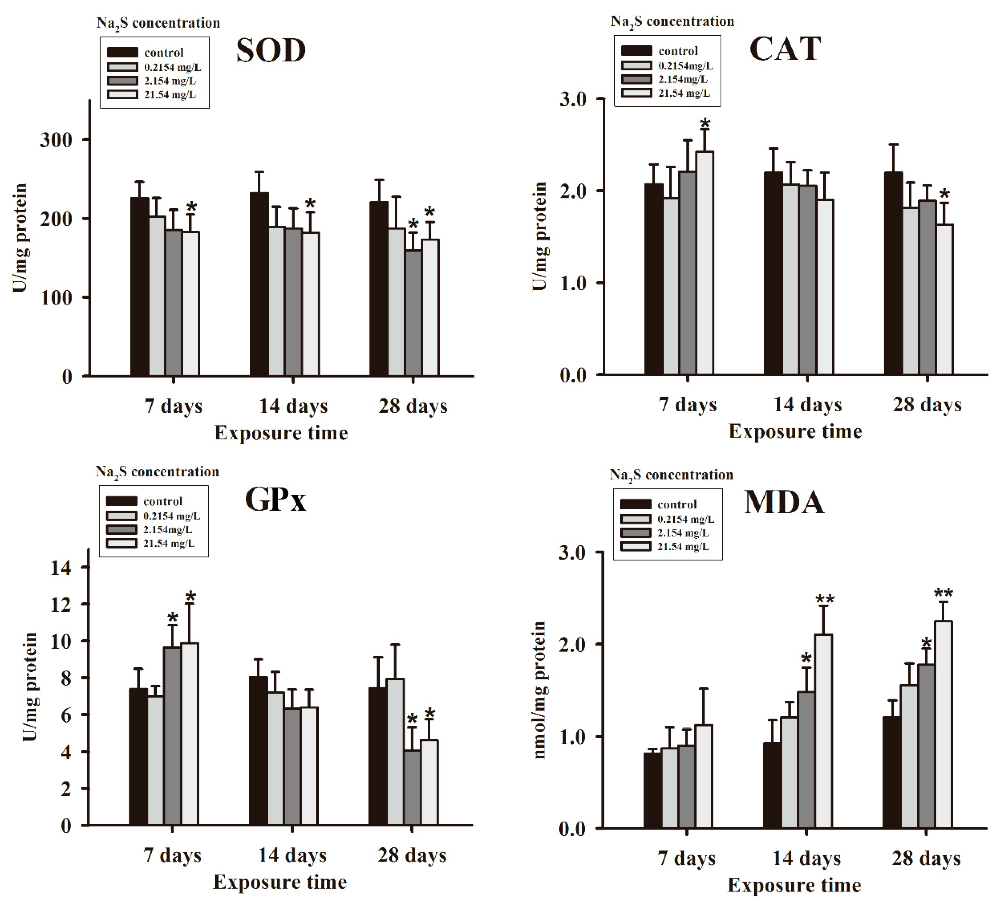
3.1. Antioxidant Parameters

The changes in the antioxidant parameters exposed to sulfide are illustrated in Figure 1. Generally, compared with the control, the activities of SOD were inhibited after exposure to various doses of Na2S. Significant inhibitions of SOD activities were presented in the 21.54 mg/L Na2S group exposed for 7 days and above and the 2.154 mg/L Na2S group exposed for 28 days. After 7 days of exposure, the CAT activity was significantly increased in the 21.54 mg/L Na2S group; when exposed for 14 and 28 days, there was a decrease trend for CAT activity in the sulfide treatment groups, and CAT activity was dramatically decreased after being exposed to 21.54 mg/L Na2S for 28 days. When exposed to a higher concentration of Na2S (2.154 mg/L and 21.54 mg/L) for a short time (7 days), the GPx activities were significantly increased while the GPx activities were significantly decreased after exposure for a long time (28 days). There was no significant difference in the MDA content after being exposed to sulfide for 7 days. An increasing trend was shown after being treated with sulfide for 14 days or above. The MDA levels were remarkably enhanced in the 2.154 mg/L and 21.54 mg/L Na2S groups after exposure for 14 or 28 days.

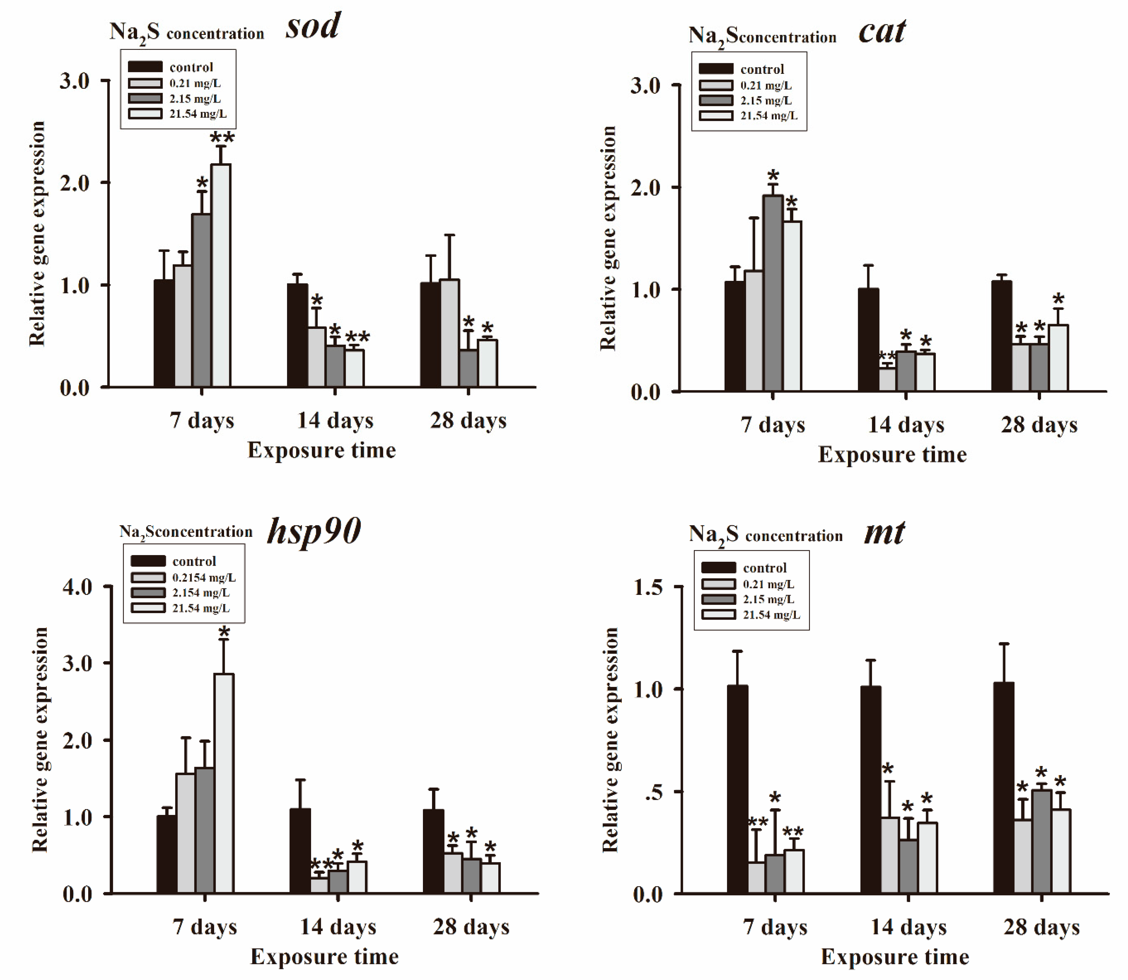
3.2. Genes Expressions

The genes expressions including sod, cat, hsp90, and mt are shown in Figure 2. Exposure to sulfide for 7 days, the sod expression was significantly upregulated in the 2.154 mg/L and 21.54 mg/L Na2S groups. While exposed for 14 days, significant downregulation of sod expression was detected in all of the sulfide treated groups. Additionally, sod expression was significantly decreased in the higher Na2S dose groups (2.154 mg/L and 21.54 mg/L) after exposure for 28 days. Similar to the expression pattern of sod for a short time (7 days) exposure to sulfide enhanced the expression of cat, and exposure to 2.154 and 21.54 mg/L Na2S significantly upregulated the cat expression level while long time (14 and 28 days) exposure in all sulfide treatment groups significantly downregulated the cat expression level. For hsp90 expression, it was dramatically upregulated after exposure to 21.54 mg/L Na2S for 7 days. With the increase in the exposure period (14 and 28 days), hsp90 expression was significantly downregulated in all sulfide exposure groups. It was shown that the expression of mt was significantly downregulated in all sulfide treated groups regardless of the exposure time.

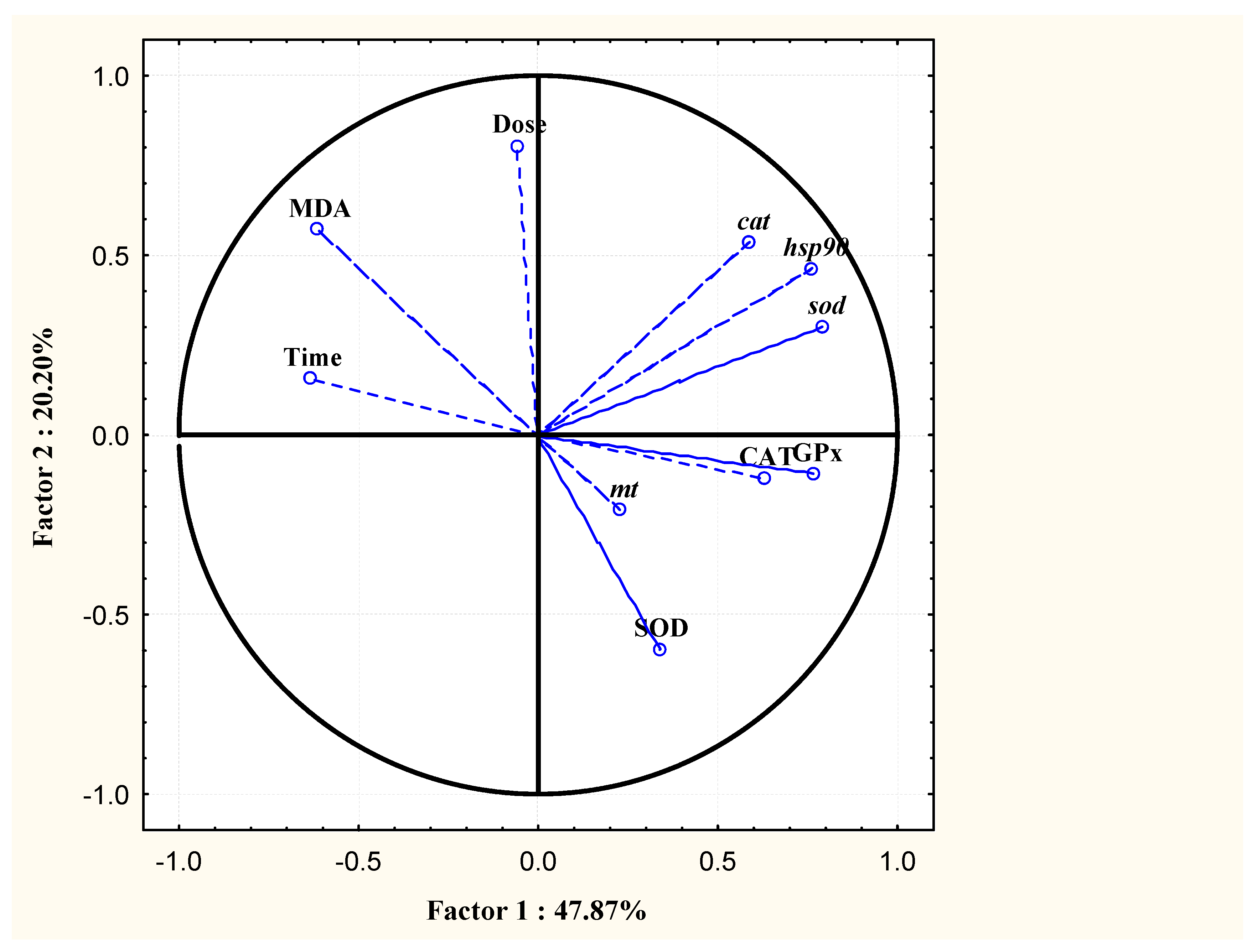
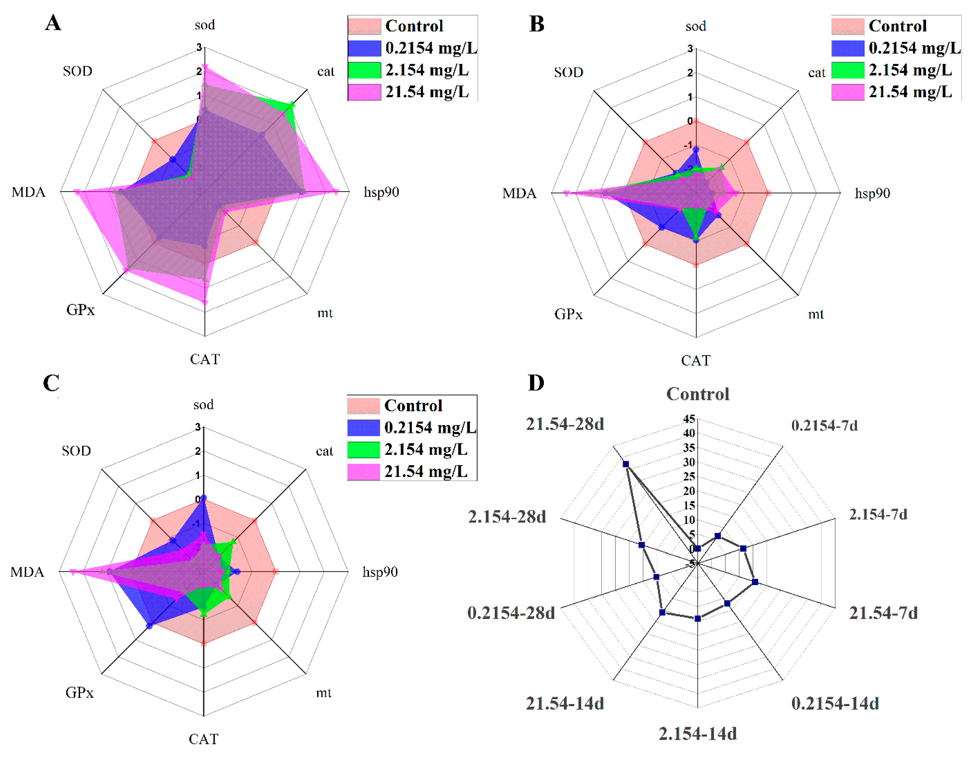
3.3. PCA and IBR Results

The PCA results are shown in Figure 3. The first two principal components (PC) explained 68.07% of the total variances. The PC1 and PC2 accounted for 47.87% and 20.2% of the total variances, respectively. The MDA level had a highly positive correlation with the Na2S concentrations as well as exposure time.
The IBR results are presented in Figure 4. Based on all parameters, there were two responsive patterns according to the exposure period. The star plot of the 7 days exposure was remarkably different from the plots of the 14 and 28 days of exposure (Figure 4A−C). After exposure to sulfide for 7 days, all biomarkers were activated or upregulated except for SOD and mt, which was inhibited and downregulated by sulfide, respectively. After sulfide exposure for 14 and 28 days, all indicators were decreased or downregulated except for the MDA content, which was increased by sulfide. The IBR values of each treatment are shown in Figure 4D, which were calculated based on all of the biomarkers detected. The high IBR value stands for high stress caused by the environmental factor. As seen from the star plot, toxic effects generated by sulfide were in dose−dependent and time−dependent manners.

4. Discussion

Generally, there is a dynamic equilibrium between oxidants and antioxidants in organisms [29], which is crucial for the health of organisms. When antioxidants are exhausted or ROS accumulates excessively, the equilibrium will be disturbed and proposes oxidative stress to the organism [30,31]. Oxidative stress was caused by multiple environmental factors such as UV exposure, environmental pollution, stress of hypoxia, pathogenic infection, etc. [32,33,34,35]. In aquatic organisms, the detoxification of ROS is controlled by the antioxidant defense system, which was composed of antioxidant enzymes (including SOD, CAT, glutathione reductase and GPx) and nonenzymatic antioxidants such as vitamin C, reduced glutathione (GSH), vitamin A, and vitamin E [17,19,36]. SOD, the first line of defense against ROS in living beings, converses superoxide radicals (O2) into hydrogen peroxide (H2O2). Thereafter, H2O2 is reduced by CAT to form molecular oxygen and water. GPx catalyzes the reduction of both hydrogen peroxide and lipid peroxide [32] and plays an antidotal role in protecting cells from oxidative stress.
The well-known toxicity of sulfide is that it can suppress the activity of cytochrome c oxidase in mitochondria, disturb electron transport, and reduce the production of ATP [1,2,37]. Exposure to sulfide also induced the generation of ROS in multiple organisms [3,38,39]. Therefore, a deduction could be made assuredly that the antioxidant defense system was affected by sulfide treatment. In aquatic organisms, alternation of antioxidant responses under sulfide stress for a short time (no more than 72 h) has been reported in several studies [10,22,23]. However, the changes in the antioxidant defense system for chronic exposure to sulfide have rarely been reported, especially in fish. In the present research, the rice field eel, an economically important aquaculture fish in China, was used to evaluate the effects of the antioxidant defense system under exposure to sulfide for 28 days.
After exposure to 2.154 and 21.54 mg/L Na2S for a short time (7 days), the gene transcription of sod and cat were significantly enhanced, coupled with significant increase in the activities of CAT and GPx. The level of MDA, which was an important indicator reflecting lipid peroxidation, did not change after short time exposure. This result indicates that the rice field eel did not suffer oxidative stress after treatment with sulfide for 7 days. This might be attributed to the capacity of antioxidant defense system being enhanced to scavenge excessive ROS by an increase in the activity of antioxidant enzymes such as CAT, GPx, etc. under short time sulfide stress. This consequence also indicated that adaptive responses to oxidative stress worked to maintain redox status in rice field eel. It is worth noting that the activities of SOD were decreased while the transcription levels of sod were significantly upregulated after exposure to 2.154 and 21.54 mg/L Na2S. This inconsistency might be attributable to there being three SOD isoforms in the organisms including Cu/Zn-SOD, Mn-SOD, and Fe-SOD [40], and only one type of SOD isoform transcription level was measured in the present study. After a prolonged exposure period (28 days), the activities of SOD, CAT, GPx, and the transcription levels of sod and cat were decreased and downregulated, respectively, in the higher sulfide dose (2.154 and 21.54 mg/L Na2S) groups. The MDA contents were remarkably increased after higher sulfide dose (2.154 and 21.54 mg/L Na2S) treatment for long time (14 and 28 days) exposure, suggesting the capacity of the antioxidant defense system failed to clear excessive ROS and the equilibrium between the oxidants and antioxidants was broken. The oxidative stress was mainly generated by the inhibition of antioxidant enzyme exposure to high doses of sulfide. Based on the time effects of sulfide, short-term exposure resulted in an increase in the activity of antioxidant enzymes and long-term exposure caused damage to the antioxidant defense system, leading to oxidative stress to the rice field eel. Although the exposure times were different in the present research, the activities of SOD, CAT, and GPx were increased after short time exposure to sulfide, and these activities were inhibited gradually for a prolonged exposure time in red swamp crayfish [10]. Aside from sulfide, similar consequences were obtained when common carp and rainbow trout were exposed to tributyltin and verapamil, respectively [41,42].
Heat shock proteins, also called stress proteins, function as molecular chaperones by helping correct the folding of the native and refolding of the denatured proteins [43,44]. Not only heat shock but also multiple stressful conditions such as oxidative stress, pollutants, and other toxic chemicals can induce the expression of HSPs [43,45]. According to the molecular weight, there are five types of HSP families, and the HSP90 is well conserved and abundant cytosolic protein in the eukaryotic cells [44]. The expression of hsp90 has been used as a biomarker for environmental assessment for its sensitivity to environmental stress [46]. In the present study, the expression of hsp90 was significantly upregulated in the highest sulfide dose group after 7 days of exposure. This situation may be attributable to hsp90 playing the role of refolding the damaged proteins to avoid cell damage under sulfide stress. When rice field eel was exposed to sulfide for 14 days and above, the transcription levels of hsp90 were remarkably downregulated in all sulfide treatment groups [44,46,47]. The expression of HSPs was changed in different tissues, pollutants, exposure concentrations, and periods [47]. Werner and Hinton [48] demonstrated that the protein level of HSP was reduced in polluted field sites, which might be due to the inhibition of protein biosynthesis and the decreased availability of energy by contaminants. Thus, we hypothesized a similar reason for the downregulated expression of hsp90 after sulfide treatment for a long time period in the present study.
MT is a family of cysteine-rich, low molecular weight cytosolic proteins that function in metal sequestration, protection against the toxicity of heavy metals, and oxidant stress [32,49]. Aside from metal, there is a high variety of factors including oxidative stress, radiation, cytokines, and growth factors [50]. MT is a useful biomarker for environmental research [32,51]. In the present study, exposure to sulfide significantly downregulated the transcriptional level of mt, and similar results were found by Guo, Ruan, Fan, Fang, Wang, Luo, and Yi [10], who reported that the expression level of mt was decreased significantly after treatment with sulfide for 48 h and 72 h in Procambarus clarkia [10]. Although further studies are needed to extend the knowledge about the role of MT under environmental stress, several reports have confirmed that there was a downregulation of MT in polluted waters [52,53].
In conclusion, the results of the present study demonstrated that the oxidative stress indices (SOD, CAT, GPx, and MDA) and related genes (sod, cat, hsp90, and mt) were altered under sulfide stress. Exposure to sub-lethal concentrations of sulfide caused oxidative stress and physiological stress in rice field eel. The toxic effects induced by sulfide were in dose-dependent and time-dependent manners.

Author Contributions

Conceptualization, L.Z. and Y.L.; methodology, F.Y., H.Z. and H.X.; software, N.W.; resources, H.R.; writing—original draft preparation, L.Z.; writing—review and editing, L.Z.; project administration, Z.N.; funding acquisition, Z.L. and Y.L. All authors have read and agreed to the published version of the manuscript.

Funding

This work was financially supported by the Central Public-interest Scientific Institution Basal Research Fund, the Chinese Academy of Fishery Sciences (CAFS) (Nos. 2020XT0801, 2020TD09, 2020TD33), the National Natural Science Foundation of China (Grant 21507162), and the China Three Gorges Project Corporation (No. 0799555).

Data Availability Statement

Not Acceptable.

Conflicts of Interest

The authors declare no conflict of interest.

References

  1. Bagarinao, T. Sulfide as an environmental factor and toxicant: Tolerance and adaptations in aquatic organisms. Aquat. Toxicol. 1992, 24, 21–62. [Google Scholar] [CrossRef]
  2. Shen, Y.Y.; Chen, J.Q.; Shen, W.L.; Chen, C.F.; Lin, Z.H.; Li, C.H. Molecular characterization of a novel sulfide: Quinone oxidoreductase from the razor clam Sinonovacula constricta and its expression response to sulfide stress. Comp. Biochem. Phys. B 2020, 239, 110367. [Google Scholar] [CrossRef]
  3. Shao, Y.; Chen, Z.L.; Wu, L.L. Oxidative Stress Effects of Soluble Sulfide on Human Hepatocyte Cell Line LO2. Int. J. Environ. Res. Public Health 2019, 16, 1662. [Google Scholar] [CrossRef] [Green Version]
  4. Petruci, J.F.D.; Cardoso, A.A. A new palladium chelate compound for determination of sulfide. Microchem. J. 2013, 106, 368–372. [Google Scholar] [CrossRef]
  5. Wu, L.L.; Shao, Y.; Hu, Z.J.; Gao, H.W. Effects of soluble sulfide on zebrafish (Danio rerio) embryonic development. Environ. Toxicol. Pharmcol. 2016, 42, 183–189. [Google Scholar] [CrossRef]
  6. Li, T.Y.; Li, E.C.; Suo, Y.T.; Xu, Z.X.; Jia, Y.Y.; Qin, J.G.; Chen, L.Q.; Gu, Z.M. Energy metabolism and metabolomics response of Pacific white shrimp Litopenaeus vannamei to sulfide toxicity. Aquat. Toxicol. 2017, 183, 28–37. [Google Scholar] [CrossRef]
  7. Forgan, L.G.; Forster, M.E. Oxygen consumption, ventilation frequency and cytochrome c oxidase activity in blue cod (Parapercis colias) exposed to hydrogen sulphide or isoeugenol. Comp. Biochem. Phys. C 2010, 151, 57–65. [Google Scholar] [CrossRef]
  8. Khan, A.A.; Schuler, M.M.; Prior, M.G.; Yong, S.; Coppock, R.W.; Florence, L.Z.; Lillie, L.E. Effects of hydrogen sulfide exposure on lung mitochondrial respiratory chain enzymes in rats. Toxicol. Appl. Pharmcol. 1990, 103, 482–490. [Google Scholar] [CrossRef]
  9. Suo, Y.T.; Li, E.C.; Li, T.Y.; Jia, Y.Y.; Qin, J.G.; Gu, Z.M.; Chen, L.Q. Response of gut health and microbiota to sulfide exposure in Pacific white shrimp Litopenaeus vannamei. Fish Shellfish Immunol. 2017, 63, 87–96. [Google Scholar] [CrossRef]
  10. Guo, K.; Ruan, G.L.; Fan, W.H.; Fang, L.; Wang, Q.; Luo, M.Z.; Yi, T.L. The effect of nitrite and sulfide on the antioxidant capacity and microbial composition of the intestines of red swamp crayfish, Procambarus clarkii. Fish Shellfish Immunol. 2020, 96, 290–296. [Google Scholar] [CrossRef]
  11. Tay, A.S.; Chew, S.F.; Ip, Y.K. The swamp eel Monopterus albus reduces endogenous ammonia production and detoxifies ammonia to glutamine during 144 h of aerial exposure. J. Exp. Biol. 2003, 206, 2473–2486. [Google Scholar] [CrossRef] [PubMed] [Green Version]
  12. Liang, X.M.; Zhang, J.; Guo, F.; Wei, L.L.; Zhou, Q.B. Oxidative stress and inflammatory responses in the liver of swamp eel (Monopterus albus) exposed to carbon tetrachloride. Aquaculture 2018, 496, 232–238. [Google Scholar] [CrossRef]
  13. Ma, X.; Hu, Y.; Wang, X.Q.; Ai, Q.H.; He, Z.G.; Feng, F.X.; Lu, X.Y. Effects of practical dietary protein to lipid levels on growth, digestive enzyme activities and body composition of juvenile rice field eel (Monopterus albus). Aquac. Int. 2014, 22, 749–760. [Google Scholar] [CrossRef]
  14. Xiang, J.H.; Qin, L.; Zhao, D.; Xiong, F.; Wang, G.T.; Zou, H.; Li, W.X.; Li, M.; Song, K.B.; Wu, S.G. Growth performance, immunity and intestinal microbiota of swamp eel (Monopterus albus) fed a diet supplemented with house fly larvae (Musca domestica). Aquac. Nutr. 2020, 26, 693–704. [Google Scholar] [CrossRef]
  15. Yue, H.M.; Huang, X.Q.; Ruan, R.; Ye, H.; Li, Z.; Li, C.J. Effects of dietary protein levels on the growth, body composition, serum biochemistry and digestive enzyme activity in Chinese rice field eel (Monopterus albus) fingerlings. Aquac. Res. 2020, 51, 400–409. [Google Scholar] [CrossRef]
  16. Schröder, P.; Krutmann, J. Environmental Oxidative Stress—Environmental Sources of ROS. In Reactions, Processes: Oxidants and Antioxidant Defense Systems; Grune, T., Ed.; Springer: Berlin/Heidelberg, Germany, 2005; pp. 19–31. [Google Scholar]
  17. Birben, E.; Sahiner, U.M.; Sackesen, C.; Erzurum, S.; Kalayci, O. Oxidative stress and antioxidant defense. World Allergy Organ. J. 2012, 5, 9–19. [Google Scholar] [CrossRef] [Green Version]
  18. Guemouri, L.; Artur, Y.; Herbeth, B.; Jeandel, C.; Cuny, G.; Siest, G. Biological variability of superoxide dismutase, glutathione peroxidase, and catalase in blood. Clin. Chem. 1991, 37, 1932–1937. [Google Scholar] [CrossRef]
  19. Aliahmat, N.S.; Noor, M.R.M.; Yusof, W.J.W.; Makpol, S.; Ngah, W.Z.W.; Yusof, Y.A.M. Antioxidant enzyme activity and malondialdehyde levels can be modulated by Piper betle, tocotrienol rich fraction and Chlorella vulgaris in aging C57BL/6 mice. Clinics 2012, 67, 1447–1454. [Google Scholar] [CrossRef]
  20. Camkurt, M.A.; Findikli, E.; Bakacak, M.; Tolun, F.I.; Karaaslan, M.F. Evaluation of Malondialdehyde, Superoxide Dismutase and Catalase Activity in Fetal Cord Blood of Depressed Mothers. Clin. Psychopharmacol. Neurosci. 2017, 15, 35–39. [Google Scholar] [CrossRef] [Green Version]
  21. Sato, M.; Bremner, I. Oxygen free radicals and metallothionein. Free Radic. Biol. Med. 1993, 14, 325–337. [Google Scholar] [CrossRef]
  22. Jiang, L.; Feng, J.X.; Ying, R.; Yin, F.M.; Pei, S.R.; Lu, J.G.; Cao, Y.T.; Guo, J.L.; Li, Z.F. Individual and combined effects of ammonia-N and sulfide on the immune function and intestinal microbiota of Pacific white shrimp Litopenaeus vannamei. Fish Shellfish Immunol. 2019, 92, 230–240. [Google Scholar] [CrossRef] [PubMed]
  23. Duan, Y.F.; Dong, H.B.; Wang, Y.; Li, H.; Liu, Q.S.; Zhang, Y.; Zhang, J.S. Intestine oxidative stress and immune response to sulfide stress in Pacific white shrimp Litopenaeus vannamei. Fish Shellfish Immunol. 2017, 63, 201–207. [Google Scholar] [CrossRef] [PubMed]
  24. Bradford. A rapid and sensitive method for the quantitation of microgam quantities of protein utilizing the principle of protein-dye binding. Anal. Biochem. 1976, 72, 248–254. [Google Scholar] [CrossRef]
  25. Zhong, L.; Yuan, L.; Rao, Y.; Li, Z.; Zhang, X.; Liao, T.; Xu, Y.; Dai, H.; Zhong, L.; Yuan, L. Distribution of vitellogenin in zebrafish (Danio rerio) tissues for biomarker analysis. Aquat. Toxicol. 2014, 149, 1–7. [Google Scholar] [CrossRef]
  26. Zhong, L.; Yuan, L.; Rao, Y.; Li, Z.; Gu, Q.; Long, Y.; Zhang, X.; Cui, Z.; Xu, Y.; Dai, H. Investigation of effect of 17α-ethinylestradiol on vigilin expression using an isolated recombinant antibody. Aquat. Toxicol. 2014, 156, 1–9. [Google Scholar] [CrossRef]
  27. Beliaeff, B.; Burgeot, T. Integrated biomarker response: A useful tool for ecological risk assessment. Environ. Toxicol. Chem. 2002, 21, 1316–1322. [Google Scholar] [CrossRef]
  28. Sanchez, W.; Burgeot, T.; Porcher, J.M. A novel “Integrated Biomarker Response” calculation based on reference deviation concept. Environ. Sci. Pollut. Res. 2013, 20, 2721–2725. [Google Scholar] [CrossRef]
  29. Jordan, J.; Galindo, M.F.; Calvo, S.; Gonzalez-Garcia, C.; Cena, V. Veratridine induces apoptotic death in bovine chromaffin cells through superoxide production. Br. J. Pharmacol. 2000, 130, 1496–1504. [Google Scholar] [CrossRef] [Green Version]
  30. Han, M.; Kim, C.Y.; Lee, J.; Lee, S.K.; Jeon, J.S. OsWRKY42 Represses OsMT1d and Induces Reactive Oxygen Species and Leaf Senescence in Rice. Mol. Cells 2014, 37, 532–539. [Google Scholar] [CrossRef]
  31. Kirschvink, N.; de Moffarts, B.; Lekeux, P. The oxidant/antioxidant equilibrium in horses. Vet. J. 2008, 177, 178–191. [Google Scholar] [CrossRef]
  32. van der Oost, R.; Beyer, J.; Vermeulen, N.P.E. Fish bioaccumulation and biomarkers in environmental risk assessment: A review. Environ. Toxicol. Pharmacol. 2003, 13, 57–149. [Google Scholar] [CrossRef]
  33. Blokhina, O.; Virolainen, E.; Fagerstedt, K.V. Antioxidants, oxidative damage and oxygen deprivation stress: A review. Ann. Bot. 2003, 91, 179–194. [Google Scholar] [CrossRef] [PubMed] [Green Version]
  34. Chen, L.G.; Lam, J.C.W.; Tang, L.Z.; Hu, C.Y.; Liu, M.Y.; Lam, P.K.S.; Zhou, B.S. Probiotic Modulation of Lipid Metabolism Disorders Caused by Perfluorobutanesulfonate Pollution in Zebrafish. Environ. Sci. Technol. 2020, 54, 7494–7503. [Google Scholar] [CrossRef]
  35. Chen, L.G.; Lam, J.C.W.; Hu, C.Y.; Tsui, M.M.P.; Wang, Q.; Giesy, J.P.; Lam, P.K.S. Perfluorobutanesulfonate Exposure Causes Durable and Transgenerational Dysbiosis of Gut Microbiota in Marine Medaka. Environ. Sci. Tech. Lett. 2018, 5, 731–738. [Google Scholar] [CrossRef]
  36. Livingstone, D.R. Contaminant-stimulated reactive oxygen species production and oxidative damage in aquatic organisms. Mar. Pollut. Bull. 2001, 42, 656–666. [Google Scholar] [CrossRef]
  37. Rubright, S.L.M.; Pearce, L.L.; Peterson, J. Environmental toxicology of hydrogen sulfide. Biol. Chem. 2017, 71, 1–13. [Google Scholar] [CrossRef]
  38. Fu, L.H.; Wei, Z.Z.; Hu, K.D.; Hu, L.Y.; Li, Y.H.; Chen, X.Y.; Han, Z.; Yao, G.F.; Zhang, H. Hydrogen sulfide inhibits the growth of Escherichia coli through oxidative damage. J. Microbiol. 2018, 56, 238–245. [Google Scholar] [CrossRef]
  39. Eghbal, M.A.; Pennefather, P.S.; O’Brien, P.J. H2S cytotoxicity mechanism involves reactive oxygen species formation and mitochondrial depolarisation. Toxicology 2004, 203, 69–76. [Google Scholar] [CrossRef]
  40. Tanaka, Y.; Hibino, T.; Hayashi, Y.; Tanaka, A.; Kishitani, S.; Takabe, T.; Yokota, S.; Takabe, T. Salt tolerance of transgenic rice overexpressing yeast mitochondrial Mn-SOD in chloroplasts. Plant Sci. 1999, 148, 131–138. [Google Scholar] [CrossRef]
  41. Li, Z.H.; Velisek, J.; Zlabek, V.; Grabic, R.; Machova, J.; Kolarova, J.; Li, P.; Randak, T. Chronic toxicity of verapamil on juvenile rainbow trout (Oncorhynchus mykiss): Effects on morphological indices, hematological parameters and antioxidant responses. J. Hazard. Mater. 2011, 185, 870–880. [Google Scholar] [CrossRef]
  42. Li, Z.H.; Li, P.; Shi, Z.C. Chronic Exposure to Tributyltin Induces Brain Functional Damage in Juvenile Common Carp (Cyprinus carpio). PLoS ONE 2015, 10, e0123091. [Google Scholar] [CrossRef] [PubMed] [Green Version]
  43. Gupta, S.C.; Sharma, A.; Mishra, M.; Mishra, R.K.; Chowdhuri, D.K. Heat shock proteins in toxicology: How close and how far? Life Sci. 2010, 86, 377–384. [Google Scholar] [CrossRef]
  44. Padmini, E.; Rani, M.U. Seasonal influence on heat shock protein 90 alpha and heat shock factor 1 expression during oxidative stress in fish hepatocytes from polluted estuary. J. Exp. Mar. Biol. Ecol. 2009, 372, 1–8. [Google Scholar] [CrossRef]
  45. Richter, K.; Haslbeck, M.; Buchner, J. The Heat Shock Response: Life on the Verge of Death. Mol. Cell 2010, 40, 253–266. [Google Scholar] [CrossRef]
  46. Liu, Q.P.; Huang, S.T.; Deng, C.A.; Xiong, L.; Gao, X.; Chen, Y.; Niu, C.Q.; Liu, Y. Molecular characterization of heat-shock protein 90 gene and its expression in Gobiocypris rarus juveniles exposed to pentachlorophenol. Fish Physiol. Biochem. 2015, 41, 1279–1291. [Google Scholar] [CrossRef]
  47. Taylor, D.A.; Thompson, E.L.; Nair, S.V.; Raftos, D.A. Differential effects of metal contamination on the transcript expression of immune- and stress-response genes in the Sydney Rock oyster, Saccostrea glomerata. Environ. Pollut. 2013, 178, 65–71. [Google Scholar] [CrossRef]
  48. Werner, I.; Hinton, D.E. Field validation of hsp70 stress proteins as biomarkers in Asian clam (Potamocorbula amurensis): Is downregulation an indicator of stress? Biomarkers 1999, 4, 473–484. [Google Scholar]
  49. Coyle, P.; Philcox, J.C.; Carey, L.C.; Rofe, A.M. Metallothionein: The multipurpose protein. Cell. Mol. Life Sci. 2002, 59, 627–647. [Google Scholar] [CrossRef]
  50. Ruttkay-Nedecky, B.; Nejdl, L.; Gumulec, J.; Zitka, O.; Masarik, M. The Role of Metallothionein in Oxidative Stress. Int. J. Mol. Sci. 2013, 14, 6044–6066. [Google Scholar] [CrossRef] [Green Version]
  51. Cosson, R.P. Bivalve metallothionein as a biomarker of aquatic ecosystem pollution by trace metals: Limits and perspectives. Cell Mol. Biol. 2000, 46, 295–309. [Google Scholar]
  52. Falfushynska, H.; Horyn, O.; Fedoruk, O.; Khoma, V.; Rzymski, P. Difference in biochemical markers in the gibel carp (Carassius auratus gibelio) upstream and downstream of the hydropower plant. Environ. Pollut. 2019, 255, 113213. [Google Scholar] [CrossRef] [PubMed]
  53. Falfushynska, H.I.; Gnatyshyna, L.L.; Stoliar, O.B.; Nam, Y.K. Various responses to copper and manganese exposure of Carassius auratus gibelio from two populations. Comp. Biochem. Phys. C 2011, 154, 242–253. [Google Scholar] [CrossRef] [PubMed]
Figure 1. The changes in the oxidative stress parameters (SOD, CAT, GPx, and MDA) in the liver of rice field eel after exposure to 0, 0.2154, 2.154, and 21.54 mg/L Na2S for 7, 14, and 28 days. The results are shown as the mean ± SD of six replicates. * p < 0.05, ** p < 0.01 indicate the statistically significant differences between the exposure and control groups.
Water 14 03230 g001
Figure 2. Quantitative RT-PCR analysis of the expression levels of sod, cat, hsp90, and mt in the liver of rice field eel after exposure to 0, 0.2154, 2.154, and 21.54 mg/L Na2S for 7, 14, and 28 days. The results are shown as the mean ± SD of six replicates. * p < 0.05, ** p < 0.01 indicate the statistically significant differences between the exposure and control groups.
Water 14 03230 g002
Figure 3. Ordination diagram of the PCA of Na2S doses, exposure time, and parameters measured in rice field eel after Na2S exposure.
Water 14 03230 g003
Figure 4. Star plots for measuring the various parameters of rice field eel after exposure to sulfide for 7 days (A), 14 days (B), 28 days (C), and IBR values for each treatment (D). Control, control group; 0.2154−7d, 0.2154 mg/L Na2S exposed for 7 days; 2.154−7d, 2.154 mg/L Na2S exposed for 7 days; 21.54−7d, 21.54 mg/L Na2S exposed for 7 days; 0.2154−14d, 0.2154 mg/L Na2S exposed for 14 days; 2.154−14d, 2.154 mg/L Na2S exposed for 14 days; 21.54−14d, 21.54 mg/L Na2S exposed for 14 days; 0.2154−28d, 0.2154 mg/L Na2S exposed for 28 days; 2.154−28d, 2.154 mg/L Na2S exposed for 28 days; 21.54−28d, 21.54 mg/L Na2S exposed for 28 days.
Water 14 03230 g004
Table 1. Primer sequences for qRT-PCR.
NameSequence of Primer (5′-3′)Product
Length (bp)
Efficiency (%)Gene Bank ID
β-actin-FGAGGTATCCTGACCCTGAAGTA10596.09XM_020621264
β-actin-RCGACTCTCAGCTCATTGTAGAAG
sod-FCTCTCTCTCTCGGCACCTATTA133104.6XM_020602040
sod-RGACTCAAGGCTACAAAGGTCAA
cat-FCTGCACAGTGTCAGGTCTAAA11192.11XM_020624985
cat-RTGACCTCTGAGACCCTCTATTC
mt-FTACACACCACTGGCTCTTTG9493.59XM_020604384
mt-RCGCAAGGGTCCATCTCTTT
hsp90-FTAAGGAGGTTGAGGAGGATGAG9595.36XM_020622865
hsp90-RCCTCAGCTGTGAAGTGGATATG
Publisher’s Note: MDPI stays neutral with regard to jurisdictional claims in published maps and institutional affiliations.

