Article

New Data on Reproductive System and Spermatozoa Confirm Macrodasys as a Model in Comparative Reproductive Analysis in Macrodasyida (Gastrotricha)

1
Department of Biomolecular Sciences, University of Urbino, 61029 Urbino, Italy
2
Fano Marine Center, The Inter-Institute Center for Research on Marine Biodiversity, Resources and Biotechnologies, 61032 Fano, Italy
3
Department of Life Sciences, University of Modena and Reggio Emilia, 41125 Modena, Italy
*
Author to whom correspondence should be addressed.
Water 2022, 14(19), 3085; https://doi.org/10.3390/w14193085
Submission received: 5 September 2022 / Revised: 23 September 2022 / Accepted: 24 September 2022 / Published: 1 October 2022
(This article belongs to the Special Issue Research on the Aquatic Species Biodiversity and Morphology)

Abstract

:
Gastrotricha Macrodasyida, aquatic worms, are primarily hermaphrodites with internal cross-fertilization and indirect sperm transfer. Insightful information on this matter dates to 1978 and derives from an illuminating work focused on two Macrodasys species. The role as a “model” of this taxon clashes, however, with the relative paucity of data concerning the ultrastructure of the accessory organs involved in the sperm transfer. Therefore, we have undertaken a new study on three additional species of Macrodasys focusing on the frontal and caudal organs and also on the spermatozoa. Our observations confirmed the structural plan of the reproductive system reported for the genus, but found some differences concerning the accessory reproductive organs. Most importantly, we found spermatozoa inside the caudal organ of several single specimens, suggestive of a more plausible hypothesis about sperm transfer modality in Macrodasys. In the spermatozoa, the position of the mitochondrion outside rather than inside the nucleus has been confirmed as a major difference between the spermatozoa of Macrodasys species and those of the other Macrodasyida. We suggest that the peculiar structure of the mature spermatozoon of Macrodasys could derive from an incomplete process of spermatogenesis and hence it would be useful to infer phylogenetic alliances.

1. Introduction

Gastrotricha is a phylum of minute, acoelomate worms well represented in benthic communities, both in freshwater and marine ecosystems. They are grouped into two orders: Macrodasyida, with approximately 381 mostly marine species, and Chaetonotida, with approximately 485 freshwater and marine species. Gastrotrichs are primarily oviparous hermaphrodites with internal cross-fertilization, although parthenogenesis occurs in several marine taxa and it is the rule in the numerous freshwater species encompassing a wide taxonomic spectrum (e.g., [1,2,3]). The reproductive system of the hermaphroditic Macrodasyida is anatomically complex and diverse, probably related to internal cross-fertilization [4]. Usually, there are paired testes, one or two ovaries, and two sexual accessory organs called, respectively, frontal and caudal, because of their reciprocal positions. The frontal organ, female in function, lies around mid-body, near the most mature egg, and may show an external pore, through which it probably receives sperm during copulation, thus functioning as a seminal receptacle and possibly as a spermatheca. The caudal organ, male in function, is in the posterior body region, opens into a ventral pore close to or within the anus, and probably acts as a copulatory organ [4].
Variations on this rather common organization of the reproductive system include taxa possessing two ovaries and/or a single testis, others that lack the caudal organ or the frontal organ, and others that have even developed a sclerotized stylet to facilitate sperm transfer, etc., see [5]. The position of the gonads and the accessory reproductive organs may also vary among taxa, and the organization of the reproductive structures (presence/absence of some organs) and their layout (position along the body and in relation to each other) has proved to be of taxonomic and phylogenetic significance (e.g., [6,7,8]).
The family Macrodasyidae probably testifies best to the ample diversity of the Macrodasyida reproductive system. This family encompasses members bearing the full set of reproductive structures (e.g., Macrodasys), and others whose reproductive system seems to be restricted to an ovary and frontal organ (e.g., Thaidasys) or solely to the ovary (e.g., Urodasys viviparous); moreover, sclerotized stylets of different shapes and sizes are found in the caudal organ of several species of Urodasys. To complete the framework of the heterogeneous reproductive system found among macrodasyids, Todaro et al. [9] have recently unveiled the existence of a new genus, Kryptodasys, possessing a full set of reproductive structures similar to those of Macrodasys, yet distinct by virtue of the different position of the frontal organ in relation to the largest oocyte: anterior in Kryptodasys, posterior in Macrodasys.
In addition to showing the full spectrum (or nearly so) of the reproductive system organization found in the entire order Macrodasyida, the family Macrodasyidae also includes the only taxa for which both mating behavior and sperm transfer have been fully documented so far (i.e., species of the genus Macrodasys, see Ruppert [10]). It should be emphasized that in most hermaphroditic Gastrotricha, deferent ducts usually do not join the copulatory organ, but often open externally, via independent pores. Consequently, for cross-fertilization to happen, individuals in copula must first load their caudal organ with autosperm before spermatozoa can be transferred to the frontal organ of the mating partner.
Given the completeness of its reproductive system and the circumstantiated description of the way spermatozoa are passed indirectly to the partner during copulation, Macrodasys has become a reference/model in the comparative analysis of the reproductive systems in macrodasyidan Gastrotricha, and also to infer potential sperm transfer modalities in these animals (e.g., [4,5,11,12]).
The importance that Macrodasys has assumed over time as a model organism concerning the reproductive biology of Gastrotricha clashes with the relative paucity of data concerning the ultrastructure of the accessory reproductive structures (frontal and caudal organs), which have already been reported to bear species-specific differences under optical microscopy (e.g., [13,14,15,16,17]).
In this study, we aim to reduce this gap by providing ultrastructural information on the accessory organ of two species of Macrodasys (M. caudatus and Macrodasys sp. 3) which, in addition to the information provided by Ruppert [10] on two other species, should deliver a clearer picture in this regard. We also provide an overview of the general organization of the entire reproductive system of these two species. Furthermore, again to increase the knowledge of the reproductive biology of Macrodasys, in this study we also describe the ultrastructure of the spermatozoon of three species (M. meristocytalis, Macrodasys sp. 2, and Macrodasys sp. 3) underlining the fact that currently the ultrastructure of the male gamete of Macrodasys is only known for two species, i.e., Macrodasys sp. II and M. caudatus [10,18].

2. Materials and Methods

Adult specimens were extracted from fully marine sandy sediments: Macrodasys caudatus and Macrodasys sp. 3 from the Tyrrhenian Sea (Punta Ala, Italy), M. meristocytalis from the Caribbean Sea (Cancun, Mexico), and Macrodasys sp. 2 from the Western Mediterranean Sea (La Marmorata, Sardinia, Italy). Specimens were extracted using the narcotization-decantation technique (using a 7% magnesium chloride solution as anesthetic), identified, and photographed under a Leitz Dialux 20 (Wetzlar, Germany) microscope equipped with Nomarski optics. Photomicrographs of M. caudatus were obtained with a Nikon Coolpix 995 (Tokyo, Japan) digital camera. After being identified, single specimens were recovered from the slides and fixed overnight in a 0.1-M phosphate buffered (pH 7.3) solution of paraformaldehyde, glutaraldehyde, and picric acid [19]. Thereafter, the animals were washed in 0.1 M phosphate buffer (PBS) at pH 7.2 and postfixed in a 2% osmium tetroxide solution. After rinsing in PBS, the specimens were dehydrated in a graded acetone series, stained en bloc in uranyl acetate in 70% acetone, and embedded in Araldite. Ultrathin sections were cut with an LKB Ultrotome 2088V, (Bromma, Sweden) contrasted with lead citrate, and observed under a Zeiss 902 (Eindhoven, Netherlands) transmission electron microscope.

3. Results

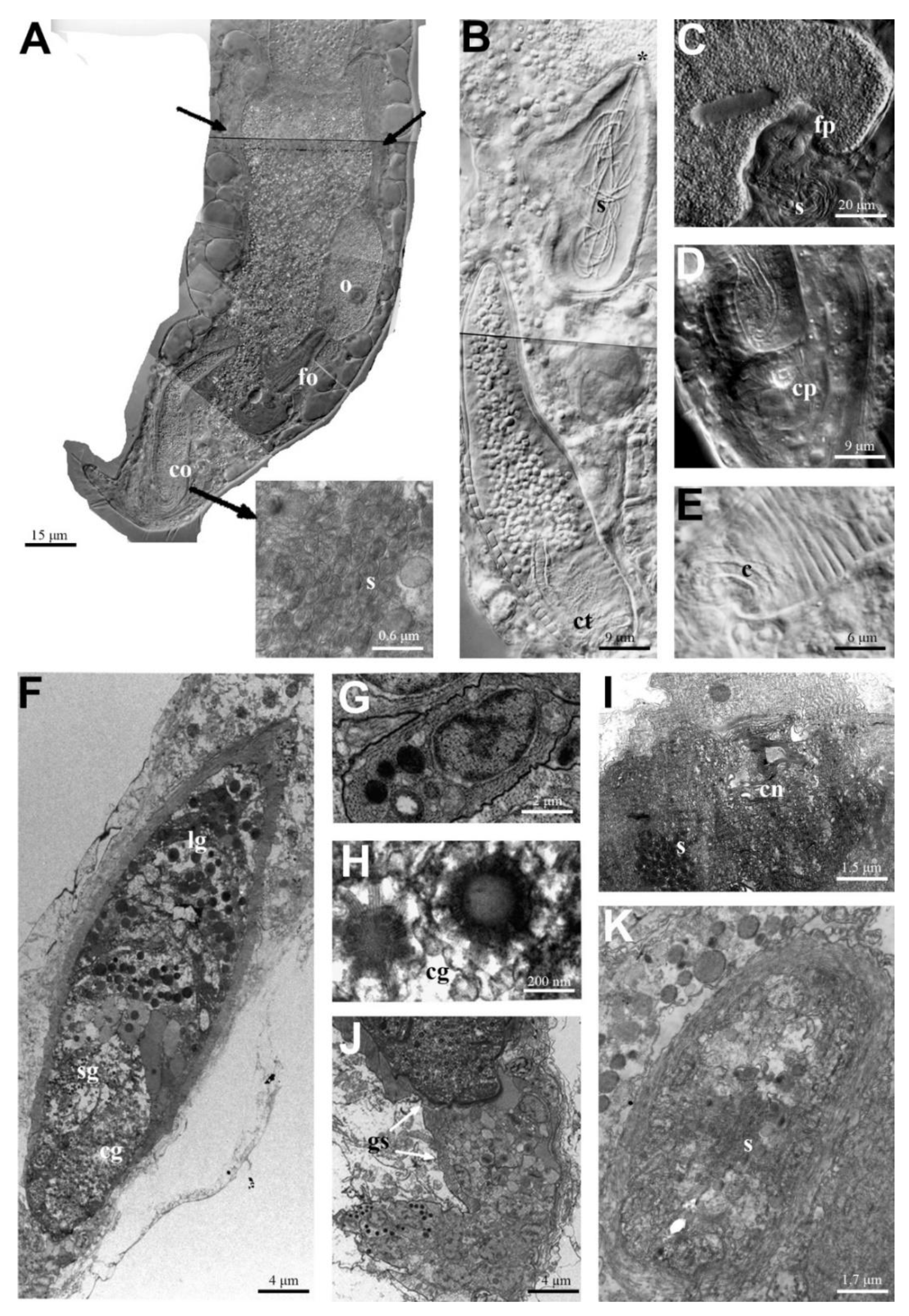
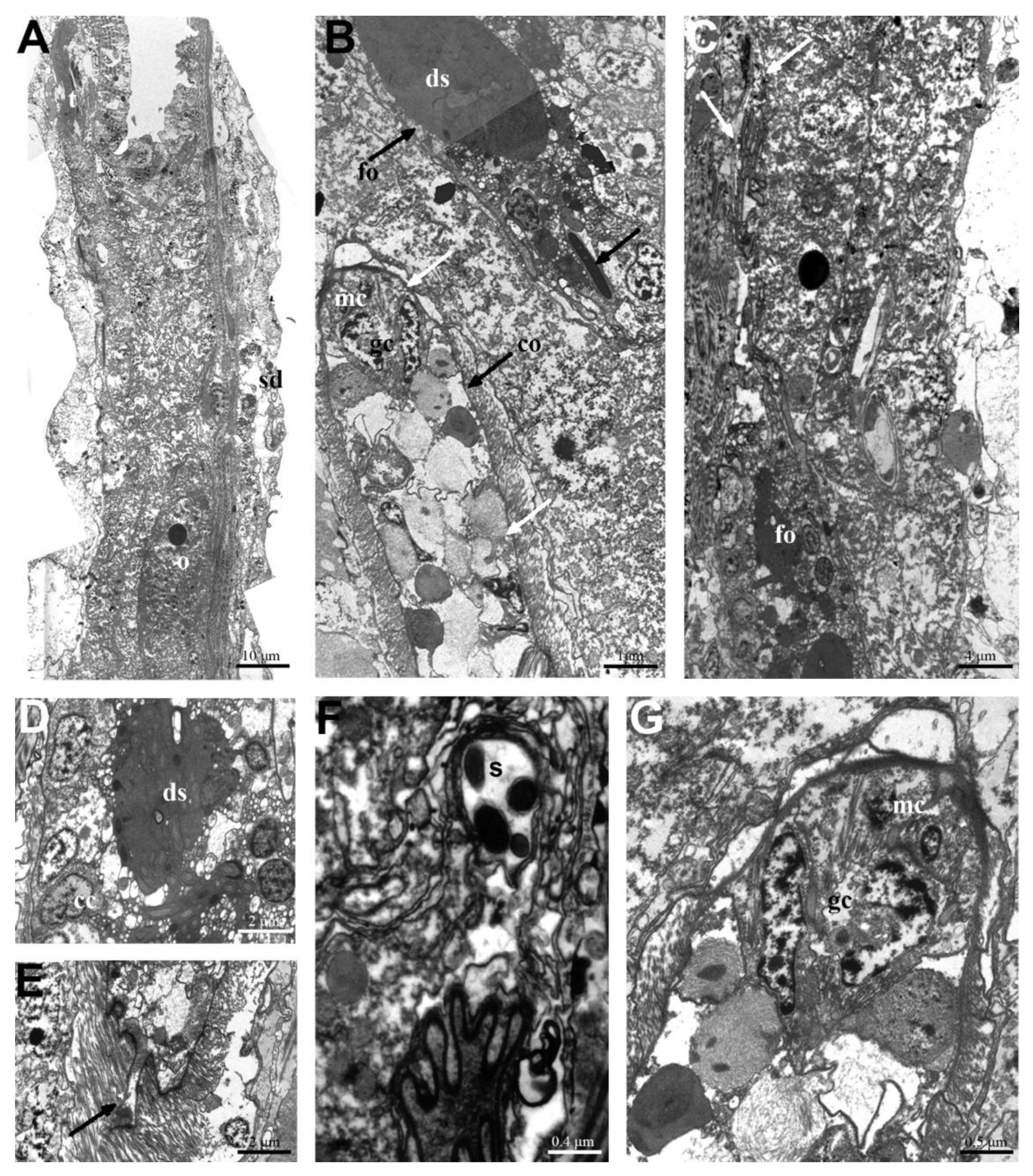
3.1. Macrodasys caudatus and Macrodasys sp. 3: Reproductive System

The following description is based on observations carried out on several individuals of M. caudatus and Macrodasys sp. 3 under both optical and electron microscopy.
The reproductive system of both species is composed of two testes, a single ovary, and two accessory reproductive structures (frontal and caudal organs). The male gonads lie anteriorly to the female ones and appear as elongated bands of germinal cells lateral to the intestine, which extends posteriorly from the level of the pharingeo-intestinal junction for about 80 µm in length (Figure 1A and Figure 2A).
The germinal male cells mature in a caudo-cephalic direction, so that the final spermatids and the mature spermatozoa are located in the anteriormost part of each testis. Here, the mature spermatozoa rotate their heads in a caudal direction and go towards the sperm duct (Figure 1A and Figure 2A).
The ovary, unpaired and compact, is located laterally to the left side of the middle gut. The oocytes grow in a caudo-cephalic direction into the central body region, and then move dorsally as they undergo growth and vitellogenesis. Therefore, the most mature oocyte is dorsolateral, with its anterior portion in contact with the posterior end of the left testicle and its posterior part in contact with the anterior part of the frontal organ (Figure 1A,C and Figure 2A,C).
In both species, the frontal organ is located on the left side of the body, posterior to the most advanced vitellogenic oocyte (Figure 1A–C and Figure 2B,C). In M. caudatus, the organ is a piriform sac with a monolayered epithelial wall consisting of irregular flat cells delimiting an inner space in which active spermatozoa were seen. Some spermatozoa were observed moving towards the anterior tip of the organ, where a small, sclerotized nozzle is present. The nozzle shows a well-defined pore through which sperm may pass to reach and fertilize the eggs (Figure 1B,C,I).
In Macrodasys sp. 3, the frontal organ has been documented only under TEM. It appears as a large ovoidal sac, made up of a monolayer of cuboidal cells delimiting a central space full of degenerating sperm packed in a large electron-dense mass (Figure 2B–D). Only a few sperm have the normal morphology (Figure 2B,F). Very long cytoplasmic protrusions of the cuboidal cells form a narrow channel that reaches the vitellogenic oocyte. In the anteriormost region of this channel, in contact with the mature oocyte, several spermatozoa were observed (Figure 2C,F). The frontal organ of Macrodasys sp. 3 lacks a sclerotized nozzle, and consequently, the internal pore of the frontal organ, through which sperm may reach the oocytes, remains elusive.
In M. caudatus, the caudal organ is located posteriorly to the frontal organ on the right side of the body. It is formed by a spindle-shaped (75 µm long) massive glandulomuscular anterior region and a short, glandular, posterior cellular sac (i.e., 10 µm long, Figure 1A,B,F,J).
The glandulomuscular region consists of a highly active secretory epithelium, coated with a well-developed muscular layer, longitudinally orientated at the anterior end of the organ and circularly at the posterior one (Figure 1B,F). Two parts of this region could be clearly distinguished by the appearance of the secretory granules: large, round, electron-dense granules were present in the anterior part (60 µm long) (Figure 1F), whereas in the posterior part small granules with an electron-dense transparent core and crystalline granules, resembling a spur with 11 rays (on the right side), were observed (Figure 1F,H). Moreover, and most importantly, in the anterior part of the glandulomuscular organ of four specimens, numerous spermatozoa were observed (Figure 1K) and inside the posterior part a straight, minute copulatory tube was visible (Figure 1B,D). The presence of spermatozoa and a copulatory tube inside the caudal organ indicated that these specimens were ready to copulate (see Discussion below).
The glandular posterior sac-like region is connected to the anterior region of the caudal organ through a collar formed by secretory cells anchored to the thickened basal lamina, which surround the posterior end of the glandulomuscular region. The sac then extends posteriorly and ventrally, folding at 90°, and opens through the body wall with a permanent ventral pore (Figure 1D). Its glandular cells are full of completely or partially electron-dense granules, are rich in disorganized muscular microfibrils, and show an evident centriole (Figure 1E,G,J).
The caudal organ of Macrodasys sp. 3 has a position and a basic structure like that of M. caudatus. The anterior part of the glandulomuscular region is formed by large and small glandular cells surrounded by longitudinal muscles and separated from one another by muscular cells. In the cytoplasm of glandular cells, granules with the same morphology as those filling the posterior region, but smaller in size, were visible (Figure 2B,G). The posterior part of the glandulomuscular region is composed of secretory cells containing many types of cytoplasmic granules: here the muscles are circular in orientation (Figure 2B,G). The copulatory tube is minute and straight and originates at the base of this part of the organ (Figure 2E). Unfortunately, we could not obtain sections of the posterior cellular sac.

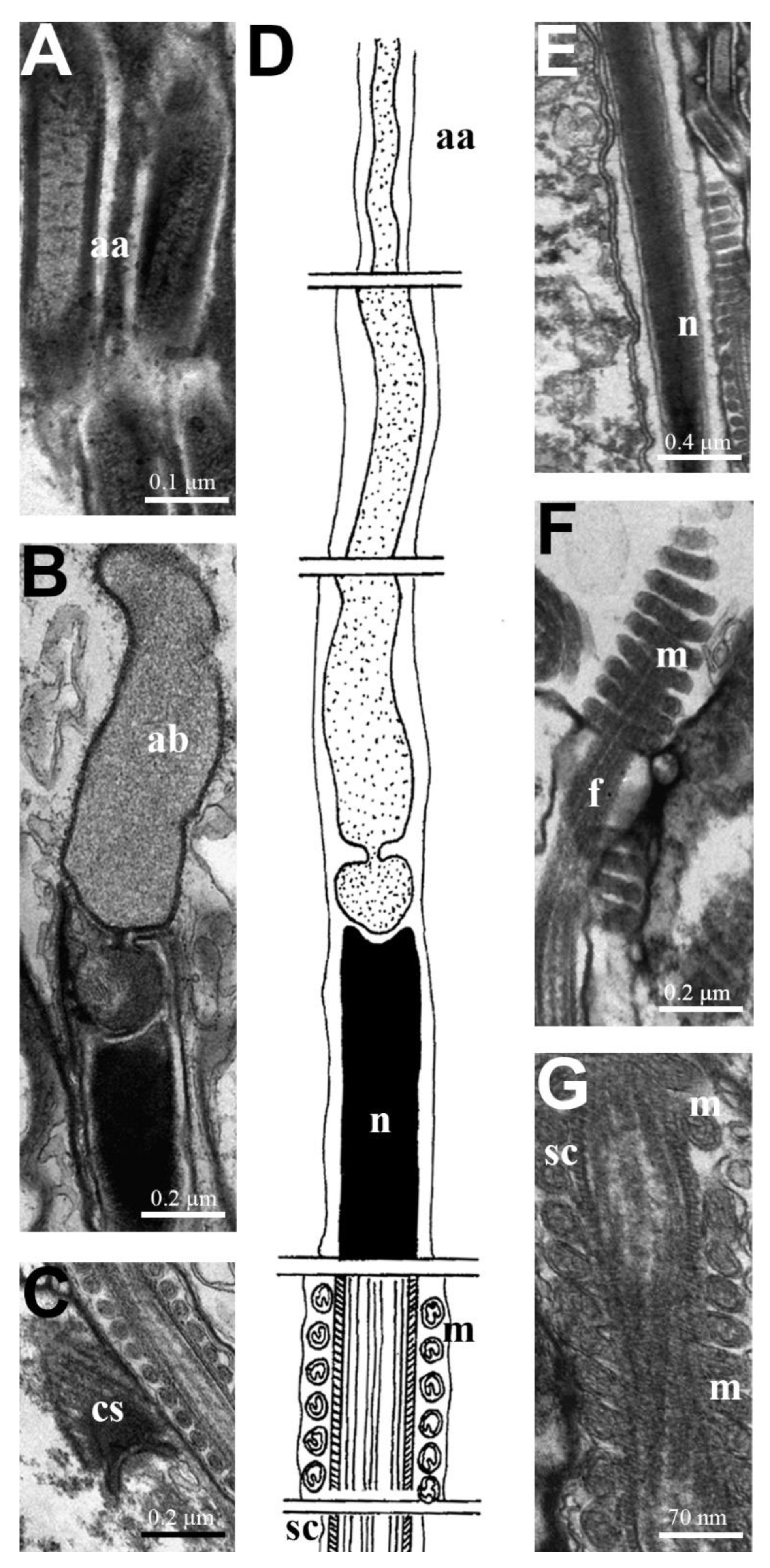
3.2. Macrodasys Spermatozoa

The spermatozoa of Macrodasys species are filiform cells, formed by an elongated head which is composed, in sequence, of the acrosome, the nucleus, and a flagellum, with a conventional 9 × 2 + 2 axoneme wrapped for its entire length with a striated cylinder, sensu Ferraguti and Balsamo [20]. A long, helical mitochondrion surrounds the nucleus and the acrosome, or the flagellum (Figure 3D and Figure 4F,G).
The acrosome consists of a long tubular structure starting from a basal nuclear cavity, and with a decreasing diameter from the base to the apex. In Macrodasys sp. 3 and M. meristocytalis, the base of the tubular structure is enlarged and roundish (average length 0.5 µm; average diameter 0.15 µm); whereas in Macrodasys sp. 2, it is enlarged and cylindrical (Figure 3A,D and Figure 4C,F,G,J).
The tubular structure of the acrosome contains a moderately electron-dense, homogeneous material for most of its length (Figure 3A,B and Figure 4A,C,H,J). In M. meristocytalis and Macrodasys sp. 2, the acrosomial apex is short and cork-screw-shaped (average length 0.8 µm), and in the first species it shows a pitch of about 72 nm and as many as 10 gyres (Figure 4A). In M. meristocytalis, the tubular structure of the acrosome is surrounded by an electron-dense layer that also extends to coat the whole nucleus (Figure 4A–C).
The nucleus is an elongated, straight or weakly helical rod of fully condensed chromatin, with an almost uniform diameter throughout its length (average diameter 0.2 µm) (Figure 3B,D,E and Figure 4B,F,G,I). At its posterior end, a hollow houses a characteristic structure connecting the nucleus to the flagellum (Figure 3C and Figure 4D,K). The flagellum contains a 9 × 2 + 2 axoneme in which the external doublets coil around the central singlets. A monolayered striated cylinder surrounds the axoneme (Figure 3D,G and Figure 4E,K,L).
In both M. meristocytalis and Macrodasys sp. 2, a single long, large, helicoidal mitochondrion surrounds the entire nucleus and the acrosome. The pitch of the mitochondrial helix is approximately 1.1 µm. In Macrodasys sp. 3, a single long, helicoidal mitochondrion surrounds the striated cylinder in the proximal tract of the flagellum. In this case, the pitch of the mitochondrial helix is lower, approximately 0.02 µm (Figure 3D,F,G and Figure 4B,C,F,G,I–K).

4. Discussion

4.1. Macrodasys caudatus and Macrodasys sp. 3: Reproductive System

Our observations of M. caudatus and Macrodasys sp. 3 confirm the basal structural plan of the reproductive system of the genus Macrodasys and support the transfer of some species from Macrodasys to Kryptodasys performed by Todaro et al. [9].
As in most species of Macrodasyida, members of Macrodasys are hermaphroditic, with separate female and male gonads. The testes are situated at the level of the pharyngeo-intestinal junction, from which each extends posteriorly into a sperm duct that opens in a ventral pore. The members of Macrodasys share with most Macrodasyida species the two separate ventral male pores [10,21,22,23,24]. By contrast, the two sperm ducts merge and open into a single median ventral pore in Crasiella, Cephalodasys, Megadasys and Turbanellidae [25,26,27]. Taxa such as Mesodasys, Diplodasys and Acanthodasys possess sperm ducts that open directly in the caudal organ [4,5]. In Macrodasys, the germinal epithelium lies posteriorly, and gamete maturation occurs in a caudo-cephalic direction, as in most macrodasyidan gastrotrichs [4,27,28,29]. Within the Macrodasyidae family, the presence and position of the frontal and caudal organs varies among the different genera. In the two species of Macrodasys investigated in this study, the two organs are close to each other, as in all other species of the genus studied so far. On the other hand, in the recently described genus Kryptodasys, the two organs are separated by the female gonad; the different positions of the frontal organ easily distinguish members of the two genera, which share a very similar general morphology of the reproductive system [9].
The frontal organ of M. caudatus and Macrodasys sp. 3 share position, but not structure, with that of the two species studied by Ruppert [10], who reported it as a muscular sac, whereas in our species it appears as a simple epithelial sac, lacking a muscular component. Moreover, among the five species studied at the ultrastructural level so far, a sclerotized nozzle is only present in M. caudatus.
Within the genus Macrodasys, a frontal organ with a muscular component and/or a nozzle has been reported for other species. For instance, in M. achradocytalis the frontal organ shows both these traits, whereas in M. ancocytalis it is epithelial, and provided with a nozzle [30]. Both these insights suggest that, in general, neither a muscular component nor the nozzle are necessary for the functionality of a frontal organ in Macrodasys. Consequently, their presence should be considered a species-specific adaptation that may be helpful also in taxonomic studies (i.e., species identification).
The presence of spermatozoa with normal morphology near the anterior internal opening of the frontal organ of M. caudatus and in the channel of Macrodasys sp. 3, as well as the presence of degenerating sperms in its more distant position, has led us to confirm the two-function hypotheses advanced for the frontal organ by Ruppert [10]: a seminal receptacle for the anterior-most region and a spermatheca for the posterior region. The seminal receptacle would receive the allosperm and route them to the mature oocyte for fertilization, while the spermatheca would be used to preserve the allosperm for some time and eventually to digest and reabsorb excess spermatozoa.
The caudal organ in M. caudatus and Macrodasys sp. 3, as in all Macrodasys species studied so far, is located posteriorly to the frontal organ, and is glandulomuscular in nature, with two well recognizable regions. The anterior region in M. caudatus, as in Macrodasys sp. II, is formed by glandular cells with large, round, electron-dense granules [10]. By contrast, in Macrodasys sp. 3, the glandular cells contain granules which are very small in size, testifying to the variability in this trait and leading to speculation that the size of these cells is not so important for the functioning of this region of the organ.
The posterior region of the glandulomuscular portion of the caudal organ in M. caudatus and Macrodasys sp. 3 contains glandular cells and possibly a copulatory tube; the two types of glandular cells described in M. caudatus are similar to two of the three cell types described in Macrodasys sp. I and Macrodasys sp. II by Ruppert [10]. The cells around the copulatory tube correspond to type II described by Ruppert [10], while those with crystalline granules, placed to their right, correspond to type I. Therefore, it can be generalized that these two types of glandular cells have a specific topographic localization in Macrodasys: the type I cells around the copulatory tube and the type II cells next to them.
It is most likely that cells of type I attend to the production of the tube, in agreement with Ruppert [10]. On the other hand, no similarity can be found between the glandular cells of the posterior part of Macrodasys sp. 3 and those of the two Macrodasys species described by Ruppert [10].
The copulatory tube, when present, is minute and straight in M. caudatus, Macrodasys sp. 3 and Macrodasys sp. II, whereas it is minute but T-shaped in Macrodasys sp. I, testifying to a certain variability of this trait among the species of the genus.
The posterior region of the caudal organ (glandular sac) in M. caudatus is connected to the anterior region through a collar formed by secretory cells, anchored to the thickened basal lamina of the caudal organ, in agreement with Ruppert [10]. We observed a ventral permanent pore, but not the internal lumen of the glandular sac. However, we think that it must be a lumen, because its position and course are identical to the description by Ruppert [10] for Macrodasys sp. I and sp. II. Our section probably refers to a superficial layer and therefore is not informative about the lumen.
Information gathered in this study may acquire relevance also in the framework of the long-hold hypothesis of fertilization in Macrodasys. In his seminal paper on sperm transfer modality in Macrodasys, Ruppert [10] reports that the copulatory tube would be everted laterally in the terminal portion of the glandulomuscular portion of the caudal organ, and once everted, would be inserted empty (without sperm) into the partner’s seminal receptacle ([10], Figure 2 pag. 212; pag. 215, line 14); the inserted tube would receive sperm soon after, and, once filled with sperm, would break free into the seminal receptacle. Ruppert argues that, since the caudal organ of each partner is loaded with autosperm solely during copulation, this would explain why spermatozoa have never been observed in the caudal organ of non-copulating individuals.
This mechanism of sperm transfer modality is certainly complicated, and hard to understand in full.
It was probably the difficulty in finding a logical explanation for some steps of the process described above, which led the author himself to modify the initial hypothesis. In fact, in a later paper [4], he wrote: “After the copulatory organ is charged with autosperm and while the organisms remain in copula the sperm are packaged in a copulatory tube that is then forcibly everted into the partner seminal receptacle” (see pag. 90, line 7).
While Ruppert’s second hypothesis seems more likely in general terms, it leaves open several questions. For instance, how is it possible that the copulatory tube is everted laterally if the posterior portion of the glandulomuscular part of the caudal organ has no opening? We think that the tube is more probably everted in the channel of the glandular sac, which has a terminal opening. Moreover, the finding for the first time of many spermatozoa inside the anterior part of the caudal organ of non-copulating individuals of M. caudatus, is clear evidence that autosperm are loaded before copulation, and not during copulation as reported by Ruppert [10].
Based on our new data, a more plausible hypothesis about sperm transfer modality in Macrodasys is that each of the potential partners first fills its caudal organ with autosperm, which pass through the canal of the glandular sac and are then packaged inside the copulatory tube during copulation. Only at this point is the copulatory tube everted by the glandular sac and inserted into the partner’s frontal organ.

4.2. Macrodasys Spermatozoa

The spermatozoa of Macrodasys species have been known for a long time only from a single micrograph of a single species (Macrodasys sp. II) provided by Ruppert [10]. This micrograph and the description provided by the author indicated that the mitochondrion coiled around the base of the acrosome and part of the nucleus. Our ultrastructural study on the spermatozoa of M. caudatus [18] confirmed Ruppert’s finding, which is also confirmed by the present observations on the spermatozoa of M. meristocytalis and Macrodasys sp. 2. In the third species examined, Macrodasys sp. 3, the single, long, helical mitochondrion is still externally coiled, but in this case around the striated cylinder in the proximal tract of the flagellum, and not around the nucleus.
The position of the mitochondrion outside instead of inside the nucleus resulted in a major difference between the spermatozoa of Macrodasys and those of the other Macrodasyida. Our ultrastructural study on the spermatozoa of M. caudatus [18] confirmed Ruppert’s finding, which was also confirmed by our study on the spermatozoa of M. meristocytalis and Macrodasys sp. 2.
The spermatozoon of Macrodasys, although different from those of the other macrodasyidans, is, however, the one that among the genera of the Macrodasyidae family is the least different. In fact, in the genus Urodasys, no mitochondria at all are present in the mature spermatozoa [31], while in the genus Kryptodasys the male gametes are aflagellate [32] and in the genus Thaidasys spermatozoa are even missing altogether [33].
It may be insightful to remember that in many other families of Macrodasyida (e.g., Lepidodasyidae, Planodasyidae, Thaumastodermatidae and Turbanellidae), the mitochondrion appears to be coiled around the nucleus at an early stage of spermiogenesis [6,34,35,36,37]. This could be an indication that in Macrodasys spermatogenesis stops at this stage, thus omitting the following phase in which the mitochondrion sinks inside the nuclear spring, as happens in the aforementioned taxa. This information could assume relevance in phylogenetic studies aimed at clarifying the evolutionary alliance within Macrodasyida.

Author Contributions

L.G., M.A.T. and M.B. conceived the ideas and drafted the original manuscript; L.G., M.A.T. and E.G., performed optical and TEM analyses; F.S. contributed to data acquisition and interpretation; the final version was written by all authors. All authors have read and agreed to the published version of the manuscript.

Funding

Maria Balsamo and Loretta Guidi were supported by Scientific Research grants from the Italian Ministry of University (MUR, art. 61, dl 73-2021).

Data Availability Statement

The data presented in this study are available only in the present study.

Acknowledgments

The authors would like to thank Marco Ferraguti for the considerable contribution given both to the drafting and in the revision of the manuscript, which greatly improved the manuscript.

Conflicts of Interest

The authors declare no conflict of interest.

References

  1. Weiss, M.J. Widespread hermaphroditism in freshwater gastrotrichs. Invertebr. Biol. 2001, 120, 308–341. [Google Scholar] [CrossRef]
  2. Leasi, F.; Todaro, M.A. Meiofaunal cryptic species revealed by confocal microscopy: The case of Xenotrichula intermedia (Gastrotricha). Mar. Biol. 2009, 156, 1335–1346. [Google Scholar] [CrossRef]
  3. Kånneby, T.; Todaro, M.A.; Jondelius, U. Phylogeny of Chaetonotidae and other Paucitubulatina (Gastrotricha: Chaetonotida) and the colonization of aquatic ecosystems. Zool. Scr. 2013, 42, 88–105. [Google Scholar] [CrossRef]
  4. Ruppert, E.E. Gastrotricha. In Microscopic Anatomy of Invertebrates; Harrison, F.W., Ruppert, R.R., Eds.; Wiley-Liss: New York, NY, USA, 1991; Volume 4, pp. 41–109. [Google Scholar]
  5. Kieneke, A.; Schmidt-Rhaesa, A. Gastrotricha. In Handbook of Zoology; Schmidt-Rhaesa, A., Ed.; De Gruyter: Berlin, Germany, 2015; pp. 1–134. [Google Scholar]
  6. Guidi, L.; Todaro, M.A.; Ferraguti, M.; Balsamo, M. Reproductive system and spermatozoa ultrastructure support the phylogenetic proximity of Megadasys and Crasiella (Gastrotricha, Macrodasyida). Contrib. Zool. 2014, 83, 119–131. [Google Scholar] [CrossRef] [Green Version]
  7. Todaro, M.A.; Dal Zotto, M.; Cesaretti, A. Marine gastrotrichs from Lanzarote, with a description of a phylogenetically relevant species of Urodasys (Gastrotricha, Macrodasyida). Mar. Biodiv. 2019, 49, 2109–2123. [Google Scholar] [CrossRef]
  8. Campos, A.; Todaro, M.A.; Garraffoni, A.R.S. A new species of Paraturbanella Remane, 1927 (Gastrotricha, Macrodasyida) from the Brazilian coast, and the molecular phylogeny of Turbanellidae Remane, 1926. Diversity 2020, 12, 42. [Google Scholar] [CrossRef] [Green Version]
  9. Todaro, M.A.; Dal Zotto, M.; Kånneby, T.; Hochberg, R. Integrated data analysis allows the establishment of a new, cosmopolitan genus of marine Macrodasyida (Gastrotricha). Sci. Rep. 2019, 9, 7989. [Google Scholar] [CrossRef] [Green Version]
  10. Ruppert, E.E. The reproductive system of gastrotrichs. II. Insemination in Macrodasys: A unique mode of sperm transfer in Metazoa. Zoomorphologie 1978, 89, 201–228. [Google Scholar]
  11. Kieneke, A.; Arbizu, P.M.; Ahlrichs, W.H. Anatomy and ultrastructure of the reproductive organs in Dactylopodola typhle (Gastrotricha: Macrodasyida) and their possible functions in sperm transfer. Invertebr. Biol. 2008, 127, 12–32. [Google Scholar]
  12. Todaro, M.A.; Leasi, F.; Hochberg, R. A new species, genus and family of marine Gastrotricha from Jamaica, with a phylogenetic analysis of Macrodasyida based on molecular data. Syst. Biodivers. 2014, 12, 473–488. [Google Scholar] [CrossRef]
  13. Remane, A. Neue Gastrotricha Macrodasyoidea. Zool. Jb. Syst. 1927, 54, 203–242. [Google Scholar]
  14. Roszczak, R. Die Psammitgastrotricha des polnischen Ostsee-strandes. Zool. Pol. 1939, 4, 1–24. [Google Scholar]
  15. Boaden, P.J.S. The interstitial fauna of some north Wales beaches. J. Mar. Biol. Assoc. U. K. 1963, 43, 79–96. [Google Scholar] [CrossRef]
  16. Ganapati, P.N.; Chandrasekhara Rao, G. On some marine interstitial gastrotrichs from the beach sands of Waltair coast. Proc. Indian Acad. Sci. 1967, 66, 214–225. [Google Scholar] [CrossRef]
  17. Thane-Fenchel, A. Interstitial gastrotrichs in some south Florida beaches. Ophelia 1970, 7, 113–137. [Google Scholar] [CrossRef]
  18. Marotta, R.; Guidi, L.; Pierboni, L.; Ferraguti, M.; Todaro, M.A.; Balsamo, M. Sperm ultrastructure of Macrodasys caudatus (Gastrotricha: Macrodasyida) and a sperm based phylogenetic analysis of Gastrotricha. Meiofauna Mar. 2005, 14, 9–21. [Google Scholar]
  19. Ermak, T.H.; Eakin, R.M. Fine structure of the cerebral and pygidial ocelli in Chone ecaudata (Polychaeta: Sabellidae). J. Ultrastruct. Res. 1976, 54, 243–260. [Google Scholar] [CrossRef]
  20. Ferraguti, M.; Balsamo, M. Sperm morphology and anatomy of the genital organs in Mesodasys laticaudatus Remane, 1951 (Gastrotricha: Macrodasyida). J. Submicrosc. Cytol. Pathol. 1994, 26, 21–28. [Google Scholar]
  21. Ruppert, E.E.; Shaw, K. The reproductive system of gastrotrichs. I. Introduction with morphological data for two new Dolichodasys species. Zool. Scr. 1977, 6, 185–195. [Google Scholar] [CrossRef]
  22. Ruppert, E.E. The reproductive system of gastrotrichs. III Genital organs of Thaumastodermatinae subfaM. n. and Diplodasyinae subfam. n. with discussion of reproduction in Macrodasyida. Zool. Scr. 1978, 7, 93–114. [Google Scholar] [CrossRef]
  23. Guidi, L.; Ferraguti, M.; Todaro, M.A.; Pierboni, L.; Balsamo, M. Unusual spermatozoa and reproductive modalities of Xenodasys eknomios (Gastrotricha: Xenodasyidae). It. J. Zool. 2009, 76, 165–172. [Google Scholar] [CrossRef]
  24. Hummon, W.D.; Todaro, M.A. Analytic taxonomy and notes on marine, brackish-water and estuarine Gastrotricha. Zootaxa 2010, 2392, 1–32. [Google Scholar] [CrossRef]
  25. Remane, A. Morphologie und Verwandschaftsbeziehungen der aberranten Gastrotrichen. I. Z. Morph. Oecol. Tiere 1926, 5, 625–754. [Google Scholar] [CrossRef]
  26. Schmidt, P. Interstitielle fauna von Galapagos. IV. Gastrotricha. Mikrofauna des Meeresbodens; Akademie der Wissenchaften und der Literatur: Mainz, Germany, 1974; pp. 1–76. [Google Scholar]
  27. Guidi, L.; Todaro, M.A.; Ferraguti, M.; Balsamo, M. Reproductive system of the genus Crasiella (Gastrotricha, Macrodasyida). Helgol. Mar. Res. 2011, 65, 175–185. [Google Scholar] [CrossRef] [Green Version]
  28. Fischer, U. Ultrastructure of penetrated spermatozoa, ovary, and oogenesis of Dactylopodola baltica (Gastrotricha, Macrodasyida). Invertebr. Reprod. Dev. 1996, 29, 71–78. [Google Scholar] [CrossRef]
  29. Balsamo, M.; Ferraguti, M.; Guidi, L.; Todaro, M.A.; Tongiorgi, P. Reproductive system and spermatozoa of Paraturbanella tessieri (Gastrotricha, Macrodasyida): Implications for the sperm transfer modality in Turbanellidae. Zoomorphology 2002, 121, 235–241. [Google Scholar] [CrossRef]
  30. Evans, W.A. Morphological variability in warm-temperate and subtropical populations of Macrodasys (Gastrotricha: Macrodasyida: Macrodasyidae) with the description of seven new species. Proc. Biol. Soc. Wash. 1994, 107, 239. [Google Scholar]
  31. Balsamo, M.; Guidi, L.; Pierboni, L.; Marotta, R.; Todaro, M.A.; Ferraguti, M. Living without mitochondria: Spermatozoa and spermatogenesis in two species of Urodasys (Gastrotricha, Macrodasyida) from dysoxic sediments. Invertebr. Biol. 2007, 126, 1–9. [Google Scholar] [CrossRef]
  32. Guidi, L.; Balsamo, M.; Ferraguti, M.; Todaro, M.A. Reproductive organs and spermatogenesis of the peculiar spermatozoa of the genus Kryptodasys (Gastrotricha, Macrodasyida), with an appraisal of the occurrence and origin of the tail-less spermatozoa in Gastrotricha. J. Zool. Syst. Evol. Res. 2021, 59, 1673–1688. [Google Scholar] [CrossRef]
  33. Todaro, M.A.; Dal Zotto, M.; Leasi, F. An integrated morphological and molecular approach to the description and systematisation of a novel genus and species of Macrodasyida (Gastrotricha). PLoS ONE 2015, 10, e0130278. [Google Scholar] [CrossRef] [Green Version]
  34. Guidi, L.; Ferraguti, M.; Pierboni, L.; Balsamo, M. Spermiogenesis and spermatozoa in Acanthodasys aculeatus (Gastrotricha, Macrodasyida): An ultrastructural study. Acta Zool. 2003, 84, 77–85. [Google Scholar] [CrossRef]
  35. Guidi, L.; Pierboni, L.; Ferraguti, M.; Todaro, M.A.; Balsamo, M. Spermatology of the genus Lepidodasys Remane, 1926 (Gastrotricha, Macrodasyida): Towards a revision of the family Lepidodasyidae Remane 1927. Acta Zool. 2004, 85, 211–221. [Google Scholar] [CrossRef]
  36. Guidi, L.; Garraffoni, A.R.S.; Semprucci, F.; Balsamo, M. Spermatozoa ultrastructure, spermatogenesis, and reproductive system of Acanthodasys australis (Gastrotricha, Macrodasyida). Zool. Anz. 2020, 286, 108–116. [Google Scholar] [CrossRef]
  37. Todaro, M.A.; Guidi, L.; Ferraguti, M.; Balsamo, M. A fresh look at Dinodasys mirabilis (Gastrotricha, Macrodasyida), with focus on the reproductive apparatus and sperm ultrastructure. Zoomorphology 2012, 131, 115–125. [Google Scholar] [CrossRef]
Figure 1. Genital system of Macrodasys caudatus. (AE) Nomarski interference optics; (FK) transmission electron microscopy. (A) Dorsal view of a living specimen. Inset: magnification of spermatozoa inside the caudal organ. (B) Frontal organ containing active spermatozoa and caudal organ with secretory cells and copulatory tube. (C) Anteriormost part of frontal organ, full of active spermatozoa moving towards the pore in contact with the mature oocyte. (D) Detail of the ventral pore of the caudal organ. (E) Collar surrounding the posterior end of caudal organ. (F) Oblique TEM section of caudal organ showing the anterior and the posterior regions of the organ, with different secretory granules, large in the anterior portion, small and cristalline in the posterior one. (G) Longitudinal section of the caudal organ: cells of the glandular sac with the cytoplasm full of electron-dense granules and muscular fibrils. (H) Longitudinal section of the caudal organ: detail of crystalline granules. (I) Oblique section of the frontal organ. The spermatozoa and the cuticularized nozzle are visible. (J) Longitudinal section of the glandular sac. (K) Longitudinal section of the caudal organ, containing spermatozoa. c-collar, cg-crystalline granules, cn-cuticularized nozzle, co-caudal organ, cp-caudal organ pore, ct-copulatory tube, fo-frontal organ, fp-frontal organ pore, gs-glandular sac, lg-large granules, o-mature oocyte, s-spermatozoa, sg-small granules.
Water 14 03085 g001
Figure 2. Genital system and spermatozoa of Macrodasys sp. 3. Transmission electron microscopy. (A) Longitudinal section of the middle region of the body. A testis, sperm duct and the mature oocyte are visible. (B). Longitudinal section of the frontal and caudal organ. In the frontal organ, the large mass with degenerating sperm is visible; only one sperm with normal morphology is clearly recognizable (black arrow). In the caudal organ the two glandulomuscular regions are easily distinguishable (white arrows). (C) Longitudinal section of the frontal organ: long protrusions (white arrows) of the cuboidal cells form a channel that extends to the vitellogenic oocyte. (D) Longitudinal section of the frontal organ: detail of degenerating sperms packed in a large mass covered with a sheath of cuboidal cells. (E) Longitudinal section of the caudal organ: detail of the origin of the copulatory tube (arrow). (F) Longitudinal section of the frontal organ and cross sections of spermatozoa inside the anterior part of the protrusion of the cuboidal cells. (G) Longitudinal section of the caudal organ; close-up of the anterior glandulomuscular region: the glandular and muscular cells are visible, co-copulatory organ, ds-degenerating sperms, fo-frontal organ, gc-glandular cells, mc-muscular cells, o-oocyte, t-testis, s-spermatozoa, sd-sperm duct.
Water 14 03085 g002
Figure 3. Mature spermatozoa of Macrodasys sp. 3. (AC) TEM Longitudinal sections. (A) Acrosome apex. (B) Acrosome base and structure connecting it to the nucleus apex. (C) Structure connecting the nucleus to the flagellum. (D) Schematic drawing of the mature sperm. (EG) TEM Longitudinal sections. (E) Detail of the nucleus. (F) Detail of the mitochondrion surrounding the anterior tract of the flagellum. (G) Detail of the mitochondrion surrounding the posterior part of the flagellum, aa-acrosome apex, ab-acrosomal base, cs-connecting structure, f-flagella, m-mitochondrion, n-nucleus, sc-striated cylinder.
Water 14 03085 g003
Figure 4. Mature spermatozoa of Macrodasys meristocytalis and Macrodasys sp. 2. (AF) Macrodasys meristocytalis. (AE) TEM Longitudinal sections. (A) Acrosome: the tubular structure, the electron-dense layer (arrow) and the cork-screw apex are visible. (B) Nucleus-mitochondrion complex. (C) Connection between the base of the acrosome and the anterior end of the nucleus: the connecting structure is indicated (black arrow). (D) Detail of the structure connecting nucleus and flagellum. (E) Detail of the flagellum: the conventional axoneme and the striated cylinder (arrows) are visible. F. Schematic drawing of the mature sperm. (GL) Macrodasys sp. 2. G. Schematic drawing of the mature sperm. (HK) TEM Longitudinal sections. (H) Acrosome apex. (I) Nucleus-mitochondrion complex. (J) Connection between the base of the acrosome and the anterior end of the nucleus. The structure connecting them is indicated (arrow). (K) Connection between the base of the nucleus and the base of the flagellum. The structure which connects them is visible (arrow). (L) TEM cross-sections of three flagella surrounded by the striated cylinder, aa-acrosome apex, f-flagellum, m-mitochondrion, n-nucleus, sc-striated cylinder, t-tubular structure.
Water 14 03085 g004
Publisher’s Note: MDPI stays neutral with regard to jurisdictional claims in published maps and institutional affiliations.

