
An Overview of Light-Mediated Impact of Graphene Oxide on Algae: Photo-Transform, Toxicity and Mechanism
 ,
, 
Abstract
:1. Introduction
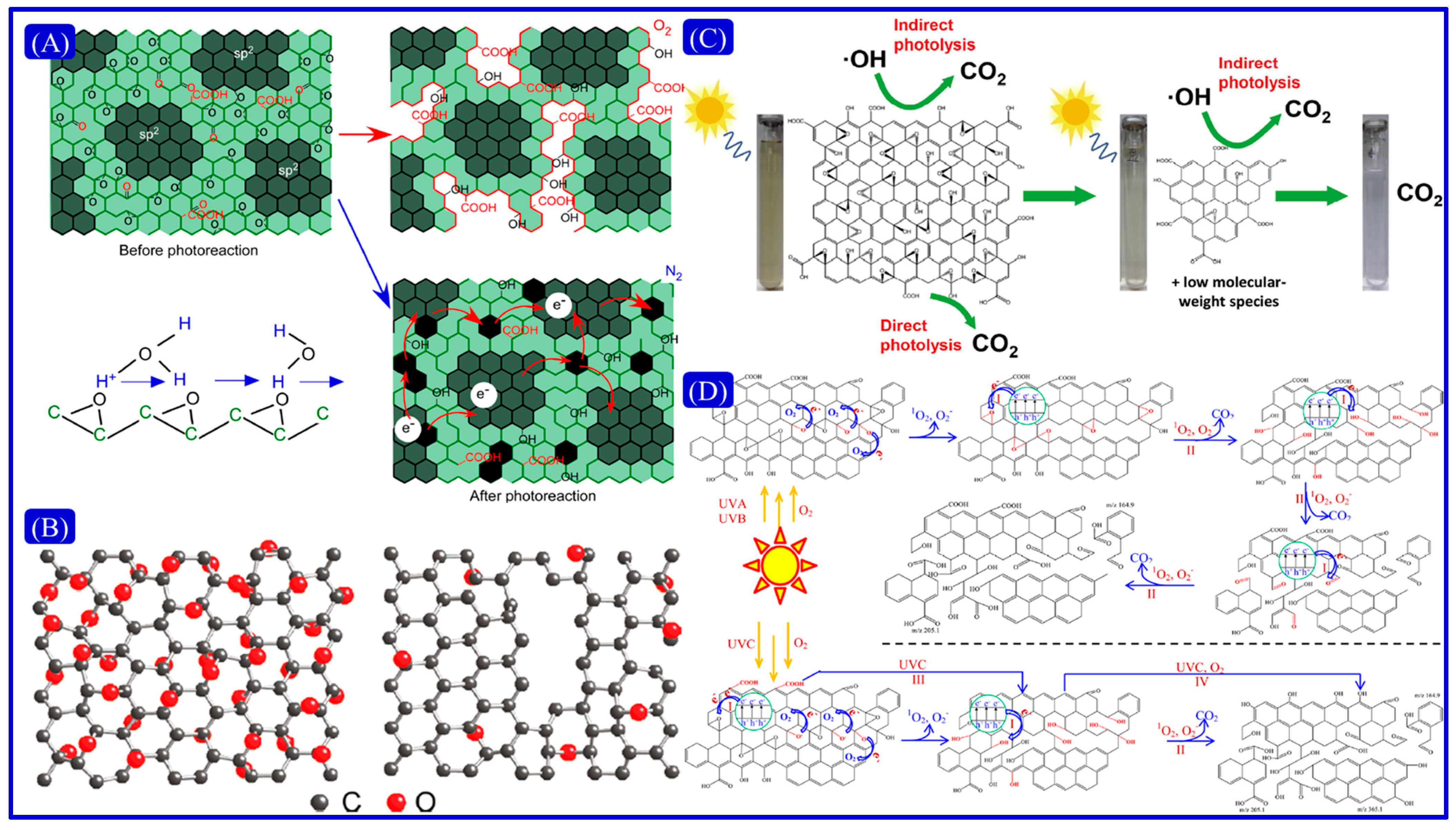
2. Effects of Different Light Conditions on Physicochemical Properties of Graphene Oxide and Photo-Transform Mechanism
2.1. UV Irradiation

2.2. Visible Light
2.3. Simulated Sunlight
3. Biotoxicity and Mechanism of Light-Mediated GO to Algae
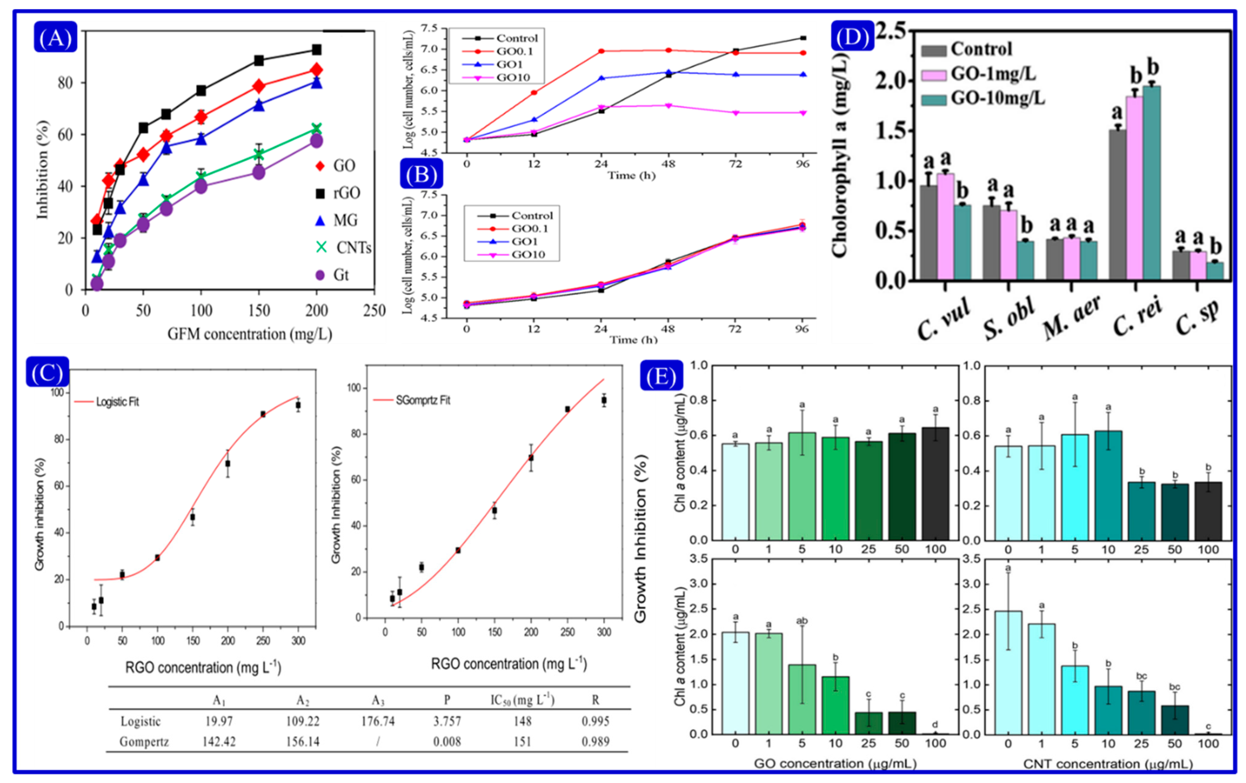
3.1. Cell Division

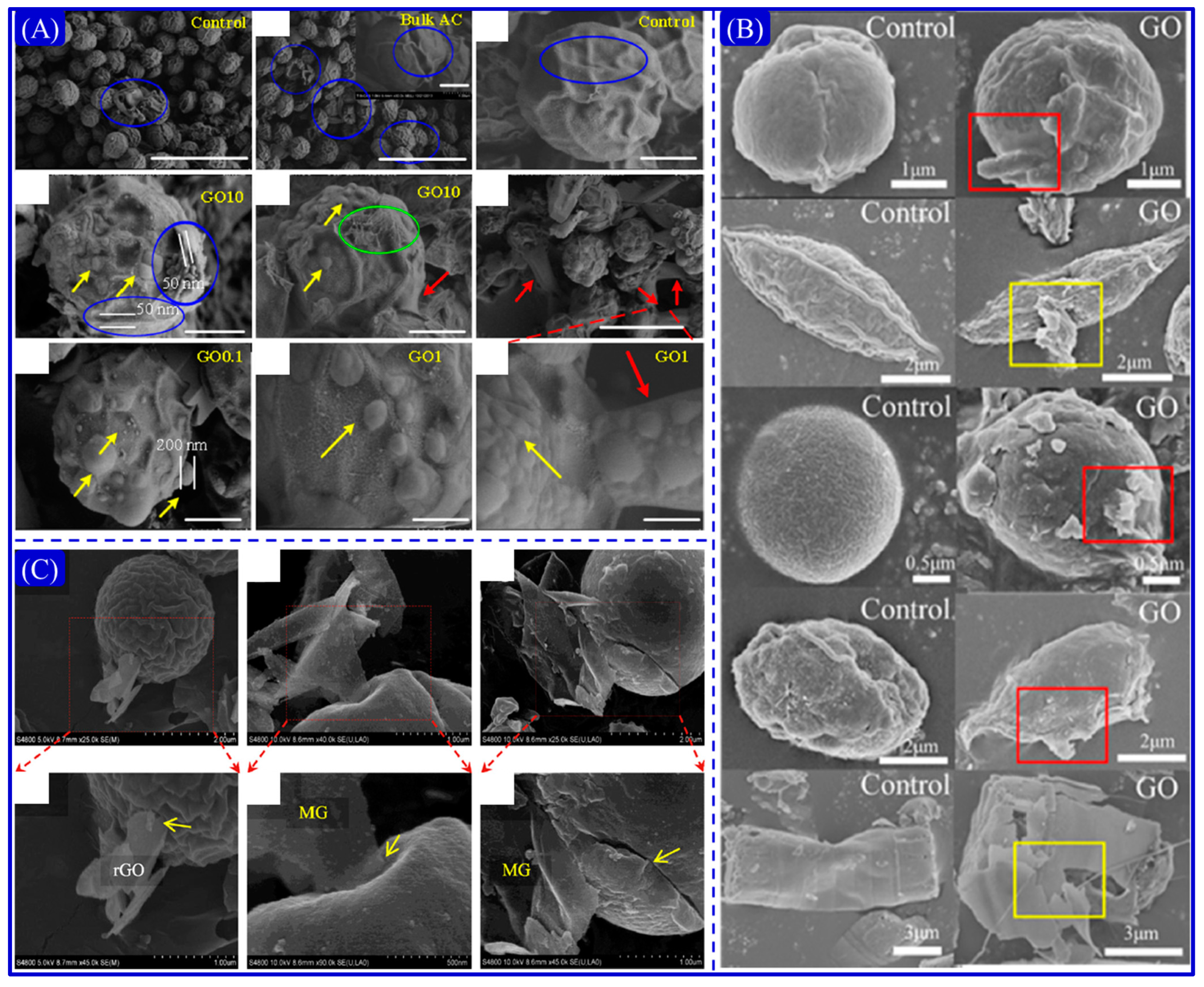
3.2. Cell Membrane Damage and Shadowing Effects
3.3. ROS Generation and Oxidative Stress
3.4. Effects of Photosynthesis and Chlorophyll Content
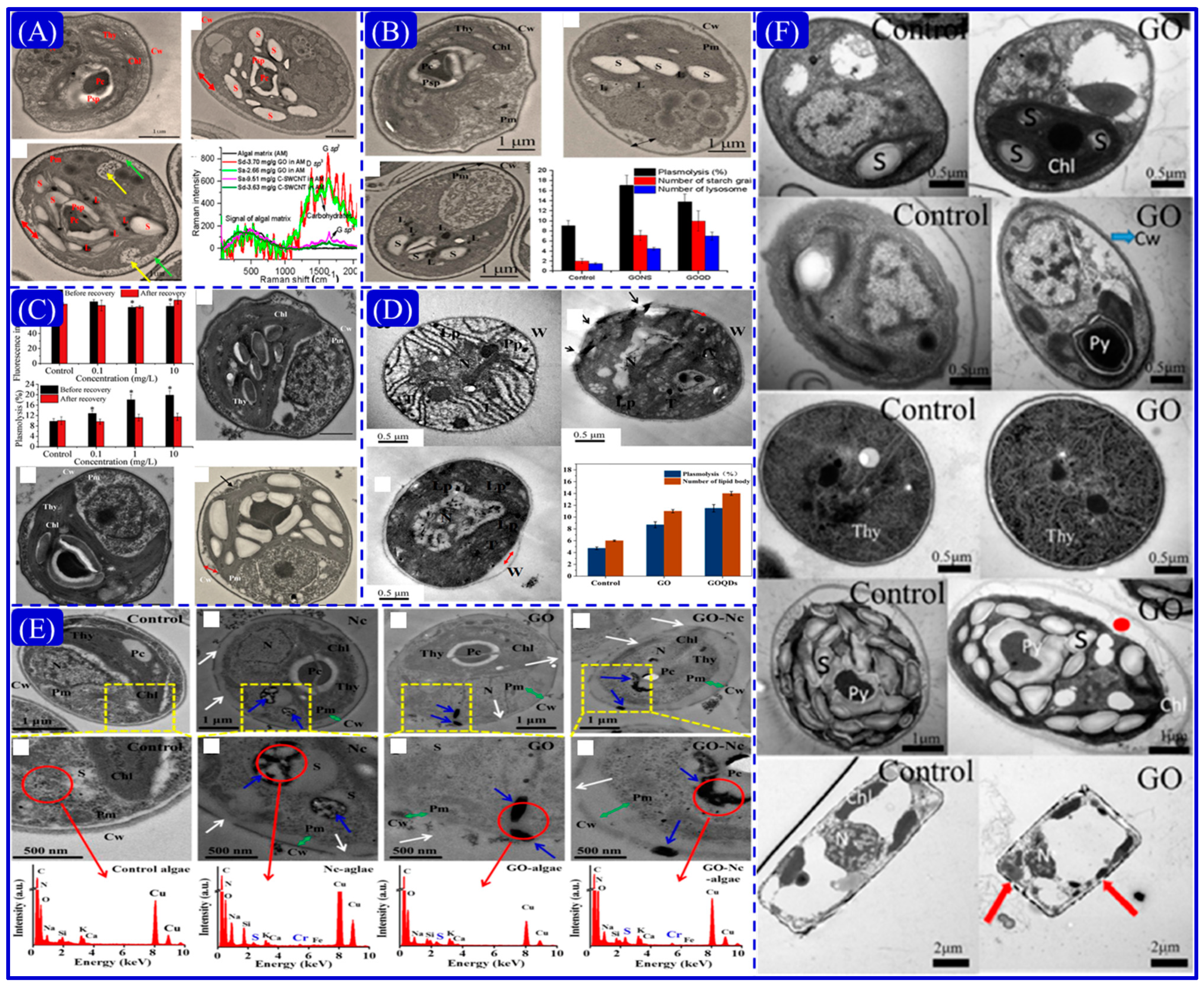
3.5. Cellular Ultrastructural Damage and Membrane Permeability
4. Biotoxicity Mechanism of Light-Mediated GO to Algae
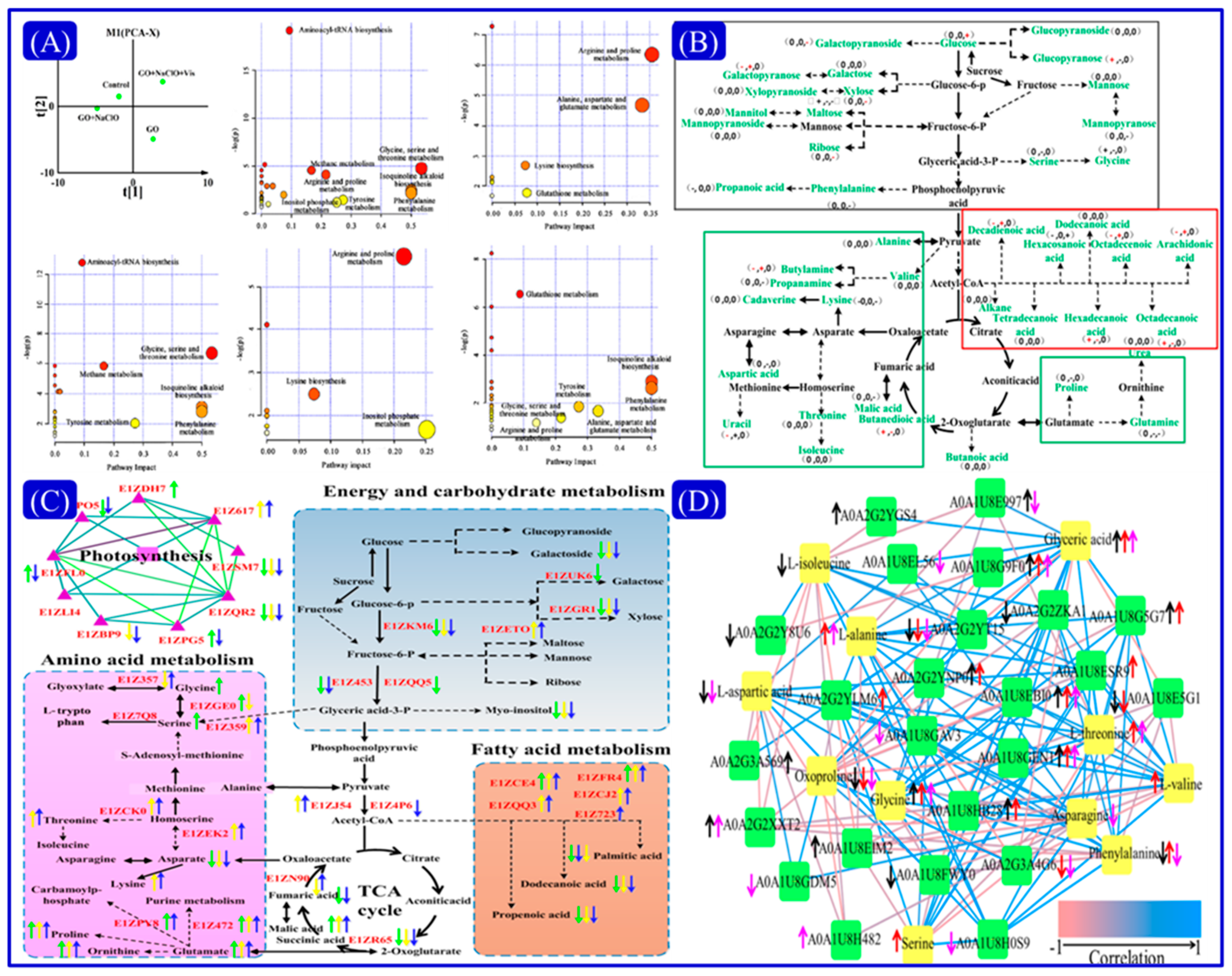
4.1. Metabolomics
4.2. Proteomics
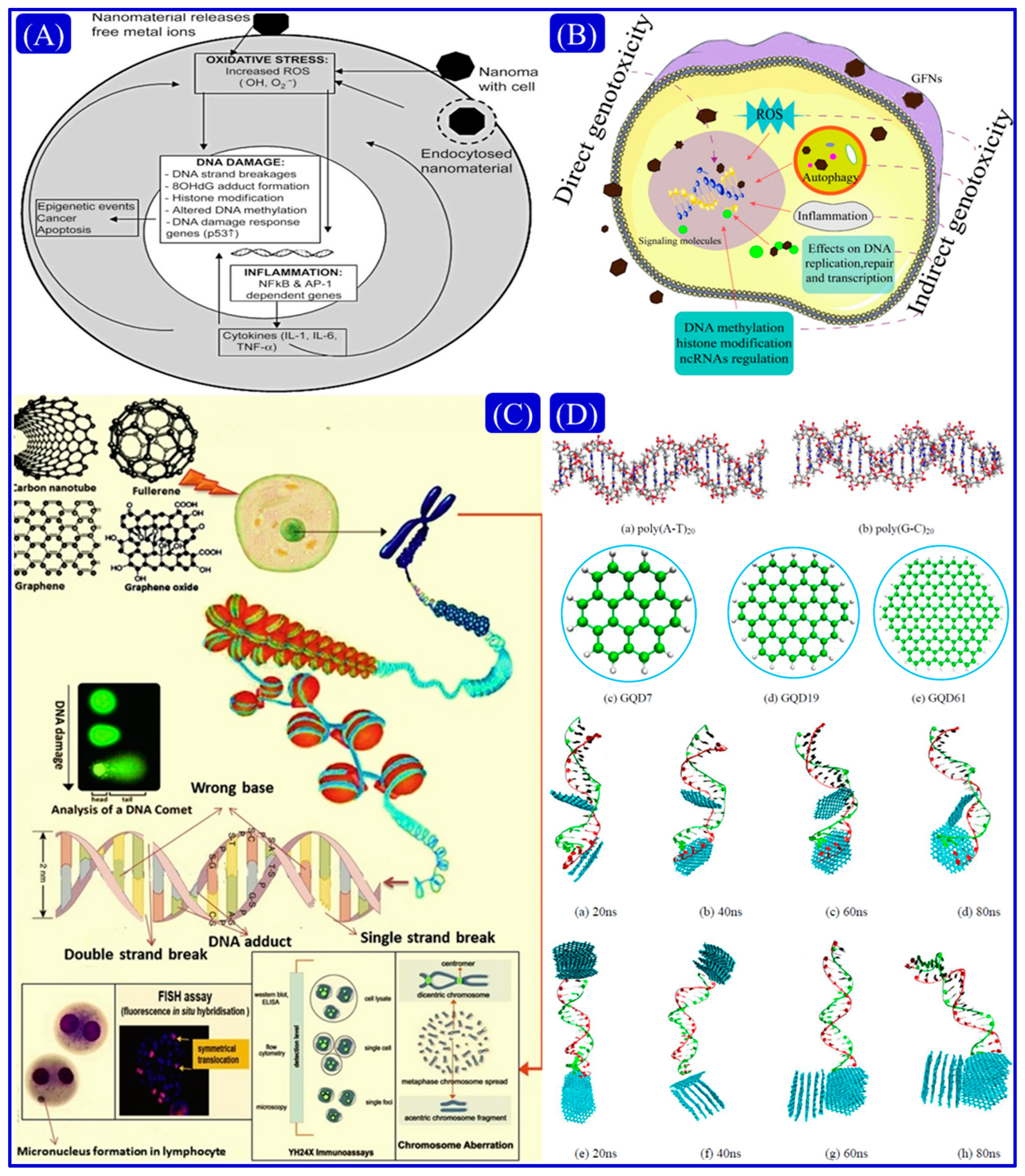
4.3. Genotoxicity
5. Challenges and Perspectives
- (1)
- Based on the unique physicochemical properties of GO, researchers have carried out extensive experimental research work. After GO is exposed to the natural environment, it is easily affected by natural environment factors, thereby affecting its physical and chemical properties. In future work, we should first study the photo-transformation mechanism under single illumination conditions, because the mechanism under single illumination will be clearer. In order to provide a theoretical foundation and research basis for studying the mechanism in natural or real environments, we must first explore the change in physical and chemical properties of GO and the mechanism of light transformation under UV and VL illumination, and then explore the influence and mechanism under simulated solar illumination.
- (2)
- The current research status is that researchers mainly focus on the biotoxicity of GO, without considering the changes in the biotoxicity of GO to aquatic organisms, such as algae, and the mechanism of interaction under light conditions. To solve this, we can set different light conditions for experimental exploration. The laboratory-related studies all include high-concentration and short-term exposure experiments. The concentration of GO used in laboratory experiments is generally high and the exposure time is short. The concentration and time difference between the actual situation and the actual situation will affect the study of its toxicity. Low-dose and long-term exposure experiments should be carried out, taking into account the complex natural environment and low-intensity light effects in the actual environment. Most of the relevant studies are carried out on algae in medium conditions, and in the follow-up study, we can add a simulated natural surface water experimental group, which can better evaluate various toxicity indicators in the real aquatic environment.
- (3)
- Proteomics, metabolomics and genotoxicity are very important elements in the study of the mechanisms of toxicity, but there are not many reports on the application of aquatic organisms such as algae. So far, there are still many shortcomings in this field, and there is a lack of detection and tracking technology specifically for studying the interaction of graphene-like nanomaterials with DNA. In addition, the evaluation database on the types of graphene-based nanomaterials, applied doses, and exposure times is incomplete. In conclusion, continued research is needed to address the above issues, combining several omics to explore the mechanism of toxicity.
Author Contributions
Funding
Conflicts of Interest
References
- Zhou, Q.; Wang, S.; Liu, J.; Hu, X.; Liu, Y.; He, Y.; He, X.; Wu, X. Geological evolution of offshore pollution and its long-term potential impacts on marine ecosystems. Geosci. Front. 2022, 13, 101427. [Google Scholar] [CrossRef]
- Dhamodharan, D.; Ghoderao, P.P.; Dhinakaran, V.; Mubarak, S.; Divakaran, N.; Byun, H.-S. A review on graphene oxide effect in energy storage devices. J. Ind. Eng. Chem. 2021, 106, 20–36. [Google Scholar] [CrossRef]
- Novoselov, K.S.; Fal′ko, V.I.; Colombo, L.; Gellert, P.R.; Schwab, M.G.; Kim, K. A roadmap for graphene. Nature 2012, 490, 192–200. [Google Scholar] [CrossRef] [PubMed]
- Liu, Y.C.; Jiao, L.F.; Wu, Q.; Zhao, Y.P.; Cao, K.Z.; Liu, H.Q.; Wang, Y.J.; Yuan, H.T. Synthesis of rGO-supported layered MoS2 for high-performance rechargeable Mg batteries. Nanoscale 2013, 5, 9562–9567. [Google Scholar] [CrossRef]
- Dreyer, D.R.; Park, S.; Bielawski, C.W.; Ruoff, R.S. The chemistry of graphene oxide. Chem. Soc. Rev. 2010, 39, 228–240. [Google Scholar] [CrossRef]
- Zhu, Y.; Murali, S.; Cai, W.; Li, X.; Suk, J.W.; Potts, J.R.; Ruoff, R.S. Graphene and graphene oxide: Synthesis, properties, and applications. Adv. Mater. 2010, 22, 3906–3924. [Google Scholar] [CrossRef]
- Glogic, E.; Adán-Más, A.; Sonnemann, G.; Montemor, M.D.F.; Guerlou-Demourgues, L.; Young, S.B. Life cycle assessment of emerging Ni–Co hydroxide charge storage electrodes: Impact of graphene oxide and synthesis route. RSC Adv. 2019, 9, 18853–18862. [Google Scholar] [CrossRef] [PubMed]
- Zhou, Q.; Hu, X. Systemic Stress and Recovery Patterns of Rice Roots in Response to Graphene Oxide Nanosheets. Environ. Sci. Technol. 2017, 51, 2022–2030. [Google Scholar] [CrossRef]
- Zhou, Q.; Li, D.; Wang, T.; Hu, X. Leaching of graphene oxide nanosheets in simulated soil and their influences on microbial communities. J. Hazard. Mater. 2021, 404, 124046. [Google Scholar] [CrossRef]
- Bai, H.; Jiang, W.; Kotchey, G.P.; Saidi, W.A.; Bythell, B.J.; Jarvis, J.M.; Marshall, A.G.; Robinson, R.A.S.; Star, A. Insight into the Mechanism of Graphene Oxide Degradation via the Photo-Fenton Reaction. J. Phys. Chem. C 2014, 118, 10519–10529. [Google Scholar] [CrossRef]
- Matsumoto, Y.; Koinuma, M.; Ida, S.; Hayami, S.; Taniguchi, T.; Hatakeyama, K.; Tateishi, H.; Watanabe, Y.; Amano, S. Photoreaction of Graphene Oxide Nanosheets in Water. J. Phys. Chem. C 2011, 115, 19280–19286. [Google Scholar] [CrossRef]
- Zhang, X.-F.; Shao, X.; Liu, S. Dual Fluorescence of Graphene Oxide: A Time-Resolved Study. J. Phys. Chem. A 2012, 116, 7308–7313. [Google Scholar] [CrossRef] [PubMed]
- Gao, Y.; Chen, C.; Tan, X.; Xu, H.; Zhu, K. Polyaniline-modified 3D-flower-like molybdenum disulfide composite for efficient adsorption/photocatalytic reduction of Cr(VI). J. Colloid Interface Sci. 2016, 476, 62–70. [Google Scholar] [CrossRef] [PubMed]
- Chowdhury, I.; Hou, W.-C.; Goodwin, D.; Henderson, M.; Zepp, R.G.; Bouchard, D. Sunlight affects aggregation and deposition of graphene oxide in the aquatic environment. Water Res. 2015, 78, 37–46. [Google Scholar] [CrossRef] [PubMed]
- Huang, C.; Xia, T.; Niu, J.F.; Yang, Y.; Lin, S.J.; Wang, X.K.; Yang, G.Q.; Mao, L.; Xing, B.S. Transformation of 14C-Labeled graphene to 14CO2 in the shoots of a rice plant. Angew Chem. Int. Ed. Engl. 2018, 57, 9759–9763. [Google Scholar] [CrossRef]
- Mitrano, D.M.; Motellier, S.; Clavaguera, S.; Nowack, B. Review of nanomaterial aging and transformations through the life cycle of nano-enhanced products. Environ. Int. 2015, 77, 132–147. [Google Scholar] [CrossRef]
- Zou, X.; Zhang, L.; Wang, Z.; Luo, Y. Mechanisms of the Antimicrobial Activities of Graphene Materials. J. Am. Chem. Soc. 2016, 138, 2064–2077. [Google Scholar] [CrossRef]
- Liu, Y.; Li, L.; Zheng, L.; Fu, P.; Wang, Y.; Nguyen, H.; Shen, X.; Sui, Y. Antioxidant responses of triangle sail mussel Hyriopsis cumingii exposed to harmful algae Microcystis aeruginosa and high pH. Chemosphere 2020, 243, 125241. [Google Scholar] [CrossRef]
- Harke, M.J.; Steffen, M.M.; Gobler, C.J.; Otten, T.G.; Wilhelm, S.W.; Wood, S.A.; Paerl, H.W. A review of the global ecology, genomics, and biogeography of the toxic cyanobacterium, Microcystis spp. Harmful Algae 2016, 54, 4–20. [Google Scholar] [CrossRef]
- He, X.; Wu, P.; Wang, S.; Wang, A.; Wang, C.; Ding, P. Inactivation of harmful algae using photocatalysts: Mechanisms and performance. J. Clean. Prod. 2021, 289, 125755. [Google Scholar] [CrossRef]
- Ouyang, S.; Zhou, Q.; Zeng, H.; Wang, Y.; Hu, X. Natural Nanocolloids Mediate the Phytotoxicity of Graphene Oxide. Environ. Sci. Technol. 2020, 54, 4865–4875. [Google Scholar] [CrossRef] [PubMed]
- Ding, X.; Wang, J.; Rui, Q.; Wang, D. Long-term exposure to thiolated graphene oxide in the range of μg/L induces toxicity in nematode Caenorhabditis elegans. Sci. Total Environ. 2018, 616–617, 29–37. [Google Scholar] [CrossRef] [PubMed]
- Akhavan, O.; Ghaderi, E. Toxicity of graphene and graphene oxide nanowalls against bacteria. ACS Nano 2010, 4, 5731–5736. [Google Scholar] [CrossRef] [PubMed]
- Crane, M.; Handy, R.D.; Garrod, J.; Owen, R. Ecotoxicity test methods and environmental hazard assessment for engineered nanoparticles. Ecotoxicology 2008, 17, 421–437. [Google Scholar] [CrossRef]
- Zhang, H.; Huang, Q.; Xu, A.; Wu, L.J. Spectroscopic probe to contribution of physicochemical transformations in the toxicity of aged ZnO NPs to Chlorella vulgaris: New insight into the variation of toxicity of ZnO NPs under aging process. Nanotoxicology 2016, 10, 1177–1187. [Google Scholar] [CrossRef]
- Gomes, T.; Xie, L.; Brede, D.; Lind, O.-C.; Solhaug, K.A.; Salbu, B.; Tollefsen, K.E. Sensitivity of the green algae Chlamydomonas reinhardtii to gamma radiation: Photosynthetic performance and ROS formation. Aquat. Toxicol. 2017, 183, 1–10. [Google Scholar] [CrossRef]
- Dauda, S.; Chia, M.A.; Bako, S.P. Toxicity of titanium dioxide nanoparticles to Chlorella vulgaris Beyerinck (Beijerinck) 1890 (Trebouxiophyceae, Chlorophyta) under changing nitrogen conditions. Aquat. Toxicol. 2017, 187, 108–114. [Google Scholar] [CrossRef]
- Ouyang, S.; Hu, X.; Zhou, Q.; Li, X.; Miao, X.; Zhou, R. Nanocolloids in Natural Water: Isolation, Characterization, and Toxicity. Environ. Sci. Technol. 2018, 52, 4850–4860. [Google Scholar] [CrossRef]
- Ahmad, F.; Yao, H.; Zhou, Y.; Liu, X. Toxicity of cobalt ferrite (CoFe2O4) nanobeads in Chlorella vulgaris: Interaction, adaptation and oxidative stress. Chemosphere 2015, 139, 479–485. [Google Scholar] [CrossRef]
- Hu, X.; Li, D.; Gao, Y.; Mu, L.; Zhou, Q. Knowledge gaps between nanotoxicological research and nanomaterial safety. Environ. Int. 2016, 94, 8–23. [Google Scholar] [CrossRef]
- Gao, Y.; Ren, X.; Zhang, X.; Chen, C. Environmental fate and risk of ultraviolet- and visible-light-transformed graphene oxide: A comparative study. Environ. Pollut. 2019, 251, 821–829. [Google Scholar] [CrossRef] [PubMed]
- Gao, Y.; Zeng, X.; Zhang, W.; Zhou, L.; Xue, W.; Tang, M.; Sun, S. The aggregation behaviour and mechanism of commercial graphene oxide in surface aquatic environments. Sci. Total Environ. 2022, 806, 150942. [Google Scholar] [CrossRef]
- Zhou, Q.; Ma, S.; Zhan, S. Superior photocatalytic disinfection effect of Ag-3D ordered mesoporous CeO2 under visible light. Appl. Catal. B Environ. 2018, 224, 27–37. [Google Scholar] [CrossRef]
- Ouyang, S.; Hu, X.; Zhou, Q. Envelopment–Internalization Synergistic Effects and Metabolic Mechanisms of Graphene Oxide on Single-Cell Chlorella vulgaris Are Dependent on the Nanomaterial Particle Size. ACS Appl. Mater. Interfaces 2015, 7, 18104–18112. [Google Scholar] [CrossRef] [PubMed]
- Zhou, Q.; Yue, Z.; Li, Q.; Zhou, R.; Liu, L. Exposure to PbSe Nanoparticles and Male Reproductive Damage in a Rat Model. Environ. Sci. Technol. 2019, 53, 13408–13416. [Google Scholar] [CrossRef]
- Zhou, Q.; Liu, Y.; Li, T.; Zhao, H.; Alessi, D.S.; Liu, W.; Konhauser, K.O. Cadmium adsorption to clay-microbe aggregates: Implications for marine heavy metals cycling. Geochim. Cosmochim. Acta 2020, 290, 124–136. [Google Scholar] [CrossRef]
- Mudiam, M.K.R.; Ratnasekhar, C.; Sonane, M.; Satish, A. Metabolomics reveals the perturbations in the metabolome of Caenorhabditis elegans exposed to titanium dioxide nanoparticles. Nanotoxicology 2015, 9, 994–1004. [Google Scholar] [CrossRef]
- Faria, A.F.; Perreault, F.; Elimelech, M. Elucidating the Role of Oxidative Debris in the Antimicrobial Properties of Graphene Oxide. ACS Appl. Nano Mater. 2018, 1, 1164–1174. [Google Scholar] [CrossRef]
- Wang, T.; Wen, J.; Guo, S.; Mu, L. Hypochlorite and visible-light irradiation affect the transformation and toxicity of graphene oxide. Sci. Total Environ. 2020, 723, 138010. [Google Scholar] [CrossRef]
- Li, X.; Mu, L.; Hu, X. Integrating proteomics, metabolomics and typical analysis to investigate the uptake and oxidative stress of graphene oxide and polycyclic aromatic hydrocarbons. Environ. Sci. Nano 2018, 5, 115–129. [Google Scholar] [CrossRef]
- Gioria, S.; Vicente, J.L.; Barboro, P.; la Spina, R.; Tomasi, G.; Urban, P.; Kinsner-Ovaskainen, A.; Francois, R.; Chassaigne, H. A combined proteomics and metabolomics approach to assess the effects of gold nanoparticles in vitro. Nanotoxicology 2016, 10, 736–748. [Google Scholar] [CrossRef] [PubMed]
- Chowdhury, I.; Duch, M.C.; Mansukhani, N.; Hersam, M.C.; Bouchard, D. Colloidal Properties and Stability of Graphene Oxide Nanomaterials in the Aquatic Environment. Environ. Sci. Technol. 2013, 47, 6288–6296. [Google Scholar] [CrossRef] [PubMed]
- Gao, Y.; Ren, X.; Song, G.; Chen, D.; Zhang, X.; Chen, C. Colloidal properties and stability of UV-transformed graphene oxide in aqueous solutions: The role of disorder degree. J. Hazard. Mater. 2020, 382, 121097. [Google Scholar] [CrossRef]
- Hou, W.-C.; Chowdhury, I.; Goodwin, J.D.G.; Henderson, W.M.; Fairbrother, D.H.; Bouchard, D.; Zepp, R.G. Photochemical Transformation of Graphene Oxide in Sunlight. Environ. Sci. Technol. 2015, 49, 3435–3443. [Google Scholar] [CrossRef] [PubMed]
- Hou, W.-C.; Henderson, W.M.; Chowdhury, I.; Goodwin, D.G.; Chang, X.; Martin, S.; Fairbrother, D.H.; Bouchard, D.; Zepp, R.G. The contribution of indirect photolysis to the degradation of graphene oxide in sunlight. Carbon 2016, 110, 426–437. [Google Scholar] [CrossRef]
- Pham, V.T.H.; Truong, V.K.; Quinn, M.D.J.; Notley, S.; Guo, Y.; Baulin, V.; Al Kobaisi, M.; Crawford, R.; Ivanova, E.P. Graphene Induces Formation of Pores That Kill Spherical and Rod-Shaped Bacteria. ACS Nano 2015, 9, 8458–8467. [Google Scholar] [CrossRef]
- Hui, L.; Piao, J.-G.; Auletta, J.; Hu, K.; Zhu, Y.; Meyer, T.; Liu, H.; Yang, L. Availability of the Basal Planes of Graphene Oxide Determines Whether It Is Antibacterial. ACS Appl. Mater. Interfaces 2014, 6, 13183–13190. [Google Scholar] [CrossRef]
- Szpyrkowicz, L.; Juzzolino, C.; Kaul, S.N. A Comparative study on oxidation of disperse dyes by electrochemical process, ozone, hypochlorite and fenton reagent. Water Res. 2001, 35, 2129–2136. [Google Scholar] [CrossRef]
- Koinuma, M.; Ogata, C.; Kamei, Y.; Hatakeyama, K.; Tateishi, H.; Watanabe, Y.; Taniguchi, T.; Gezuhara, K.; Hayami, S.; Funatsu, A.; et al. Photochemical Engineering of Graphene Oxide Nanosheets. J. Phys. Chem. C 2012, 116, 19822–19827. [Google Scholar] [CrossRef]
- Andryushina, N.S.; Stroyuk, O.L.; Yanchuk, I.B.; Yefanov, A.V. A dynamic light scattering study of photochemically reduced colloidal graphene oxide. Colloid Polym. Sci. 2013, 292, 539–546. [Google Scholar] [CrossRef]
- Zhao, Y.; Liu, Y.; Zhang, X.; Liao, W. Environmental transformation of graphene oxide in the aquatic environment. Chemosphere 2021, 262, 127885. [Google Scholar] [CrossRef] [PubMed]
- Taniguchi, T.; Kurihara, S.; Tateishi, H.; Hatakeyama, K.; Koinuma, M.; Yokoi, H.; Hara, M.; Ishikawa, H.; Matsumoto, Y. pH-driven, reversible epoxy ring opening/closing in graphene oxide. Carbon 2015, 84, 560–566. [Google Scholar] [CrossRef]
- Schwenzer, B.; Kaspar, T.C.; Shin, Y.; Gotthold, D.W. Spectroscopic Study of Graphene Oxide Membranes Exposed to Ultraviolet Light. J. Phys. Chem. C 2016, 120, 12559–12567. [Google Scholar] [CrossRef]
- Szabó, T.; Tombácz, E.; Illés, E.; Dékány, I. Enhanced acidity and pH-dependent surface charge characterization of successively oxidized graphite oxides. Carbon 2006, 44, 537–545. [Google Scholar] [CrossRef]
- Du, T.; Adeleye, A.S.; Keller, A.A.; Wu, Z.; Han, W.; Wang, Y.; Zhang, C.; Li, Y. Photochlorination-induced transformation of graphene oxide: Mechanism and environmental fate. Water Res. 2017, 124, 372–380. [Google Scholar] [CrossRef]
- Yuan, X.; Peng, D.; Jing, Q.; Niu, J.; Cheng, X.; Feng, Z.; Wu, X. Green and Effective Removal of Aqueous Graphene Oxide under UV-Light Irradiation. Nanomaterials 2018, 8, 654. [Google Scholar] [CrossRef]
- Cao, X.; Zhao, J.; Wang, Z.; Xing, B. New insight into the photo-transformation mechanisms of graphene oxide under UV-A, UV-B and UV-C lights. J. Hazard. Mater. 2021, 403, 123683. [Google Scholar] [CrossRef]
- Zhang, S.; Li, B.; Wang, X.; Zhao, G.; Hu, B.; Lu, Z.; Wen, T.; Chen, J.; Wang, X. Recent developments of two-dimensional graphene-based composites in visible-light photocatalysis for eliminating persistent organic pollutants from wastewater. Chem. Eng. J. 2020, 390, 124642. [Google Scholar] [CrossRef]
- Hu, X.; Mu, L.; Kang, J.; Lu, K.; Zhou, R.; Zhou, Q. Humic Acid Acts as a Natural Antidote of Graphene by Regulating Nanomaterial Translocation and Metabolic Fluxes in Vivo. Environ. Sci. Technol. 2014, 48, 6919–6927. [Google Scholar] [CrossRef]
- Hu, X.; Mu, L.; Lu, K.; Kang, J.; Zhou, Q. Green Synthesis of Low-Toxicity Graphene-Fulvic Acid with an Open Band Gap Enhances Demethylation of Methylmercury. ACS Appl. Mater. Interfaces 2014, 6, 9220–9227. [Google Scholar] [CrossRef]
- Adeleye, A.S.; Wang, X.; Wang, F.; Hao, R.; Song, W.; Li, Y. Photoreactivity of graphene oxide in aqueous system: Reactive oxygen species formation and bisphenol A degradation. Chemosphere 2018, 195, 344–350. [Google Scholar] [CrossRef] [PubMed]
- Du, T.; Adeleye, A.S.; Zhang, T.; Jiang, C.; Zhang, M.; Wang, H.; Li, Y.; Keller, A.A.; Chen, W. Influence of light wavelength on the photoactivity, physicochemical transformation, and fate of graphene oxide in aqueous media. Environ. Sci. Nano 2018, 5, 2590–2603. [Google Scholar] [CrossRef]
- Zhou, X.; Zhang, Y.; Wang, C.; Wu, X.; Yang, Y.; Zheng, B.; Wu, H.; Guo, S.; Zhang, J. Photo-Fenton Reaction of Graphene Oxide: A New Strategy to Prepare Graphene Quantum Dots for DNA Cleavage. ACS Nano 2012, 6, 6592–6599. [Google Scholar] [CrossRef] [PubMed]
- Adeleye, A.S.; Ho, K.T.; Zhang, M.; Li, Y.; Burgess, R.M. Fate and Transformation of Graphene Oxide in Estuarine and Marine Waters. Environ. Sci. Technol. 2019, 53, 5858–5867. [Google Scholar] [CrossRef] [PubMed]
- Shams, M.; Guiney, L.M.; Huang, L.; Ramesh, M.; Yang, X.; Hersam, M.C.; Chowdhury, I. Influence of functional groups on the degradation of graphene oxide nanomaterials. Environ. Sci. Nano 2019, 6, 2203–2214. [Google Scholar] [CrossRef]
- Zhao, Y.; Jafvert, C.T. Environmental photochemistry of single layered graphene oxide in water. Environ. Sci. Nano 2015, 2, 136–142. [Google Scholar] [CrossRef]
- Kuang, Y.; Shang, J.; Zhu, T. Photoactivated Graphene Oxide to Enhance Photocatalytic Reduction of CO2. ACS Appl. Mater. Interfaces 2020, 12, 3580–3591. [Google Scholar] [CrossRef]
- Hou, W.-C.; Wang, Y.-S. Photocatalytic Generation of H2O2 by Graphene Oxide in Organic Electron Donor-Free Condition under Sunlight. ACS Sustain. Chem. Eng. 2017, 5, 2994–3001. [Google Scholar] [CrossRef]
- Chong, Y.; Ge, C.; Fang, G.; Wu, R.; Zhang, H.; Chai, Z.; Chen, C.; Yin, J.-J. Light-Enhanced Antibacterial Activity of Graphene Oxide, Mainly via Accelerated Electron Transfer. Environ. Sci. Technol. 2017, 51, 10154–10161. [Google Scholar] [CrossRef]
- Gao, Y.; Wu, J.; Ren, X.; Tan, X.; Hayat, T.; Alsaedi, A.; Cheng, C.; Chen, C. Impact of graphene oxide on the antibacterial activity of antibiotics against bacteria. Environ. Sci. Nano 2017, 4, 1016–1024. [Google Scholar] [CrossRef]
- Chen, Y.; Ren, C.; Ouyang, S.; Hu, X.; Zhou, Q. Mitigation in Multiple Effects of Graphene Oxide Toxicity in Zebrafish Embryogenesis Driven by Humic Acid. Environ. Sci. Technol. 2015, 49, 10147–10154. [Google Scholar] [CrossRef] [PubMed]
- Li, X.; Li, F.; Gao, Z.; Fang, L. Toxicology of Graphene Oxide Nanosheets Against Paecilomyces catenlannulatus. Bull. Environ. Contam. Toxicol. 2015, 95, 25–30. [Google Scholar] [CrossRef] [PubMed]
- Zhao, J.; Wang, Z.; White, J.C.; Xing, B. Graphene in the Aquatic Environment: Adsorption, Dispersion, Toxicity and Transformation. Environ. Sci. Technol. 2014, 48, 9995–10009. [Google Scholar] [CrossRef] [PubMed]
- Schwab, F.; Bucheli, T.D.; Lukhele, L.P.; Magrez, A.; Nowack, B.; Sigg, L.; Knauer, K. Are Carbon Nanotube Effects on Green Algae Caused by Shading and Agglomeration? Environ. Sci. Technol. 2011, 45, 6136–6144. [Google Scholar] [CrossRef]
- Markovic, M.; Andelkovic, I.; Shuster, J.; Janik, L.; Kumar, A.; Losic, D.; McLaughlin, M.J. Addressing challenges in providing a reliable ecotoxicology data for graphene-oxide (GO) using an algae (Raphidocelis subcapitata), and the trophic transfer consequence of GO-algae aggregates. Chemosphere 2020, 245, 125640. [Google Scholar] [CrossRef]
- Zhao, J.; Cao, X.; Wang, Z.; Dai, Y.; Xing, B. Mechanistic understanding toward the toxicity of graphene-family materials to freshwater algae. Water Res. 2017, 111, 18–27. [Google Scholar] [CrossRef]
- Hu, X.; Gao, Y.; Fang, Z. Integrating metabolic analysis with biological endpoints provides insight into nanotoxicological mechanisms of graphene oxide: From effect onset to cessation. Carbon 2016, 109, 65–73. [Google Scholar] [CrossRef]
- Du, S.; Zhang, P.; Zhang, R.; Lu, Q.; Liu, L.; Bao, X.; Liu, H. Reduced graphene oxide induces cytotoxicity and inhibits photosynthetic performance of the green alga Scenedesmus obliquus. Chemosphere 2016, 164, 499–507. [Google Scholar] [CrossRef]
- Yin, J.; Fan, W.; Du, J.; Feng, W.; Dong, Z.; Liu, Y.; Zhou, T. The toxicity of graphene oxide affected by algal physiological characteristics: A comparative study in cyanobacterial, green algae, diatom. Environ. Pollut. 2020, 260, 113847. [Google Scholar] [CrossRef]
- Cruces, E.; Barrios, A.C.; Cahue, Y.P.; Januszewski, B.; Gilbertson, L.M.; Perreault, F. Similar toxicity mechanisms between graphene oxide and oxidized multi-walled carbon nanotubes in Microcystis aeruginosa. Chemosphere 2021, 265, 129137. [Google Scholar] [CrossRef]
- Hu, X.; Lu, K.; Mu, L.; Kang, J.; Zhou, Q. Interactions between graphene oxide and plant cells: Regulation of cell morphology, uptake, organelle damage, oxidative effects and metabolic disorders. Carbon 2014, 80, 665–676. [Google Scholar] [CrossRef]
- Hu, X.; Ouyang, S.; Mu, L.; An, J.; Zhou, Q. Effects of Graphene Oxide and Oxidized Carbon Nanotubes on the Cellular Division, Microstructure, Uptake, Oxidative Stress, and Metabolic Profiles. Environ. Sci. Technol. 2015, 49, 10825–10833. [Google Scholar] [CrossRef]
- Lammel, T.; Boisseaux, P.; Fernández-Cruz, M.-L.; Navas, J.M. Internalization and cytotoxicity of graphene oxide and carboxyl graphene nanoplatelets in the human hepatocellular carcinoma cell line Hep G2. Part. Fibre Toxicol. 2013, 10, 1–21. [Google Scholar] [CrossRef] [PubMed]
- Bianco, A. Graphene: Safe or Toxic? The Two Faces of the Medal. Angew. Chem. Int. Ed. 2013, 52, 4986–4997. [Google Scholar] [CrossRef] [PubMed]
- Sanchez, V.C.; Jachak, A.; Hurt, R.H.; Kane, A.B. Biological Interactions of Graphene-Family Nanomaterials: An Interdisciplinary Review. Chem. Res. Toxicol. 2012, 25, 15–34. [Google Scholar] [CrossRef] [PubMed]
- Nogueira, P.; Nakabayashi, D.; Zucolotto, V. The effects of graphene oxide on green algae Raphidocelis subcapitata. Aquat. Toxicol. 2015, 166, 29–35. [Google Scholar] [CrossRef]
- Petersen, E.J.; Henry, T.B.; Zhao, J.; MacCuspie, R.I.; Kirschling, T.L.; Dobrovolskaia, M.A.; Hackley, V.; Xing, B.; White, J.C. Identification and Avoidance of Potential Artifacts and Misinterpretations in Nanomaterial Ecotoxicity Measurements. Environ. Sci. Technol. 2014, 48, 4226–4246. [Google Scholar] [CrossRef]
- Long, Z.; Ji, J.; Yang, K.; Lin, D.; Wu, F. Systematic and Quantitative Investigation of the Mechanism of Carbon Nanotubes’ Toxicity toward Algae. Environ. Sci. Technol. 2012, 46, 8458–8466. [Google Scholar] [CrossRef]
- Akhavan, O.; Ghaderi, E.; Hashemi, E.; Akbari, E. Dose-dependent effects of nanoscale graphene oxide on reproduction capability of mammals. Carbon 2015, 95, 309–317. [Google Scholar] [CrossRef]
- Chang, Y.; Yang, S.-T.; Liu, J.-H.; Dong, E.; Wang, Y.; Cao, A.; Liu, Y.; Wang, H. In vitro toxicity evaluation of graphene oxide on A549 cells. Toxicol. Lett. 2011, 200, 201–210. [Google Scholar] [CrossRef]
- Begum, P.; Fugetsu, B. Induction of cell death by graphene in Arabidopsis thaliana (Columbia ecotype) T87 cell suspensions. J. Hazard. Mater. 2013, 260, 1032–1041. [Google Scholar] [CrossRef] [PubMed]
- Li, F.M.; Liang, Z.; Zheng, X.; Zhao, W.; Wu, M.; Wang, Z.Y. Toxicity of nano-TiO2 on algae and the site of reactive oxygen species production. Aquat. Toxicol. 2015, 158, 1–13. [Google Scholar] [CrossRef] [PubMed]
- Zhang, M.; Yu, Q.; Liang, C.; Liu, Z.; Zhang, B.; Li, M. Graphene oxide induces plasma membrane damage, reactive oxygen species accumulation and fatty acid profiles change in Pichia pastoris. Ecotoxicol. Environ. Saf. 2016, 132, 372–378. [Google Scholar] [CrossRef]
- Zhao, F.-F.; Wang, S.-C.; Zhu, Z.-L.; Wang, S.-G.; Liu, F.-F.; Liu, G.-Z. Effects of oxidation degree on photo-transformation and the resulting toxicity of graphene oxide in aqueous environment. Environ. Pollut. 2019, 249, 1106–1114. [Google Scholar] [CrossRef]
- Melegari, S.; Perreault, F.; Moukha, S.; Popovic, R.; Creppy, E.E.; Matias, W.G. Induction to oxidative stress by saxitoxin investigated through lipid peroxidation in Neuro 2A cells and Chlamydomonas reinhardtii alga. Chemosphere 2012, 89, 38–43. [Google Scholar] [CrossRef] [PubMed]
- Banchi, E.; Carniel, F.C.; Montagner, A.; Bosi, S.; Bramini, M.; Crosera, M.; Leon, V.; Martin, C.; Pallavicini, A.; Vazquez, E.; et al. Graphene-based materials do not impair physiology, gene expression and growth dynamics of the aeroterrestrial microalga Trebouxia gelatinosa. Nanotoxicology 2019, 13, 492–509. [Google Scholar] [CrossRef] [PubMed]
- Tang, Y.; Tian, J.; Li, S.; Xue, C.; Xue, Z.; Yin, D.; Yu, S. Combined effects of graphene oxide and Cd on the photosynthetic capacity and survival of Microcystis aeruginosa. Sci. Total Environ. 2015, 532, 154–161. [Google Scholar] [CrossRef] [PubMed]
- Hu, C.; Hu, N.; Li, X.; Zhao, Y. Graphene oxide alleviates the ecotoxicity of copper on the freshwater microalga Scenedesmus obliquus. Ecotoxicol. Environ. Saf. 2016, 132, 360–365. [Google Scholar] [CrossRef]
- Yan, Z.; Yang, X.; Lynch, I.; Cui, F. Comparative evaluation of the mechanisms of toxicity of graphene oxide and graphene oxide quantum dots to blue-green algae Microcystis aeruginosa in the aquatic environment. J. Hazard. Mater. 2022, 425, 127898. [Google Scholar] [CrossRef]
- Kang, W.; Li, X.; Sun, A.; Yu, F.; Hu, X. Study of the Persistence of the Phytotoxicity Induced by Graphene Oxide Quantum Dots and of the Specific Molecular Mechanisms by Integrating Omics and Regular Analyses. Environ. Sci. Technol. 2019, 53, 3791–3801. [Google Scholar] [CrossRef]
- Luo, C.; Li, Y.; Yang, L.; Wang, X.; Long, J.; Liu, J. Superparamagnetic iron oxide nanoparticles exacerbate the risks of reactive oxygen species-mediated external stresses. Arch. Toxicol. 2015, 89, 357–369. [Google Scholar] [CrossRef] [PubMed]
- Jiang, H.-S.; Qiu, X.-N.; Li, G.-B.; Li, W.; Yin, L.-Y. Silver nanoparticles induced accumulation of reactive oxygen species and alteration of antioxidant systems in the aquatic plant Spirodela polyrhiza. Environ. Toxicol. Chem. 2014, 33, 1398–1405. [Google Scholar] [CrossRef] [PubMed]
- Martín-de-Lucía, I.; Campos-Mañas, M.C.; Agüera, A.; Leganés, F.; Fernández-Piñas, F.; Rosal, R. Combined toxicity of graphene oxide and wastewater to the green alga Chlamydomonas reinhardtii. Environ. Sci. Nano 2018, 5, 1729–1744. [Google Scholar] [CrossRef]
- Li, Y.; Yuan, H.; Bussche, A.V.D.; Creighton, M.; Hurt, R.H.; Kane, A.B.; Gao, H. Graphene microsheets enter cells through spontaneous membrane penetration at edge asperities and corner sites. Proc. Natl. Acad. Sci. USA 2013, 110, 12295–12300. [Google Scholar] [CrossRef]
- Wang, X.-Y.; Lei, R.; Huang, H.-D.; Wang, N.; Yuan, L.; Xiao, R.-Y.; Bai, L.-D.; Li, X.; Li, L.-M.; Yang, X.-D. The permeability and transport mechanism of graphene quantum dots (GQDs) across the biological barrier. Nanoscale 2015, 7, 2034–2041. [Google Scholar] [CrossRef]
- Treuel, L.; Brandholt, S.; Maffre, P.; Wiegele, S.; Shang, L.; Nienhaus, G.U. Impact of Protein Modification on the Protein Corona on Nanoparticles and Nanoparticle–Cell Interactions. ACS Nano 2015, 8, 503–513. [Google Scholar] [CrossRef]
- Paget, V.; Moche, H.; Kortulewski, T.; Grall, R.; Irbah, L.; Nesslany, F.; Chevillard, S. Human Cell Line-Dependent WC-Co Nanoparticle Cytotoxicity and Genotoxicity: A Key Role of ROS Production. Toxicol. Sci. 2014, 143, 385–397. [Google Scholar] [CrossRef]
- Watson, C.; Ge, J.; Cohen, J.; Pyrgiotakis, G.; Engelward, B.P.; Demokritou, P. High-Throughput Screening Platform for Engineered Nanoparticle-Mediated Genotoxicity Using CometChip Technology. ACS Nano 2014, 8, 2118–2133. [Google Scholar] [CrossRef]
- Mu, Q.; Jiang, G.; Chen, L.; Zhou, H.; Fourches, D.; Tropsha, A.; Yan, B. Chemical Basis of Interactions Between Engineered Nanoparticles and Biological Systems. Chem. Rev. 2014, 114, 7740–7781. [Google Scholar] [CrossRef]
- Tan, W.; Peralta-Videa, J.R.; Gardea-Torresdey, J.L. Interaction of titanium dioxide nanoparticles with soil components and plants: Current knowledge and future research needs—A critical review. Environ. Sci. Nano 2018, 5, 257–278. [Google Scholar] [CrossRef]
- Zenobi, R. Single-Cell Metabolomics: Analytical and Biological Perspectives. Science 2013, 342, 1243259. [Google Scholar] [CrossRef] [PubMed]
- Perreault, F.; Samadani, M.; Dewez, D. Effect of soluble copper released from copper oxide nanoparticles solubilisation on growth and photosynthetic processes of Lemna gibba L. Nanotoxicology 2014, 8, 374–382. [Google Scholar] [CrossRef] [PubMed]
- Besseling, E.; Wang, B.; Lürling, M.; Koelmans, A.A. Correction to Nanoplastic Affects Growth of S. obliquus and Reproduction of D. magna. Environ. Sci. Technol. 2014, 48, 14065. [Google Scholar] [CrossRef]
- Toubiana, D.; Batushansky, A.; Tzfadia, O.; Scossa, F.; Khan, A.; Barak, S.; Zamir, D.; Fernie, A.R.; Nikoloski, Z.; Fait, A. Combined correlation-based network and mQTL analyses efficiently identified loci for branched-chain amino acid, serine to threonine, and proline metabolism in tomato seeds. Plant J. 2015, 81, 121–133. [Google Scholar] [CrossRef] [PubMed]
- Lee, Y.-H.; Cheng, F.-Y.; Chiu, H.-W.; Tsai, J.-C.; Fang, C.-Y.; Chen, C.-W.; Wang, Y.-J. Cytotoxicity, oxidative stress, apoptosis and the autophagic effects of silver nanoparticles in mouse embryonic fibroblasts. Biomaterials 2014, 35, 4706–4715. [Google Scholar] [CrossRef]
- Singh, A.V.; Mehta, K.K.; Worley, K.; Dordick, J.S.; Kane, R.S.; Wan, L.Q. Carbon Nanotube-Induced Loss of Multicellular Chirality on Micropatterned Substrate Is Mediated by Oxidative Stress. ACS Nano 2014, 8, 2196–2205. [Google Scholar] [CrossRef]
- Li, X.; Sun, S.; Guo, S.; Hu, X. Identifying the Phytotoxicity and Defense Mechanisms Associated with Graphene-Based Nanomaterials by Integrating Multiomics and Regular Analysis. Environ. Sci. Technol. 2021, 55, 9938–9948. [Google Scholar] [CrossRef]
- Prasad, R.Y.; Wallace, K.; Daniel, K.M.; Tennant, A.H.; Zucker, R.M.; Strickland, J.; Dreher, K.; Kligerman, A.D.; Blackman, C.F.; DeMarini, D.M. Effect of Treatment Media on the Agglomeration of Titanium Dioxide Nanoparticles: Impact on Genotoxicity, Cellular Interaction, and Cell Cycle. ACS Nano 2013, 7, 1929–1942. [Google Scholar] [CrossRef]
- Nissen, P.; Hansen, J.; Ban, N.; Moore, P.B.; Steitz, T.A. The Structural Basis of Ribosome Activity in Peptide Bond Synthesis. Science 2000, 289, 920–930. [Google Scholar] [CrossRef]
- You, M.T.; You, X.Q.; Yang, X.; Hu, J.R.; Sun, W.L. Adsorption of antibiotics onto graphene oxide imparts their antagonistic effects on Synechocystis sp.: Model development and proteomic analysis. Environ. Sci. Nano 2022, 9, 243–253. [Google Scholar] [CrossRef]
- Liu, Q.; Zhang, Y.; Wang, Y.; Wang, W.; Gu, C.; Huang, S.; Yuan, H.; Dhankher, O.P. Quantitative proteomic analysis reveals complex regulatory and metabolic response of Iris lactea Pall. var. chinensis to cadmium toxicity. J. Hazard. Mater. 2020, 400, 123165. [Google Scholar] [CrossRef] [PubMed]
- Sun, J.; Zhou, Q.; Hu, X. Integrating multi-omics and regular analyses identifies the molecular responses of zebrafish brains to graphene oxide: Perspectives in environmental criteria. Ecotoxicol. Environ. Saf. 2019, 180, 269–279. [Google Scholar] [CrossRef] [PubMed]
- Singh, N.; Manshian, B.; Jenkins, G.J.; Griffiths, S.M.; Williams, P.M.; Maffeis, T.G.; Wright, C.; Doak, S.H. NanoGenotoxicology: The DNA damaging potential of engineered nanomaterials. Biomaterials 2009, 30, 3891–3914. [Google Scholar] [CrossRef]
- Wu, K.; Zhou, Q.; Ouyang, S. Direct and Indirect Genotoxicity of Graphene Family Nanomaterials on DNA—A Review. Nanomaterials 2021, 11, 2889. [Google Scholar] [CrossRef]
- Bohne, J.; Cathomen, T. Genotoxicity in gene therapy: An account of vector integration and designer nucleases. Curr. Opin. Mol. Ther. 2008, 10, 214–223. [Google Scholar] [PubMed]
- Huang, R.; Zhou, Y.; Hu, S.; Zhou, P.-K. Targeting and non-targeting effects of nanomaterials on DNA: Challenges and perspectives. Rev. Environ. Sci. Bio./Technol. 2019, 18, 617–634. [Google Scholar] [CrossRef]
- Burgum, M.J.; Clift, M.J.D.; Evans, S.J.; Hondow, N.; Tarat, A.; Jenkins, G.J.; Doak, S.H. Few-layer graphene induces both primary and secondary genotoxicity in epithelial barrier models in vitro. J. Nanobiotechnology 2021, 19, 24. [Google Scholar] [CrossRef]
- Samadian, H.; Salami, M.S.; Jaymand, M.; Azarnezhad, A.; Najafi, M.; Barabadi, H.; Ahmadi, A. Genotoxicity assessment of carbon-based nanomaterials; Have their unique physicochemical properties made them double-edged swords? Mutat. Res. Rev. Mutat. Res. 2020, 783, 108296. [Google Scholar] [CrossRef]
- Kong, Z.; Hu, W.; Jiao, F.; Zhang, P.; Shen, J.-W.; Cui, B.; Wang, H.; Liang, L. Theoretical Evaluation of DNA Genotoxicity of Graphene Quantum Dots: A Combination of Density Functional Theory and Molecular Dynamics Simulations. J. Phys. Chem. B 2020, 124, 9335–9342. [Google Scholar] [CrossRef]
- Tu, Y.; Lv, M.; Xiu, P.; Huynh, T.; Zhang, M.; Castelli, M.; Liu, Z.; Huang, Q.; Fan, C.; Fang, H.; et al. Destructive extraction of phospholipids from Escherichia coli membranes by graphene nanosheets. Nat. Nanotechnol. 2013, 8, 594–601. [Google Scholar] [CrossRef]
- Ema, M.; Gamo, M.; Honda, K. A review of toxicity studies on graphene-based nanomaterials in laboratory animals. Regul. Toxicol. Pharmacol. 2017, 85, 7–24. [Google Scholar] [CrossRef] [PubMed]
- Magdolenova, Z.; Collins, A.; Kumar, A.; Dhawan, A.; Stone, V.; Dusinska, M. Mechanisms of genotoxicity. A review of in vitro and in vivo studies with engineered nanoparticles. Nanotoxicology 2014, 8, 233–278. [Google Scholar] [CrossRef] [PubMed]


Publisher’s Note: MDPI stays neutral with regard to jurisdictional claims in published maps and institutional affiliations. |
© 2022 by the authors. Licensee MDPI, Basel, Switzerland. This article is an open access article distributed under the terms and conditions of the Creative Commons Attribution (CC BY) license (https://creativecommons.org/licenses/by/4.0/).
Share and Cite
Gao, Y.; Chen, L.; Cheng, S.; Zhu, L.; Liu, L.; Wen, P.; Zhou, L.; Xue, W.; Lu, S.; Zhang, W.; et al. An Overview of Light-Mediated Impact of Graphene Oxide on Algae: Photo-Transform, Toxicity and Mechanism. Water 2022, 14, 2997. https://doi.org/10.3390/w14192997
Gao Y, Chen L, Cheng S, Zhu L, Liu L, Wen P, Zhou L, Xue W, Lu S, Zhang W, et al. An Overview of Light-Mediated Impact of Graphene Oxide on Algae: Photo-Transform, Toxicity and Mechanism. Water. 2022; 14(19):2997. https://doi.org/10.3390/w14192997
Chicago/Turabian StyleGao, Yang, Li Chen, Shenghua Cheng, Ling Zhu, Lijuan Liu, Peihuan Wen, Letao Zhou, Wenjing Xue, Songhua Lu, Wei Zhang, and et al. 2022. "An Overview of Light-Mediated Impact of Graphene Oxide on Algae: Photo-Transform, Toxicity and Mechanism" Water 14, no. 19: 2997. https://doi.org/10.3390/w14192997