Article

Effects of Temperature on Growth, Molting, Feed Intake, and Energy Metabolism of Individually Cultured Juvenile Mud Crab Scylla paramamosain in the Recirculating Aquaculture System

1
Key Laboratory of Aquacultural Biotechnology, Ningbo University, Chinese Ministry of Education, Ningbo 315211, China
2
Collaborative Innovation Center for Zhejiang Marine High-Efficiency and Healthy Aquaculture, Ningbo 315211, China
3
Marine Economic Research Center, Donghai Academy, Ningbo University, Ningbo 315211, China
4
Key Laboratory of Environment Controlled Aquaculture (KLECA), Ministry of Education, 52 Heishijiao Street, Dalian 116023, China
5
College of Marine Technology and Environment, Dalian Ocean University, Dalian 116023, China
*
Author to whom correspondence should be addressed.
Water 2022, 14(19), 2988; https://doi.org/10.3390/w14192988
Submission received: 20 August 2022 / Revised: 11 September 2022 / Accepted: 12 September 2022 / Published: 23 September 2022
(This article belongs to the Special Issue Advances in Aquaculture Ecology Research)

Abstract

:
An eight-week experiment was conducted to investigate the effects of temperature (20, 25, 30, and 35 °C) on growth performance, feed intake, energy metabolism, antioxidant capacity, and the stress response of juvenile Scylla paramamosain in a recirculating aquaculture system. The results showed that the survival rate of the 35 °C group was 80.36 ± 5.92%, significantly lower than that of the other three groups (100%). The high molt frequency of mud crabs was observed in high-temperature groups, accompanied by a higher ecdysone level and ecdysone receptor gene expression but lower molt inhibitory hormone gene expression. However, the molt increment (73.58 ± 2.18%), food intake, and feed conversion efficiency showed a parabolic trend, with the lowest value found in the 35 °C group. Oxygen consumption rate and ammonia excretion rate increased with the increasing temperature, and oxygen-nitrogen ratio, lactic acid, triglyceride, total cholesterol, glucose, and cortisol peaked at 35 °C. Temperature also significantly affected the antioxidant system of S. paramamosain. Crabs in the 25 °C and 30 °C had a significantly higher total antioxidant capacity and lower malondialdehyde compared with the 35 °C group (p < 0.05). Although the high temperature promoted molting, it decreased the feeding rate and growth performance, leading to oxidative stress and functional hypoxia. The quadratic function model demonstrated the optimum temperature for the specific growth rate of juvenile S. paramamosain was 28.5–29.7 °C.

1. Introduction

Throughout tropical and temperate waters, the mud crab (Scylla paramamosain) is a common and economically important marine crab [1,2]. There is a long history of mud crab farming in China, Japan, and the Philippines [3]. China’s 2020 mud crab production is 159,433 tonnes [4]. Mud crab is mainly cultured in ponds, but the unit output is low due to serious cannibalism. In recent years, aquaculturists have tried to culture the mud crab in recirculating aquaculture system (RAS). Unlike traditional pond culture, RAS can prevent cannibalism from the early developing stages and effectively improve the survival rate of mud crabs, even in the nursery. Although researchers have studied the factors such as tank bottom area [5] and tank color [6], they are still insufficient compared to the rapid development of the industry.
Temperature is a ubiquitous factor in the life history of aquatic animals, and the high specific heat and heat conduction of water create a challenging thermal environment for aquatic animals [7]. The temperature of the water plays a major part in the survival and growth of crustaceans, according to a number of studies. For example, the mortality rate of ornamental red cherry shrimp (Neocaridina heteropoda heteropoda) at 32 °C was significantly higher than at 24 and 28 °C [8]. The survival rate of Macrobrachium amazonicum was also affected by temperature, and the survival rate at 28 °C was higher than that at 30 and 32 °C [9]. On growth, the red king crab (Paralithodes camtschaticus) grows exponentially with temperature [10]. Crustaceans achieve faster growth by increasing the molt increment (MI) or molt frequency (MF). Synthesized in the Y organ, ecdysone diffuses across the cell membrane and releases into the hemolymph, where it is converted to 20-HE and binds to the EcR-RXR-ecdysone complex to regulate molting [11]. However, the mih gene inhibits molting by inhibiting ecdysone in the hemolymph [12].
According to the principle of thermodynamics, the increase in temperature stimulates the metabolic process of the organism [13]. Ambient temperature can significantly affect aquatic animals’ metabolic levels and physiological regulation mechanisms. The optimal temperature strongly supports the physiological and biochemical processes of the organisms. At the same time, it can provide maximum energy efficiency [14,15]. The Oxygen consumption rate (OCR) of most crustaceans in the thermophilic range increases gradually with increasing temperature [16]. When the temperature is too high, the metabolic level of crustaceans decreases [17]. For example, in the southern rock lobster (Jasus edwardsii), the OCR positively correlates to the temperature at 18–22 °C. However, when the temperature increased further, the metabolic level decreased instead [17]. Furthermore, since ammonia production is the result of amino acid deamination, ammonia excretion rate (AER) can be used to estimate protein utilization by aquatic organisms [18]. Compared with fishes, crustaceans had an open-vessel circulatory system and transported nutrients through the hemolymph [19]. Hemolymph metabolites could reflect the morphological and physiological adaptation of crustaceans to the environment [20]. Total cholesterol (T-CHO), triglyceride (TG), and glucose (GLU) in hemolymph can evaluate the energy metabolism of crustaceans [21].
An increase in temperature is associated with a higher metabolic rate (Q10 effect), resulting in increased oxygen consumption, increased flux at the electron transport chain level, and more reactive oxygen species (ROS) [22]. ROS can oxidize surrounding molecules, impair cellular functions, and lead to oxidative stress [23,24]. Oxidative enzymes and non-enzymatic antioxidants are used by aquatic organisms to remove excess ROS. The antioxidant enzymes superoxide dismutase (SOD), catalase (CAT), and glutathione peroxidase (GPX) are among them. Non-enzymatic antioxidants include fat-soluble vitamins (e.g., alpha-tocopherol) and small water-soluble molecules such as glutathione (GSH) [25]. Total antioxidant capacity (T-AOC) reflects the metabolic ability of antioxidant enzymatic and non-enzymatic systems under external stress [26]. Malondialdehyde (MDA), as the end product of lipid peroxidation, reflects the degree of cellular oxidative damage [27]. Studies have shown that long-term stress in crustaceans inhibits antioxidant enzyme activity and produces more MDA [5,28,29], accompanied by reduced food intake (FI) [30].
Therefore, this study aimed to evaluate the optimal temperature range for juvenile mud crabs in RAS in terms of growth, molting, energy metabolism, antioxidant capacity, and stress response. This study could help define the management protocol of mud crab and support the design of crab RAS.

2. Material and Methods

2.1. Experimental Design

Four temperatures, i.e., 20, 25, 30, and 35 °C, were set with four independent RAS. Each RAS consisted of 7 square tanks (0.5 × 0.3 × 0.2 m3) with six compartments (0.1 × 0.1 × 0.13 m3) in each tank. Thus, each RAS had 42 compartments with one crab per compartment. The crabs were distributed to three replicates for each treatment, with 14 crabs per replicate. The experiment lasted eight weeks at Ningbo University.

2.2. Experimental Animal and Rearing Conditions

The experiment was conducted in July–August 2021 in the Intelligent Aquaculture Laboratory (Ningbo City, Zhejiang province, China). Selection of healthy and uniformly sized juvenile mud crabs bought from mud crab nursery farm (Ningbo City, Zhejiang province, China). Before the experiment, the crabs were domesticated in the laboratory for one week. During this period, the commercial feed was overfed daily at 20:00 (Table 1). The excessive commercial feed was siphoned, and 1/3 of isothermal seawater was exchanged at 18:00 every day.
When the experiment started, the 168 juvenile S. paramamosain with complete appendages and good vitality were weighed and then distributed to 4 RAS (weight: 0.36 ± 0.09 g). Starting from the room temperature of 28 °C, the RAS was adjusted to 20 °C (20.11 ± 0.43 °C), 25 °C (24.98 ± 0.23 °C), 30 °C (30.08 ± 0.11 °C), and 35 °C (34.88 ± 0.39 °C) at a rate of 1 °C d−1.
During the experiment, 20 and 25 °C were achieved with a refrigerating machine (AO LING HENG YE, LA-160, China). 30 and 35 °C groups were achieved by the heater (SUN SUN, AR-450, China). The same feeding protocol was used as described above, and the residual feed was counted daily. The seawater salinity was 25 ppt, and the photoperiod was 14L:10D.

2.3. Sampling

At the end of the 8-week breeding experiment, the crabs were starved for 24 h and then sampled. The crabs were anesthetized on ice, weighed, and then dissected. The eyestalk, hepatopancreas, and muscle were isolated with tweezers quickly. The hemolymph was collected with a disposable syringe from the pericardial sinus of the crab and kept at 4 °C overnight, then centrifuged at 3500 RCF for 15 min. The supernatant and all the other tissues were stored at −80 °C.

2.4. Ecdysone Content and Molting-Related Genes

Six crabs in each group were randomly selected to extract hemolymph to determine ecdysone content using a crab ecdysone-specific enzyme-linked immunosorbent assay kit (Enzyme Link Biotechnology, Shanghai, China). Every two crabs as a repeat. Most of the mud crab’s hormones were secreted and synthesized by the eyestalk. Therefore, the eyestalks of 2 crabs were selected in each replicate of each treatment to examine the relative expression of molting-related genes (total of 6/treatment). After adding liquid nitrogen to the mortar, the eyestalk was ground into powder, and the powder was added to 1 ml of Trizol reagent (Invitrogen, Waltham, MA, USA). After rapid shaking and mixing, put into liquid nitrogen flash freezing, and the total RNA was extracted after. The total RNA product was aspirated and subjected to 1% agarose denaturing gel electrophoresis to detect RNA integrity. Synthesis of cDNA using HiFiScript cDNA synthesis kit (CW Biotech. Co. Lid., Shanghai, China) with total RNA as a template by reverse transcription. The expression of mih and ecr were detected by real-time PCR (LightCycler480 II, ROCHE, Basel, Switzerland) with β-actin as an internal reference gene. The relative expression levels of ecr and mih gene were calculated by 2ΔΔCT method [31].

2.5. OCR and AER

The OCR and AER were measured on the sampling day. The water used in the experiments was fully aerated to saturation and was recorded as initial dissolved oxygen by YSI Pross handheld multiparameter water quality analyzer (USA). Six juvenile crabs of similar body weight and intact appendages from each treatment were carefully transferred to conical flasks with 0.1 L of aerated water. To prevent gas exchange, the mouth of the conical flasks was utterly sealed with plastic film. The conical flasks were sufficiently immersed in each RAS to keep a constant temperature. To exclude the interference of water respiration, a control group without crab was set for each treatment group. The experiment lasted for 60 min, and the final dissolved oxygen was measured [32]. The experimental method of AER is the same as OCR. The experiment lasted for 6 h. The AER was calculated according to the ammonia nitrogen concentration change before and after the experiment (HACH, 2604545) [33].

2.6. Antioxidant Capacity

Six crabs were randomly selected from each group, and every two crabs were used as one replicate for hepatopancreas antioxidant capacity measurement. The samples were centrifuged at 3500 rcf at 4 °C for 15 min after averaging in ice-cold physiological saline. T-AOC (A015-2-1), MDA (A003-1-2), SOD (A007-1-1), CAT (007-1-1), and GSH (A006-2-1) were analyzed using commercial kits (Nanjing Jiancheng Bioengineering Institute, Nanjing, China) according to the manufacturer’s instructions [28].

2.7. Hemolymph Indexes

Six crabs were randomly selected from each group, the hemolymph samples were determined, and every two crabs were used as one replicate. The determination of cortisol in crab hemolymph with a crab’s specific cortisol ELISA kit (Enzyme-linked Biotechnology, Shanghai, China). T-CHO (A111-1-1), UA (C012-2-1), GLU (A154-1-1), TG (A110-1-1), and LD (A109-2-1) content were determined by commercial kits (Nanjing Jiancheng Bioengineering Institute, Nanjing, China) [34].

2.8. Data Collection and Calculation

Survival rate (%) = 100 × (final number of crabs)/(initial number of crabs)
Specific growth rate (SGR, % day−1) = 100 × (Ln Wf − Ln Wi)/t
Feed conversion efficiency (FCE, %) = (Wf − Wi) × 100/FC
Molt frequency (MF) = Σ((Cn − 1) × Nn )/Nt
OCR = [(O1 − O2) × V]/(Wf × T)
AER = [(N1 − N2) × V]/(Wf × T)
O: N ratio = (OCR/16)/(AER/14)
FI (% body weight d−1) = FC/[T × (Wf + Wi)/2] × 100
Wf, final weight (g); Wi, initial weight (g); t, duration of the experiment (d); Cn, the developmental stage of crab; Nn, the number of molting stages; Nt, total number of survival crabs; O1, dissolved oxygen in the blank group (mg L−1); O2, test group dissolved oxygen (mg L−1); V, the volume of water in a beaker (L); T, metabolism time (h); N1, ammonia nitrogen in the control group (mg L−1); N2, ammonia nitrogen in the experimental group (mg L−1); FC, the weight of food ingested during the experiment (dry weight, g).

2.9. Statistics

Using SPSS 25.0 statistical software, all results were expressed as mean ± standard deviation (n = 3). Before analysis, the Kolmogorov - Smirnov and Levene tests were used to test the distribution normality and homogeneity of variance of the original data. One-way ANOVA and Tukey’s multiple comparison post hoc test were used to determine whether significant variation existed between the treatments. The ecr gene expression and CAT could not achieve normality and homogeneity and were analyzed with the nonparametric Kruskal-Wallis test. A quadratic model was used to fit the SGR to obtain the optimal growth temperature of the juvenile mud crabs. To determine the relationship between temperature, growth, ingestion, OCR, AER, molting, and the expression of mih and ecr, Pearson correlation analysis and t-test were used. A statistically significant level of p < 0.05 was applied in the present study.

3. Results

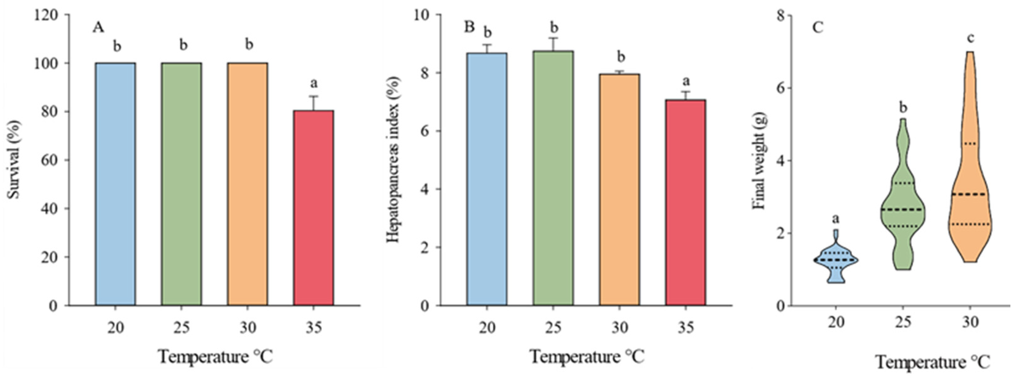
3.1. Survival, Growth Performance, Molting, and Feeding

The survival rate and hepatopancreatic index of the 35 °C group were significantly lower than the other groups (p < 0.05) (Figure 1A,B). The temperature significantly affected the growth, and SGR showed a parabolic trend (Figure 1C and Figure 2A). Juvenile mud crabs had the best growth performance between 28.5 °C and 29.7 °C in accordance with the quadratic regression model analysis (Figure 2).
The MF of the 30 °C (19.80 ± 1.33) group was significantly higher than the 20 °C (8.40 ± 1.05), 25 °C (13.21 ± 0.51), and 35 °C (14.00 ± 1.54) (p < 0.05). However, the MI of the 35 °C groups (73.58 ± 2.18%) was significantly lower than that of the 20 °C (87.13 ± 9.10%), 25 °C (93.44 ± 5.16%), and 30 °C (86.58 ± 4.36%) groups (p < 0.05) (Figure 3).
With the increase in temperature, the average daily food intake showed a trend of first increase and then decrease, and the difference was significant (p < 0.05) (Figure 4A). The feed conversion efficiency at 25 °C and 30 °C was significantly higher than that in the 20 and 35 °C groups (p < 0.05), the 35 °C group was significantly lower than the other three groups (p < 0.05), and the FCE in the 30 °C group was the highest (45.48 ± 1.21%), the FCE of the 35 °C group was the lowest (21.00 ± 3.00%) (Figure 4B).

3.2. Ecdysone Content and Expression of Molting-Related Genes

The content of ecdysone in crab hemolymph increased significantly with the increasing temperature (p < 0.05) (Figure 5A). The expression level of the ecr gene in the 35 °C group was the highest and was significantly higher than that in the other three groups (p < 0.05) (Figure 5B). The expression of the mih gene decreased with the increasing temperature (Figure 5C).

3.3. OCR, AER, and O: N

Between 20–30 °C, the OCR increased significantly with the increasing temperature (p < 0.05), but no significant difference was found between the 30 °C and 35 °C groups (p > 0.05) (Figure 6A). The AER peaked at 35 °C and was significantly higher than the 20–30 °C group (p < 0.05) (Figure 6B). The O: N showed a parabolic trend and peaked at 30 °C (Figure 6C).

3.4. Antioxidant Capacity

T-AOC activity was the highest at 25 and 30 °C (Figure 7A). The activity of CAT showed a significant upward trend with the increasing temperature (p < 0.05) (Figure 7B). The GSH and SOD activities peaked at 30 °C (Figure 7C,D). Moreover, the MDA content was significantly lower in the 25 and 30 °C groups compared to the 35 °C group (Figure 7E).

3.5. Hemolymph Index

In comparison with other groups, the hemolymph of the 35 °C group contained significantly more cortisol and LD (p < 0.05), and there was no significant difference between the 20–30 °C groups (p > 0.05) (Figure 8A,B). The uric acid content increased with the increasing temperature (Figure 8F). Glucose and total cholesterol showed a parabolic trend (Figure 8C,E). Significantly higher TG content was found in the 35 °C group than in either the 30 °C or 20 °C groups (p < 0.05) (Figure 8D).

3.6. Correlation Analysis

Correlations between temperature and SGR, MF, MI, OCR, AER, FI, FCE, and relative expression of mih and ecr parameters were analyzed by Pearson correlation coefficient (Figure 9). Overall, ecr gene expression, AER, OCR, and MF were positively correlated with temperature, while mih gene expression and MI were negatively correlated. Interestingly, the mih gene expression was also negatively correlated with SGR, MF, OCR, AER, and FI.

4. Discussion

Temperature significantly affected the survival and growth of the mud crab. For crabs reared at 20–30 °C, the survival rate was 100%, while high water temperature (35 °C) caused mortality. The same trend was also observed in growth performance. According to the quadratic regression analysis, the optimal water temperature for the growth of mud crab was 28.5–29.7 °C. A similar study also suggested that the most suitable temperature in Crablet 1 to Crablet 2 phase was 28–32 °C [35]. However, another recent research observed that the SGR of the juvenile S. paramamosain in the 36–37 °C (14.65 ± 0.23% day−1) group was significantly higher than the 27–28 °C (12.30 ± 0.42% day−1), 30–31 °C (11.58 ± 0.14% day−1), and 33–34 °C (10.11 ± 0.06% day−1) groups [35]. The difference could be partly attributed to the fact that the mud crabs were cultured in groups of 30 individuals per tank [36]; thus, temperature-induced cannibalism could potentially contribute to the growth performance. In addition, the speculation was also supported by the markedly lower survival rate (28.9 ± 2.94%) and higher SGR (14.65 ± 0.23% day−1) at 36–37 °C [35] compared to the present study (80.36 ± 5.92% and 3.00 ± 0.26% day−1 at 35 °C).
The growth of organisms was closely related to feeding [37]. Generally, FI increases with increasing temperature and decreases when the temperature exceeds the optimum temperature range [38]. In this study, both low and high temperatures inhibited FI. Low temperatures inhibit the metabolic capacity of crabs, thereby affecting their appetite and energy balance. On the contrary, long-term high-temperature stress caused heat stress and decreased FI. In the present study, the FCE and FI had a consistent trend, indicating that a suitable temperature not only stimulates FI but also effectively improves the assimilation of food. This result was also observed in turbot (Scophthalmus maximus) [39], Penaeus japonicus [40], and Atlantic salmon (Salmo salar) [41]. Therefore, the decline in FI and FCE could be one of the reasons for the suboptimum growth performance for the 20 and 35 °C group.
Molting is essential in crustacean growth [42]. High water temperature inhibited the expression of the mih gene but consequently promoted ecdysone synthesis and ecr gene expression. These factors could jointly promote the synthesis of the EcR-RXR-ecdysone complex, resulting in a significantly increasing MF. Crustacean growth and molting are highly correlated in most studies [5,43,44,45]. Interestingly, in this study, both MI and MF dropped simultaneously in the 35 °C group compared with the 30 °C group. A similar phenomenon was observed in Penaeus japonicus [40]. Nonetheless, the underlying physiological process is poorly understood. A possible explanation is crab’s metabolic capacity is temperature-dependent.
The metabolic rate of animals, measured by OCR and AER, varies directly with temperature [46,47]. O: N could represent the ratio of protein, fat, and carbohydrate catabolism of aquatic organisms [48]. O: N < 10 suggests the respiratory substrate mainly consisted of protein. On the contrary, a high O: N means the substrate is mainly provided by fats and carbohydrates [49]. The OCR of crabs at 30–35 °C was similar, but AER raised dramatically at 35 °C, leading to a lower O: N (105.66 ± 16.90) compared with 30 °C (143.46 ± 7.97). Though the value is much higher than 10, the results suggest a higher proportion of proteins consumed by respiration than tissue growth at 35 °C. The conjecture could further be confirmed by the high hemolymph uric acid level.
The stress response of an organism can be divided into three levels [50]. First-order stress responses include elevated cortisol levels [51]. Secondary stress includes increased energy mobilization, manifested primarily by increases in blood glucose and circulating lipids, which provide the organism with the energy necessary to resist stress [52]. Primary and secondary stress responses trigger tertiary stress responses that ultimately affect animal growth and survival [53]. In this study, a significant increase in cortisol, GLU, TG, and T-CHO in the hemolymph at high temperatures was observed. The results indicate that elevated temperature caused cortisol secretion, increased protein catabolism, and plasma cholesterol, and promoted GLU by inhibiting GLU breakdown. Furthermore, mud crabs raised at 35 °C also had higher lactic acid and significantly lower T-AOC, SOD, and GSH, suggesting an inferior thermal resistance [54,55] and antioxidant capacity, leading to down-regulated survival rates.

5. Conclusions

The effects of rearing temperature on mud crab S. paramamosain were estimated by growth, molting, feeding intake, energy metabolism, and stress responses. The result showed that the crabs at high temperature (35 °C) had a significantly higher ecdysone level and expression of the ecr gene but lower MI, MF, and survival rate. The effect could be mediated through respiration, energy metabolism, and antioxidant pathways. It was found that 28.5–29.7 °C provided the best conditions for mud crab growth. These findings could help regulate the temperature in mud crab RAS and provide a thread for the thermal adaptation of crustaceans.

Author Contributions

Conceptualization, C.S. and J.L.; methodology, J.L.; formal analysis, J.L.; investigation, J.L.; Funding acquisition, C.S.; writing—original draft preparation, J.L.; writing—review and editing, C.S., Y.Y., Z.R., Q.W., Z.M., C.M.; project administration, C.W. All authors have read and agreed to the published version of the manuscript.

Funding

The research was sponsored by the National Natural Science Foundation of China (Grant Nos. 32172994 and 31972783), the National Key Research and Development Program of China (Project No. 2019YFD0901000), the Province Key Research and Development Program of Zhejiang (2021C02047), Key Scientific and Technological Grant of Zhejiang for Breeding New Agricultural Varieties (2021C02069-6), 2025 Technological Innovation for Ningbo (2019B10010), China Agriculture Research System of MOF and MARA, SanNongLiuFang Zhejiang Agricultural Science and Technology Cooperation Project (2021SNLF029), K. C. Wong Magna Fund in Ningbo University and the Scientific Research Foundation of Graduate School of Ningbo University (IF2020145).

Institutional Review Board Statement

Our study did not involve endangered or protected species. In China, breeding and catching mud crabs, Scylla paramamosain, does not require specific permits. All efforts were made to minimize animal suffering and discomfort. The animal study protocol was approved by the Animal Ethics Committee of Ningbo University.

Data Availability Statement

The data presented in this study are not publicly available but are available upon request from the corresponding author.

Acknowledgments

Thanks to Qingsong Zhao for providing the site for this experiment.

Conflicts of Interest

The authors declare no conflict of interest.

References

  1. Keenan, C.P. Aquaculture of the mud crab, genus Scylla-past, present and future. In Aciar Proceedings; Australian Centre for International Agricultural: Washington, DC, USA, 1999; pp. 9–13. [Google Scholar]
  2. Le Vay, L. Ecology and Management of Mud Crab Scylla spp. Asian Fish. Sci. 2001, 14, 101–112. [Google Scholar] [CrossRef]
  3. Cowan, L. Crab farming in Japan, Taiwan and the Philippines. Qld. Dep. Prim. Ind. Inf. Ser. Q 1984, 85, 184009. [Google Scholar]
  4. Liu, X. Fisheries Bureau of Agriculture Ministry of China; China Agriculture Press: Beijing, China, 2021. [Google Scholar]
  5. Yu, K.; Shi, C.; Liu, X.; Ye, Y.; Wang, C.; Mu, C.; Song, W.; Ren, Z. Tank bottom area influences the growth, molting, stress response, and antioxidant capacity of juvenile mud crab Scylla paramamosain. Aquaculture 2022, 548, 737705. [Google Scholar] [CrossRef]
  6. Ma, X.; Zhang, G.; Gao, G.; Wang, C.; Mu, C.; Ye, Y.; Shi, C. Effects of culture container color on growth, stress and carapace color of juvenile mud crabs Scylla paramamosain. J. Ningbo Univ. Sci. Technol. Ed. 2021, 34, 43–49. [Google Scholar]
  7. Beitinger, T.L.; Fitzpatrick, L.C. Physiological and Ecological Correlates of Preferred Temperature in Fish. Am. Zool. 1979, 19, 319–329. [Google Scholar] [CrossRef]
  8. Tropea, C.; Stumpf, L.; Greco, L.S.L. Effect of Temperature on Biochemical Composition, Growth and Reproduction of the Ornamental Red Cherry Shrimp Neocaridina heteropoda heteropoda (Decapoda, Caridea). PLoS ONE 2015, 10, e0119468. [Google Scholar] [CrossRef]
  9. Bastos, A.M.; Lima, J.F.; Tavares-Dias, M. Effect of increase in temperature on the survival and growth of Macrobrachium amazonicum (Palaemonidae) in the Amazon. Aquat. Living Resour. 2018, 31, 21. [Google Scholar] [CrossRef]
  10. Stoner, A.W.; Ottmar, M.L.; Copeman, L.A. Temperature effects on the molting, growth, and lipid composition of newly-settled red king crab. J. Exp. Mar. Biol. Ecol. 2010, 393, 138–147. [Google Scholar] [CrossRef]
  11. Hyde, C.J.; Elizur, A.; Ventura, T. The crustacean ecdysone cassette: A gatekeeper for molt and metamorphosis. J. Steroid Biochem. Mol. Biol. 2019, 185, 172–183. [Google Scholar] [CrossRef] [PubMed]
  12. Zhang, X.; Huang, D.; Jia, X.; Zou, Z.; Wang, Y.; Zhang, Z. Functional analysis of the promoter of the molt-inhibiting hormone (mih) gene in mud crab Scylla paramamosain. Gen. Comp. Endocrinol. 2018, 259, 131–140. [Google Scholar] [CrossRef]
  13. Lushchak, V.I. Environmentally induced oxidative stress in aquatic animals. Aquat. Toxicol. 2011, 101, 13–30. [Google Scholar] [CrossRef]
  14. Hernández-Sandoval, P.; Díaz-Herrera, F.; Díaz-Gaxiola, J.M.; Martínez-Valenzuela, C.; García-Guerrero, M. Effect of temperature on growth, survival, thermal behavior, and critical thermal maximum in the juveniles of Macrobrachium occidentale (Holthuis, 1950) (Decapoda: Caridea: Palaemonidae) from Mexico. J. Crustacean Biol. 2018, 38, 483–488. [Google Scholar] [CrossRef] [Green Version]
  15. Re, A.D.; Diaz, F.; Sierra, E.; Rodríguez, J.; Perez, E. Effect of salinity and temperature on thermal tolerance of brown shrimp Farfantepenaeus aztecus (Ives) (Crustacea, Penaeidae). J. Therm. Biol. 2005, 30, 618–622. [Google Scholar] [CrossRef]
  16. González, R.A.; Díaz, F.; Licea, A.; Re, A.D.; Sánchez, L.N.; García-Esquivel, Z. Thermal preference, tolerance and oxygen consumption of adult white shrimp Litopenaeus vannamei (Boone) exposed to different acclimation temperatures. J. Therm. Biol. 2010, 35, 218–224. [Google Scholar] [CrossRef]
  17. Thomas, C.W.; Crear, B.J.; Hart, P.R. The effect of temperature on survival, growth, feeding and metabolic activity of the southern rock lobster, Jasus edwardsii. Aquaculture 2000, 185, 73–84. [Google Scholar] [CrossRef]
  18. Frisk, M.; Steffensen, J.F.; Skov, P.V. The effects of temperature on specific dynamic action and ammonia excretion in pikeperch (Sander lucioperca). Aquaculture 2013, 404, 65–70. [Google Scholar] [CrossRef]
  19. Hu, Y.; Tan, B.; Mai, K.; Ai, Q.; Zheng, S.; Cheng, K. Growth and body composition of juvenile white shrimp, Litopenaeus vannamei, fed different ratios of dietary protein to energy. Aquac. Nutr. 2008, 14, 499–506. [Google Scholar] [CrossRef]
  20. Pascual, C.; Gaxiola, G.; Rosas, C. Blood metabolites and hemocyanin of the white shrimp, Litopenaeus vannamei: The effect of culture conditions and a comparison with other crustacean species. Mar. Biol. 2003, 142, 735–745. [Google Scholar] [CrossRef]
  21. Ciaramella, M.A.; Battison, A.L.; Horney, B. Measurement of tissue lipid reserves in the American lobster (Homarus americanus): Hemolymph metabolites as potential biomarkers of nutritional status. J. Crustac. Biol. 2014, 34, 629–638. [Google Scholar] [CrossRef]
  22. Halliwell, B.; Gutteridge, J.M. Free Radicals in Biology and Medicine; Oxford University Press: New York, NY, USA, 2015. [Google Scholar]
  23. Madeira, D.; Narciso, L.; Cabral, H.N.; Vinagre, C.; Diniz, M.S. Influence of temperature in thermal and oxidative stress responses in estuarine fish. Comp. Biochem. Physiol. Part A Mol. Integr. Physiol. 2013, 166, 237–243. [Google Scholar] [CrossRef] [PubMed]
  24. Paital, B.; Chainy, G.B.N. Effects of temperature on complexes I and II mediated respiration, ROS generation and oxidative stress status in isolated gill mitochondria of the mud crab Scylla serrata. J. Therm. Biol. 2014, 41, 104–111. [Google Scholar] [CrossRef] [PubMed]
  25. Kong, X.; Wang, G.; Li, S. Effects of low temperature acclimation on antioxidant defenses and ATPase activities in the muscle of mud crab (Scylla paramamosain). Aquaculture 2012, 370–371, 144–149. [Google Scholar] [CrossRef]
  26. Feng, G.; Shi, X.; Huang, X.; Zhuang, P. Oxidative Stress and Antioxidant Defenses after Long-term Fasting in Blood of Chinese Sturgeon (Acipenser sinensis). Procedia Environ. Sci. 2011, 8, 469–475. [Google Scholar] [CrossRef]
  27. Viarengo, A.; Canesi, L.; Pertica, M.; Poli, G.; Moore, M.N.; Orunesu, M. Heavy metal effects on lipid peroxidation in the tissues of mytilus gallopro vincialis lam. Comp. Biochem. Physiol. Part C Comp. Pharmacol. 1990, 97, 37–42. [Google Scholar] [CrossRef]
  28. Chen, S.; Migaud, H.; Shi, C.; Song, C.; Wang, C.; Ye, Y.; Mu, C. Light intensity impacts on growth, molting and oxidative stress of juvenile mud crab Scylla paramamosain. Aquaculture 2021, 545, 737159. [Google Scholar] [CrossRef]
  29. Gao, Y.; He, Z.; Zhao, B.; Li, Z.; He, J.; Lee, J.Y.; Chu, Z. Effect of stocking density on growth, oxidative stress and HSP 70 of pacific white shrimp Litopenaeus vannamei. Turk. J. Fish. Aquat. Sci. 2017, 17, 877–884. [Google Scholar] [CrossRef]
  30. Maniam, J.; Morris, M.J. The link between stress and feeding behaviour. Neuropharmacology 2012, 63, 97–110. [Google Scholar] [CrossRef] [PubMed]
  31. Chen, S.; Shi, C.; Migaud, H.; Song, C.; Mu, C.; Ye, Y.; Wang, C.; Ren, Z. Light Spectrum Impacts on Growth, Molting, and Oxidative Stress Response of the Mud Crab Scylla paramamosain. Front. Mar. Sci. 2022, 9, 840353. [Google Scholar] [CrossRef]
  32. Shi, C.; Wang, J.; Peng, K.; Mu, C.; Ye, Y.; Wang, C. The effect of tank colour on background preference, survival and development of larval swimming crab Portunus trituberculatus. Aquaculture 2019, 504, 454–461. [Google Scholar] [CrossRef]
  33. Luo, H.; Wu, Q.; Lin, J.; He, L.; Chen, X.; Niu, W.; Wang, Q.; Zheng, L. Interactive effects of temperature and body weight on oxygen consumption rate, ammonia excretion rate and asphyxiation point of Hippocampus erectus. Fish. Sci. Technol. Inf. 2022, 49, 67–72. [Google Scholar]
  34. Zhu, S.; Long, X.; Turchini, G.M.; Deng, D.; Cheng, Y.; Wu, X. Towards defining optimal dietary protein levels for male and female sub-adult Chinese mitten crab, Eriocheir sinensis reared in earthen ponds: Performances, nutrient composition and metabolism, antioxidant capacity and immunity. Aquaculture 2021, 536, 736442. [Google Scholar] [CrossRef]
  35. Nur Syafaat, M.; Mohammad, S.; Nor Azra, M.; Ma, H.; Abol-Munafi, A.B. Effect of water temperature on survival, growth and molting cycle during early crablet instar of mud crab, Scylla paramamosain (Estampador, 1950). Thalass. Int. J. Mar. Sci. 2020, 36, 543–551. [Google Scholar] [CrossRef]
  36. Phuong, N.T.; Ha, N.T.K.; Nguyen, T.E.; Do Thi, T.H. Effects of different temperatures on the growth, survival and digestive enzyme activities of mud crab Scylla paramamosain at juvenile stage. Aquac. Aquar. Conserv. Legis. 2021, 14, 2741–2750. [Google Scholar]
  37. Warren, C.E.; Davis, G.E. Laboratory Studies on the Feeding, Bioenergetics, and Growth of Fish. 1967. Available online: https://ir.library.oregonstate.edu/concern/administrative_report_or_publications/x920g185p?locale=en (accessed on 19 August 2022).
  38. Volkoff, H.; Rønnestad, I. Effects of temperature on feeding and digestive processes in fish. Temperature 2020, 7, 307–320. [Google Scholar] [CrossRef] [PubMed]
  39. Imsland, A.K.; Foss, A.; Gunnarsson, S.; Berntssen, M.H.; FitzGerald, R.; Bonga, S.W.; Stefansson, S.O. The interaction of temperature and salinity on growth and food conversion in juvenile turbot (Scophthalmus maximus). Aquaculture 2001, 198, 353–367. [Google Scholar] [CrossRef]
  40. Hewitt, D.R.; Duncan, P.F. Effect of high water temperature on the survival, moulting and food consumption of Penaeus (Marsupenaeus) japonicus (Bate, 1888). Aquac. Res. 2001, 32, 305–313. [Google Scholar] [CrossRef]
  41. Handeland, S.O.; Imsland, A.K.; Stefansson, S.O. The effect of temperature and fish size on growth, feed intake, food conversion efficiency and stomach evacuation rate of Atlantic salmon post-smolts. Aquaculture 2008, 283, 36–42. [Google Scholar] [CrossRef]
  42. Zhou, L.; Li, S.; Wang, Z.; Li, F.; Xiang, J. An eclosion hormone-like gene participates in the molting process of Palaemonid shrimp Exopalaemon carinicauda. Dev. Genes Evol. 2017, 227, 189–199. [Google Scholar] [CrossRef]
  43. Tian, H.; Yang, C.; Yu, Y.; Yang, W.; Lu, N.; Wang, H.; Liu, F.; Wang, A.; Xu, X. Dietary cholesterol level affects growth, molting performance and ecdysteroid signal transduction in Procambarus clarkii. Aquaculture 2020, 523, 735198. [Google Scholar] [CrossRef]
  44. Liu, T.; Xu, H.; Han, T.; Wang, J.; Yin, F.; Wang, C. Effect of dietary egg yolk lecithin levels on survival, growth, lipid metabolism, and antioxidant capacity of early juvenile green mud crab Scylla paramamosain. Aquaculture 2021, 540, 736706. [Google Scholar] [CrossRef]
  45. Yuan, Q.; Qian, J.; Ren, Y.; Zhang, T.; Li, Z.; Liu, J. Effects of stocking density and water temperature on survival and growth of the juvenile Chinese mitten crab, Eriocheir sinensis, reared under laboratory conditions. Aquaculture 2018, 495, 631–636. [Google Scholar] [CrossRef]
  46. Cai, Y.; Summerfelt, R.C. Effects of temperature and size on oxygen consumption and ammonia excretion by walleye. Aquaculture 1992, 104, 127–138. [Google Scholar] [CrossRef]
  47. Clarke, A.; Fraser, K.P.P. Why does metabolism scale with temperature? Funct. Ecol. 2004, 18, 243–251. [Google Scholar] [CrossRef]
  48. Mayzaud, P.; Conover, R.J. O:N atomic ratio as a tool to describe zooplankton metabolism. Mar. Ecol. Prog. Ser. 1988, 45, 289–302. [Google Scholar] [CrossRef]
  49. Brito, R.; Chimal, M.E.; Gaxiola, G.; Rosas, C. Growth, metabolic rate, and digestive enzyme activity in the white shrimp Litopenaeus setiferus early postlarvae fed different diets. J. Exp. Mar. Biol. Ecol. 2000, 255, 21–36. [Google Scholar] [CrossRef]
  50. Wendelaar Bonga, S.E. The stress response in fish. Physiol. Rev. 1997, 77, 591–625. [Google Scholar] [CrossRef]
  51. Mommsen, T.P.; Vijayan, M.M.; Moon, T.W. Cortisol in teleosts: Dynamics, mechanisms of action, and metabolic regulation. Rev. Fish Biol. Fish. 1999, 9, 211–268. [Google Scholar] [CrossRef]
  52. Jia, Y.; Fan, Y.S.; Wang, Y.Z.; Chai, Y.H.; Gao, J.W.; Dou, Y.; Peng, S. Physiological and energy metabolism responses of chinese Loach Paramisgurnus dabryanus (Dabry de Thiersant, 1872) to waterless preservation during transport. Turk. J. Fish. Aquat. Sci. 2018, 19, 279–287. [Google Scholar]
  53. Schreck, C.B. Stress and fish reproduction: The roles of allostasis and hormesis. Gen. Comp. Endocrinol. 2010, 165, 549–556. [Google Scholar] [CrossRef]
  54. Pörtner, H.O. Climate variations and the physiological basis of temperature dependent biogeography: Systemic to molecular hierarchy of thermal tolerance in animals. Comp. Biochem. Physiol. Part A Mol. Integr. Physiol. 2002, 132, 739–761. [Google Scholar] [CrossRef]
  55. Pörtner, H.-O. Oxygen- and capacity-limitation of thermal tolerance: A matrix for integrating climate-related stressor effects in marine ecosystems. J. Exp. Biol. 2010, 213, 881–893. [Google Scholar] [CrossRef] [PubMed] [Green Version]
Figure 1. Effects of different temperatures (20–35 °C) on survival rate (A), hepatopancreas index (B), and final weight (C) of juvenile mud crabs. Values are expressed as mean ± SD (n = 3). Different superscripts indicate significant differences between treatments (p < 0.05).
Water 14 02988 g001
Figure 2. Relationship between the temperature and the SGR of initial to 1st molt (A), 1st to 2nd molt (B), 2nd to 3rd molt (C), and initial to final (D), respectively. Where Xopt means the optimal temperature for the maximum SGR.
Water 14 02988 g002
Figure 3. Effects of different temperatures (20–35 °C) on the molt frequency and molt increment of juvenile mud crabs. Values are expressed as mean ± SD (n = 3). Different superscripts indicate significant differences between treatments (p < 0.05).
Water 14 02988 g003
Figure 4. Effects of different temperatures (20–35 °C) on the average daily food intake (A) and feed conversion efficiency (B) of juvenile mud crabs. Values are expressed as mean ± SD (n = 3). Different superscripts indicate significant differences between treatments (p < 0.05).
Water 14 02988 g004
Figure 5. The content of ecdysone (A) in the hemolymph. Expression of the molting-related gene (ecr) (B) and molt-inhibiting hormone (mih) (C) in eyestalks. Values are presented as mean ± SD (n = 3). Different superscripts indicate significant differences between treatments (p < 0.05).
Water 14 02988 g005
Figure 6. Oxygen consumption rate (OCR) (A), ammonia excretion rate (AER) (B), and oxygen-nitrogen ratio (O: N ratio) (C) of juvenile mud crabs at different temperatures (20–35 °C). Values are expressed as mean ± SD (n = 3). Different superscripts indicate significant differences between treatments (p < 0.05).
Water 14 02988 g006
Figure 7. Effects of different temperatures on the activities total of antioxidant capacity (T-AOC) (A), catalase (CAT) (B), superoxide dismutase (SOD) (C), glutathione (GSH) (D), and malondialdehyde (MDA) (E) content of juvenile mud crabs. Values are expressed as mean ± SD (n = 3). Different superscripts indicate significant differences between treatments (p < 0.05).
Water 14 02988 g007
Figure 8. Effects of different temperatures on cortisol (A), lactic acid (LD) (B), glucose (GLU) (C), triglyceride (TG) (D), total cholesterol (T-CHO) (E), and uric acid (UA) (F) content in juvenile mud crabs. Values are presented as mean ± SD (n = 3). Different superscripts indicate significant differences between treatments (p < 0.05).
Water 14 02988 g008
Figure 9. Correlation analyses among temperature SGR, MF, MI, OCR, AER, FI, FCE, and relative expression of mih and ecr gene (n = 3, 2 individuals per replicate for AER, OCR, ecr, and mih; n = 3 replicate, and 31, 42, 42, and 42 individuals per treatment of SGR, MF, FI, and FCE). “*” indicates p < 0.05.
Water 14 02988 g009
Table 1. Specific primers were used for real-time PCR in this study.
GeneSequence (5′ to 3′)Size (bp)Access Number
ecrF: TAAGTGATGACGACTCGGATGC150KC354381
R: ACGAGCAAGCCTTTAGCAGTG
mihF: TATCAAGTGCAGGAACTCAG110EU869539
R: GGAACATACAAGCCTAAACA
β-actinF: CGAAACCTTCAACACTCCCG154FJ641977
R: GATAGCGTGAGGAAGGGCATA
ecr: Ecdysone receptor, mih: Molt-inhibiting hormone.
Publisher’s Note: MDPI stays neutral with regard to jurisdictional claims in published maps and institutional affiliations.

