
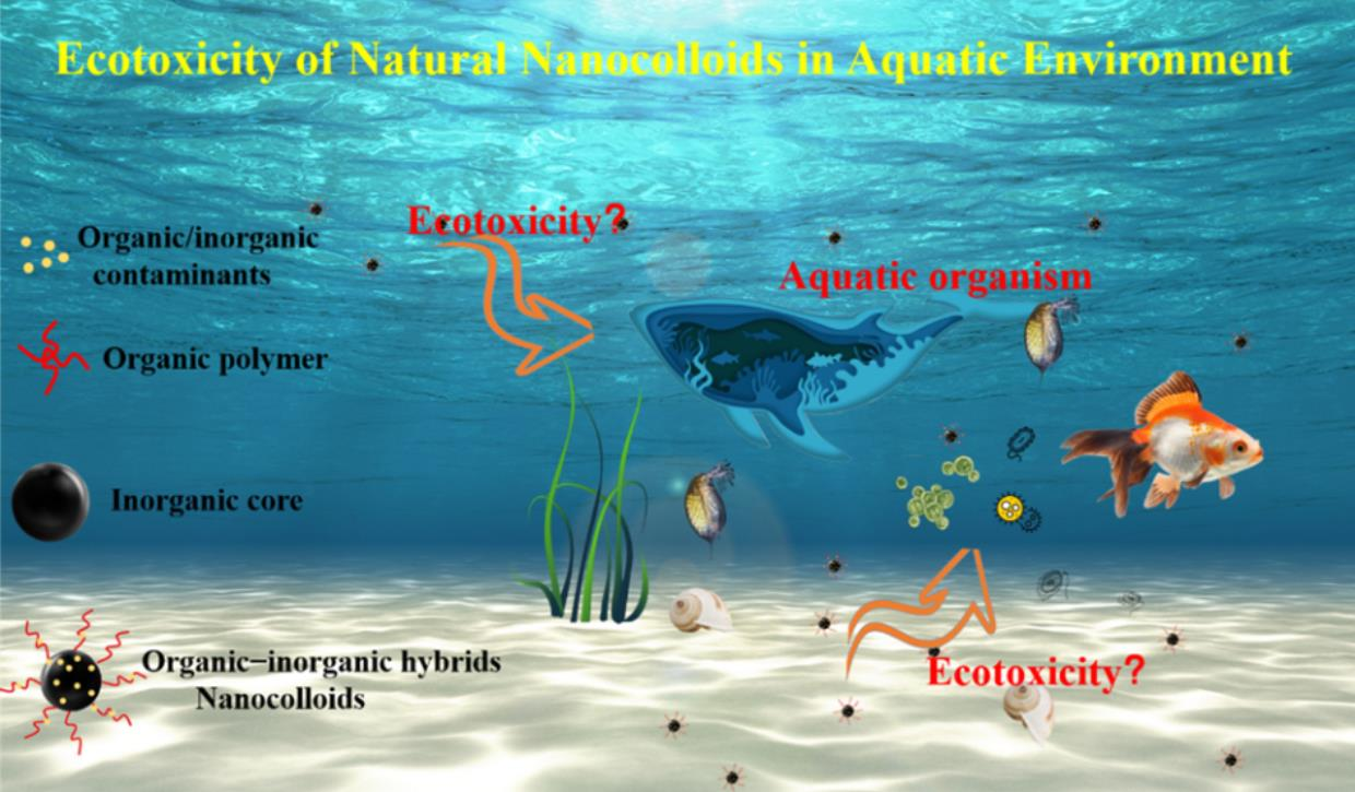
Ecotoxicity of Natural Nanocolloids in Aquatic Environment
Key Laboratory of Pollution Processes and Environmental Criteria (Ministry of Education)/Tianjin Key Laboratory of Environmental Remediation and Pollution Control, College of Environmental Science and Engineering, Nankai University, Tianjin 300350, China
*
Author to whom correspondence should be addressed.
†
These authors contributed equally to this work.
Water 2022, 14(19), 2971; https://doi.org/10.3390/w14192971
Submission received: 22 August 2022
/
Revised: 14 September 2022
/
Accepted: 19 September 2022
/
Published: 22 September 2022
(This article belongs to the Special Issue Nanocolloids in Water and Human Health)
Abstract
:Nanocolloids (Ncs) are highly dispersed mixtures of nanoscale (1–100 nm) heterogeneous systems, which are ubiquitous in aquatic environments. Ncs are considered a vital pollutant carrier due to their special surface properties and unique hydrodynamic characteristics. They play an essential role in the process of promoting pollutant migration and transformation. In recent years, with the increase in chemicals in the environment and the complexity of environmental pollution, the health threats of Ncs in ecological systems are arousing great concerning. Therefore, recent work to characterize the ecotoxicity of Ncs has focused on the potential environmental health implications, including exploration of toxicity to aquatic organisms from a wide range of the ecosystem food webs. Herein, we summarize the formation, distribution, and characterization of natural Ncs in the marine environments. Moreover, we highlight the adverse impacts of Ncs on representatives of various trophic levels aquatic organisms (e.g., algae, bacteria, invertebrates, and fish). The mechanisms of Ncs ecotoxicity at the cellular level are reviewed, and the remaining unclear points on toxic tools such as oxidative damage and metabolic disorder are presented. We also discuss the research challenges and future developments within the field of ecotoxicity. This study will bridge our knowledge gap on the ecotoxicity of Ncs.

1. Introduction
Natural nanocolloids (Ncs) represent a specific type of matter, defined as a highly dispersed multiphase heterogeneous system of nanometer magnitude (1–100 nm), also known as nano-colloidal dispersions or mixtures [1,2,3]. Like engineered nanoparticles, natural Ncs have an extensive specific surface area and can absorb other harmful pollutants in a large capacity [2,4]. For example, our previous studies showed that natural Ncs contain dangerous contaminants such as polycyclic aromatic hydrocarbons (PAHs, 14.2–50.5 μg/kg) and toxic heavy metals such as Cr [5]. Due to the high activity and good stability, Ncs can readily attach environmental pollutants (e.g., trace heavy metals or persistent organic substances) and exist in a water environment for a long time, which play an important role in the migration and transformation of pollutants [5,6].
In recent years, with the continuous entry of chemicals into the aquatic environment and the complexity of water pollution, the concentration and types of Ncs in aquatic environmental system are gradually increasing and receiving significant attention [7,8]. Many studies have confirmed that Ncs exist widely in the environment, and their concentration is at the level of mg/L, which has very important environmental significance [5,9]. Upon exposure to aquatic environments, Ncs may have adverse impacts on aquatic organisms (e.g., bacteria, algae, plants, invertebrates, and fish) [10,11,12], even leading to changes in the expression levels of endogenous metabolites and genetic materials. The fate and potential environmental impacts of Ncs in the aquatic ecological systems have attracted increasing attention.
Currently, there have been a lot of studies on engineered nanoparticles, and a clear understanding of nanotoxicity, molecular mechanism of action, and environmental effects has been obtained. However, the existing literature on the ecotoxicity of Ncs remains limited. Furthermore, studies on Ncs in the environment mainly focus on the separation, enrichment and characterization of Ncs [13,14]. In contrast, the process and mechanism of inducing adverse effects of Ncs in the aquatic environment are relatively few [14,15,16]. In aquatic environments, Ncs can interact with system components (e.g., inorganic ions and natural organic matter (NOM)), which can change environmental behaviors and ecotoxicity of Ncs [17]. In addition, the properties and toxic effects of Ncs from different sources may differ. Environmental Ncs can interact with pollutants under other environmental factors, and form Ncs–pollutants composites, which may produce or enhance nanotoxicity and induce various diseases, thus increasing the threat to the ecosystem and human health [18]. On the contrary, it has also been shown that some Ncs have beneficial biomedical effects [19].
Therefore, given the wide spread of Ncs and their important role in biogeochemical processes, the purposes of the present review are to (1) summarize the latest progress related to the formation, distribution, and characterization of Ncs; (2) highlight the toxic ecological effect and molecular mechanisms of Ncs on various representatives; and (3) discuss the important knowledge gaps on the ecotoxicity of natural Ncs, thus providing suggestions for future research.
2. Formation, Distribution and Characterization of Natural Ncs in Aquatic Environment
Ncs are commonly formed spontaneously due to human activities and naturally biogeochemical processes, which are usually mixtures and consist of various elements [20]. Ncs have a high adsorption capacity and strong affinity for trace metal elements or organic matter, and this process will affect the migration and biological activity of these pollutants in water environments [21]. Moreover, the surface of Ncs also undergoes modifications through biotic and abiotic processes in aquatic environments, for example, chemical reactions, recrystallization, and oriented aggregation [22]. Given that Ncs have a significant impact on the migration, transformation, and ecotoxicity of pollutants, the behaviors of Ncs in aquatic environments have attracted more and more attention.
2.1. Formation
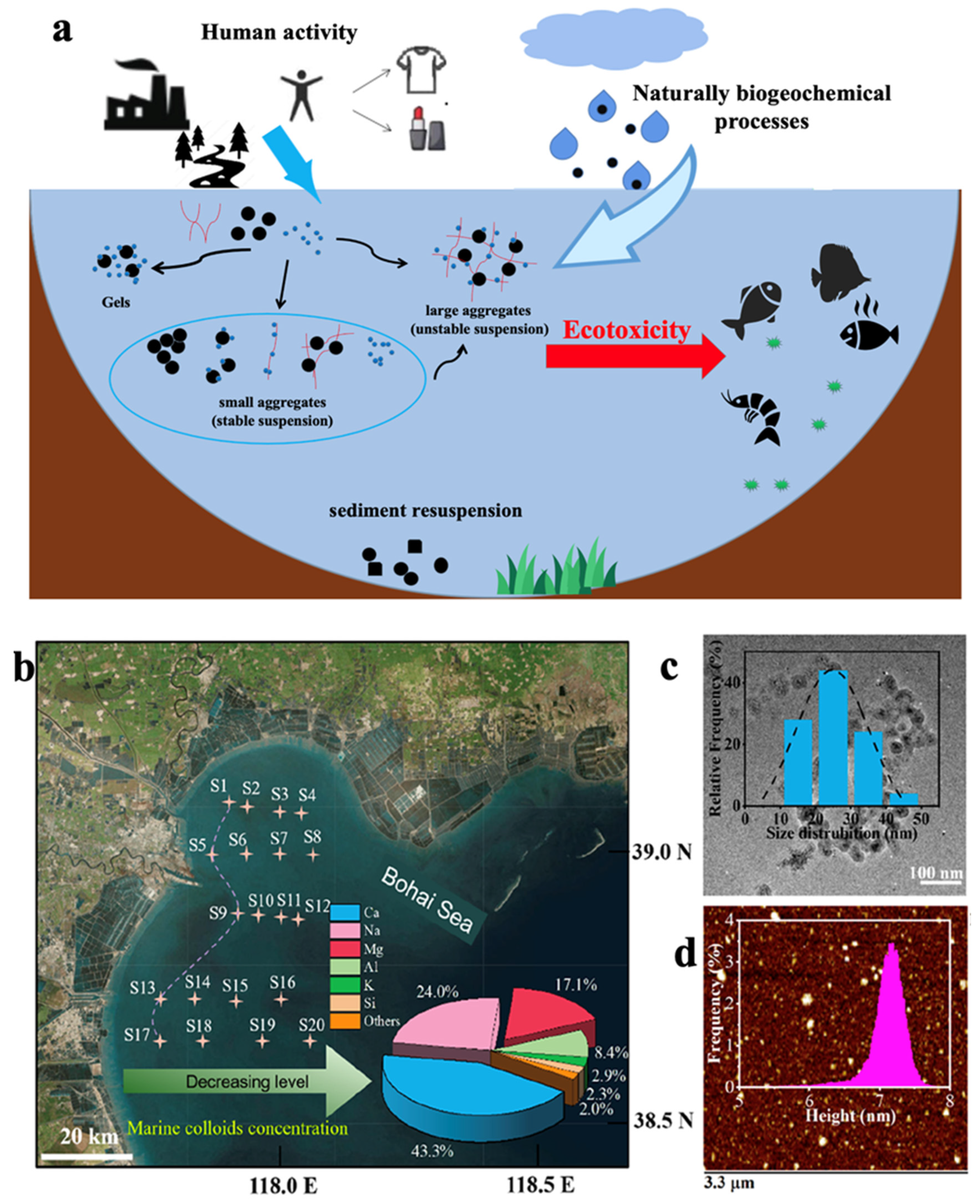
Research on the formation of Ncs is a massive task as it is complex in composition and covers diverse mechanisms, processes, and conditions. Although models of Ncs formation have been proposed by Buffle et al. (Figure 1) [23], there is still a lack of more universal models to describe the morphology and structure of aquatic colloids. Notably, the main composition of Ncs is derived from nanoparticles, Ncs are not a renaming of the concept of nanoparticles. Compared to the composition of nanoparticles, Ncs are composed of some macromolecules such as irregular network organic matter and humic acid. Moreover, Ncs are divided into two different states, the dispersed phase and the continuous phase, and are also formed by some macromolecules or particles dispersed in a heterogeneous system. Therefore, Ncs are often referred to as a nanocolloidal system, and show more stable behavior than nanoparticles in the aquatic environment [9]. Furthermore, Ncs can be divided into artificial Ncs and natural Ncs according to their sources. Artificial Ncs are generated by human activities such as microplastics and nanomaterials through sewage discharge, fishing activities, surface runoff, atmospheric deposition, and other ways into the water. On the other hand, natural Ncs are produced by chemical, photo-chemical, mechanical, thermal, and biological processes in aquatic environments. Similarly, the release of sediments from the bottom of the water, the fragmentation of large particles, photochemical degradation, and other processes lead to the formation of colloids in the water environment [8,22]. Moreover, parts of Ncs are synthesized by biological activities, which are mainly the metabolism of plants, animals, and microorganisms in the water environment, with the main biochemical processes including the physiological activities of phytoplankton and the dissolution and secretion of cells [2,24]. Most colloids in the aqueous environment are predominantly organic colloids, whose main components are dissolved organic matter. Natural hydrocolloid materials are composed of inorganic colloids (e.g., iron, aluminum, manganese oxide, hydrate or mineral), heterogeneous organic materials, and large biopolymers [24,25]. However, there is a lack of systematically detailed work on the nature and origin of natural Ncs [5].
2.2. Distribution
Ncs distribute widely in oceans, surface-, ground- and pore water, as well as in the atmosphere [26]. In an investigation of natural nanoparticles and colloidal organic matter in soils and rivers in Germany, iron oxide colloids are found to be efficiently transported [27]. However, quantitative data for natural Ncs are scarce. The sampling and transport processes have an uncontrollable effect on the stable state of the Ncs, thus reduce the confidence of the data. Moreover, the high-precision separation and quantification equipment of Ncs is also lacking. The concentrations and sizes of Ncs in the aquatic environment are influenced by pH, ionic strength, concentrations, and water sample depth. For instance, the particle size distribution of Ncs in the Haihe river ranged from 1.4 to 99.4 nm, with an average value of 17.0 nm, and the concentration of Ncs ranged from 3.7 to 7.2 mg/L [5]. The equivalent diameter of colloids in groundwater at the northwestern edge of the Sichuan Basin is 347 ± 188 nm (mean ± standard deviation, n = 300) [28]. Ncs in Cigar Lake, Canada, are measured at sizes ranging from 10 to 400 nm, at a concentration up to 1 mg·L−1. The particle size and concentration of colloids can also affect the binding characteristics to pollutants, and induce metabolic abnormalities and toxicity to aquatic animals [10,18]. Baker et al. reveal that the concentration of Ncs is a key metric in explaining the environmental behavior of hydrophobic organic pollutants [29]. Similarly, a strong relationship is found between organic carbon and copper, which further indicates that colloid plays a critical role in the migration and transformation of copper [30]. Because of the potential physiological toxicity of colloids, gaps in the quantitative characterization of colloids in the different aquatic environments should be added to future studies.
2.3. Characterization
The tangential flow ultrafiltration fractionation, cross-flow filtration, dialysis, field flow fractionation, and centrifugation are common methods, which are usually used to extract, separate, and enrich Ncs from the aquatic environments. It is also possible to use split-flow thin-cell fractionation to separate particles smaller than 1 micron in size as a pre-separation method for colloid fractionation and analysis [31]. Ncs have some characteristics common to nanomaterials (e.g., namely, small size effect, surface effect, quantum size effect and macroscopic quantum tunnel effect). Given that Ncs are heterogeneous mixtures of metallic and organic components, a multi-method approach is applied for their characterization [32]. According to the principle of analysis, the characterization methods of Ncs can be broadly divided into three categories: microscopy (e.g., scanning electron microscopy (SEM) and atomic force microscopy (AFM)), scattering spectroscopy (e.g., X-ray and fluorescence) and mass spectrometry (e.g., GC-MS and ICP-MS).
The microscopic processes can be applied for the qualitative analysis of Ncs. AFM, with its extremely high resolution, makes it possible to determine the size of natural nanoparticles with low concentration and complex conformation. It also satisfies the condition of ensuring that natural Ncs are undisturbed during the characterization process [33,34]. AFM investigates the surface structure and properties of a substance by detecting the feeble interatomic forces between the surface of sample and a miniature force-sensitive element. In the image of AFM, the shape of Ncs mainly included fibrils (10–100 nm), near-spherical Ncs (30–50 nm), and a surface film in the river [33]. Transmission electron microscopy (TEM) with a point resolution of up to 0.1 nm allows observation of surface morphology, particle size distribution, and crystal structure at the atomic scale. As shown in Figure 1, the morphology and size of the marine colloids are recorded by their TEM and AFM images. The TEM images results revealed that the lateral diameters of the isolated Ncs are mainly ranged from 20 to 80 nm, and the thicknesses of Ncs are around 1–8 nm based on the AFM images results [9]. Near-field scanning light microscopy is also used to analyze imaging of Ncs [35]. SEM is mainly applied to observe the surface morphology of the sample and measure the particle size distribution by secondary electron signal imaging. In the image of SEM images, it was observed that near-spherical Ncs were the most common nanocolloidal morphologies in the lake. Compared to SEM, environmental scanning electron microscopy (ESEM) in the measurement of the particle size of colloids in the aqueous environment can observe the hydrated sample in its natural state without the need to coat the sample with a non-conductive model [36].
Scattering spectroscopy is used to study the properties of Ncs, and mass spectrometry is a reliable method for quantifying Ncs. Energy-dispersive X-ray spectroscopy (EDS) is used as an accessory to electron microscopy, usually in combination with TEM or SEM, to analyze the morphology and elemental composition of Ncs. Generally, the elemental composition of the Ncs is identified in situ using the TEM-EDX method, the metal elements and the surface functional groups of Ncs are detected by ICP-MS and FTIR spectroscopy, respectively [10]. The ICP-MS results confirm that Ncs are consisted of various of metal elements (e.g., Ca, Mg, Na, Fe, Mg), and the abundance of Ca is the most [10]. The aromatic protein and humic acid-like fractions were detected in Ncs by FTIR [10]. Other spectroscopic techniques such as Raman, X-ray diffraction, and UV-vis spectroscopy are also widely used to characterize the functional groups of Ncs [37]. The organic carbon content and form present in Ncs, the type and content of inorganic elements can also be analyzed by fluorescence spectroscopy. The complex environmental characteristics of Ncs and their ecological effects should be addressed by combining multiplexes of existing techniques and developing new high-precision, low-interference detection methods. As shown above, Ncs are organic–inorganic hybrids.
3. Toxicity of Natural Ncs towards the Different Aquatic Organisms
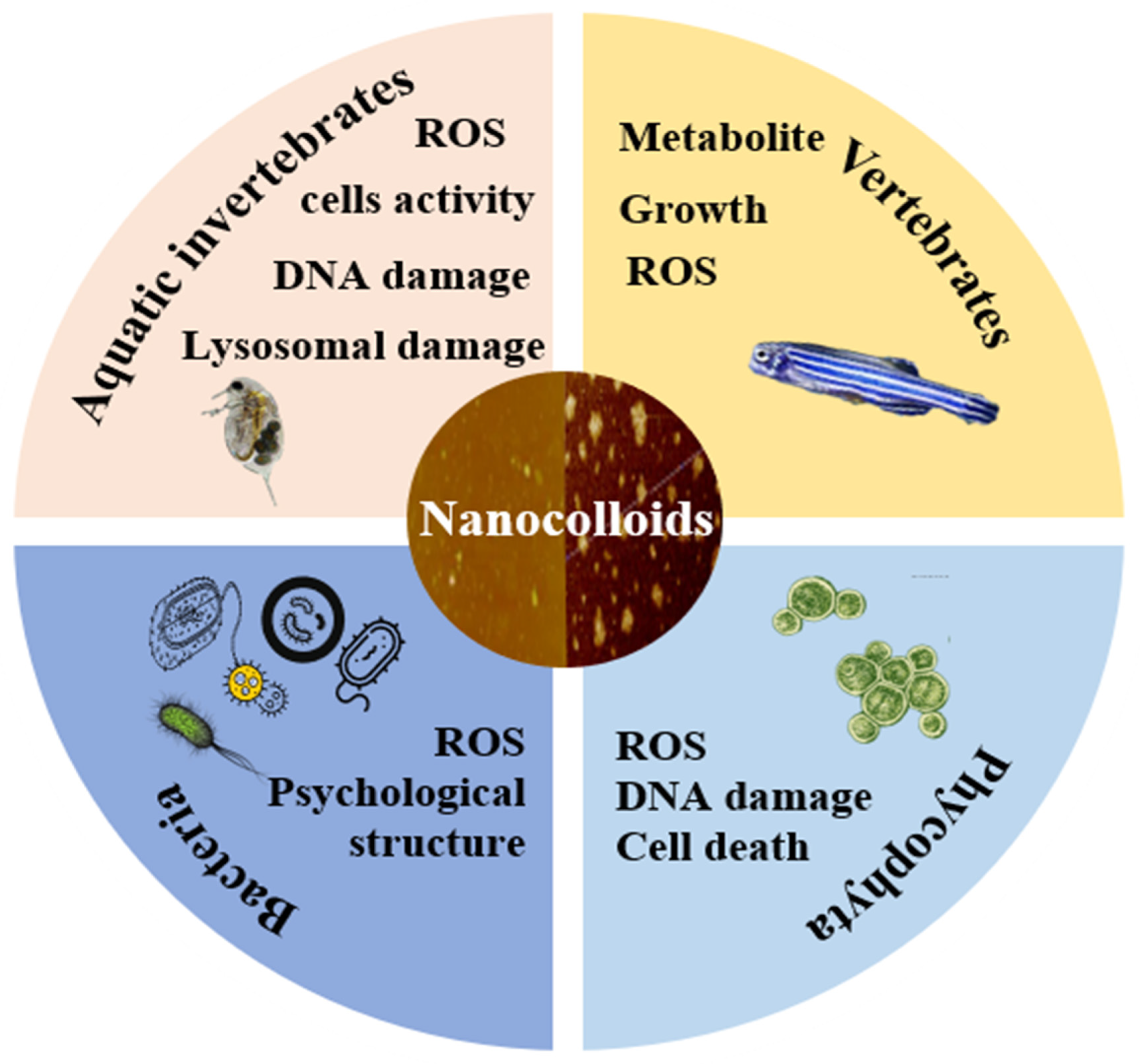
Ncs contribute to enormous environmental processes, but the understanding of Ncs’ ecotoxicity remains crucial in complex environmental matrices due to the profound complexity and heterogeneity of colloids. Research on plant nanotoxicity of nanometals has focused almost exclusively on artificial nanomaterials, while significant work on the nanotoxicity of natural Ncs is still lacking. Thus, this section provides insights into the ecotoxicity of natural Ncs on aquatic organisms (Figure 2).
3.1. Algae
The uptake of Ncs by plants can be via active transport. For example, nano-silver Ncs (10–50 nm) entering the plant through cell wall pores, and the larger nano-silver Ncs (>50 nm) entering the plant via endocytosis [38]. Smaller particle-size nanoparticles are more accessible to plant cells and have a high degree of mobility and internalization within the plant [39]. However, there is a lack of more generalized verification of whether their smaller particle size enhances toxicity in the plants, although these findings have been confirmed in human cells [40]. When stressed by nanoparticles, plants produce a variety of ROS byproducts when detoxified, leading to oxidative stress and cell death [41,42].
Algae play an important role in aquatic systems, and are also used as generic model organisms to assess the toxicity of substances [43]. In both exposure experiments, Ncs were observed to wrap around the surface of algal cells, reducing cell permeability and impeding the uptake of nutrients into the cells and thus inhibiting cell division and growth. Our previous research revealed that the Ncs entered the algal cells and clustered mainly near the starch grains, and Ncs exhibited stronge plasmolysis, chloroplast damage and more starch grains in algal cells [5]. Moreover, the sharp surface of Ncs can induce the physical damage to the surface of the algal cells, which affects the synthesis of chlorophyll, leading to an inhibition of photosynthesis [44,45]. After the internalization of Ncs in Chlorella vulgaris, a decrease in amino acids (such as serine, aspartic acid, and arginine) and increase in fatty acids could explain the metabolic abnormalities at a molecular level [45]. Jing et al. also mentioned that it might be the size effect that causes the toxicity of nano-ZnO at concentrations greater than 50 mg/L, and toxicity increases with concentration increasing [43]. Regarding genotoxicity, nanoscale zerovalent iron Ncs affect cell division in metaphase/anaphase breaks and anaphase/telophase, and form adhesive bridges to induce the production of binucleated and multinucleated cells. Oxygen and hydrogen peroxide produced by nanoparticles also cause DNA damage and cell death [46]. Although more attention has been paid to the complex toxicity of Ncs because of their large specific surface area, less research has been performed on the toxicity of natural aquatic colloids. There is no quantitative conclusion about the trapping effect of Ncs, the elimination of adverse effects caused by the culture medium in experiments and the induced oxidative stress and cell ultrastructure damage by Ncs.
3.2. Aquatic Invertebrates
Invertebrates are linked as primary producers, secondary consumers, and most of them are sensitive to pollutants. Ncs can rapidly adsorb onto dead organic matter, which affects microscopic species communities that depend on dead organic matter for food, and thus indirectly affecting invertebrates. The presence of these processes has been demonstrated for silver Ncs at ambient concentrations [47]. The bridging effect of multi-particles in Ncs gives enhanced enrichment properties to individual nanoparticles that hardly ever adsorb to soluble natural organic matter [48].
As a model organism for invertebrates, in addition to being sensitive to pollutants and having a fast reproduction and short growth cycle, the identification of the genome of Daphnia magna (D. magna) is widely applied, which has dramatically reduced the difficulty of investigating genotoxicity [49]. The shape and charge of the Ncs have a marked impact on the toxicity of D. magna. The positively charged Ncs would bind to negatively charged phospholipid membranes, thus inducing high toxic effects [50]. The production of ROS upon entry of Ncs into cells is considered as one of the hallmarks of their toxicity. Accumulation of ROS could cause cell membrane damage, lipid peroxidation, and reduced the cell division levels as well as the cell death [51]. Ncs could induce the immunotoxicity of invertebrates by reducing the phagocytic activity of blood cells, which are also the first line of defense of the bivalve [52]. In addition, the genotoxicity of Ncs has been of great concern. Nano-silver Ncs cause DNA breaks in Daphnia magna, further leading to increased cell damage and mortality, with more complex and severe effects at the population level [53]. The lysosomal damage caused by nano-silver Ncs also could affect the immune system of marine worms [54]. Natural nano-clay (2:1 layered silicate) has also been shown to have a significant decrease in the population growth rate of cladoceran species, which is lower toxic effects than the artificially modified nano-clay [55]. The toxicity exposure pathways and mechanisms of the engineered nanoparticles are well established. However, the exposure pathways for natural Ncs are unknown. Ncs may enter the circulatory system via the digestive system, but the metabolic transformation pathways and toxicity mechanisms of Ncs in invertebrates at environmental concentrations are not yet experimentally demonstrated [56]. Toxic effects in aquatic invertebrates can also be used as an emerging criterion to measure the toxicity of nanoparticles in the water column [53].
The low level of testing of Ncs toxicity to invertebrates may be due to the belief that invertebrates are fully adapted to the constant presence of Ncs in the water column, especially as some experiments have shown that short-term exposure does not cause mortality. However, because of the rapid increase in Ncs concentrations due to anthropogenic activities, it is necessary to explore toxicity-limiting concentrations such as sub-lethal concentrations of Ncs to reduce the enrichment of the food chain for Ncs.
3.3. Vertebrates
Vertebrates have the highest evolutionary status in aquatic ecosystems, where the toxicity of Ncs can be characterized more fully and in more detail. Because animal cells lack the protective structure of a cell wall, Ncs may enter the animal’s body more efficiently. Hence, Ncs may cause much more serious harm to animals than plants [57]. With its short cycle and high fecundity, transparent embryos are sensitivity to most environmental pollutants and 70% homology with human DNA. Moreover, zebrafish embryos are of interest as a model organism for toxicity studies to extrapolate the toxic effects of vertebrates in the water column [58,59]. After the exposure of environmental concentrations of Ncs (0.45, 4.5, 45 mg/L), the zebrafish embryos showed varying degrees of pericardial/yolk sac edema, uninflated swim bladder, tail flexure and spinal curvature. Metabolomics analysis result indicates the down-regulation of trimethylamine/terephthalic acid/sucrose, some amino acid metabolism, and an increase in urea, glycine and cholesterol metabolism might contribute to the above toxic effects [10]. The simultaneous observation of the non-monotonic variation in toxicity and concentration poses a greater challenge for the quantitative analysis of Ncs toxicity. As in plants, oxidative stress in zebrafish due to Ncs generates large amounts of ROS, which can be explained by a reduction in proline, resulting in a disruption of the oxidative/antioxidant system [60]. Pericardial oedema and increased ROS were also observed in zebrafish embryos exposed to titanium dioxide nanoparticles. Inhibition of amino acid metabolism promotes nanotoxicity and reduces resistance to xenobiotic stimuli [61]. The ROS produced directly by the nanoparticles also attack the genetic material, the cell membrane, and thus damage the cell structure [62].
The effect of toxicity of Ncs is similar to the artificial nanoparticles [63]. Compared with the natural Ncs, the ecotoxicity of artificial nanoparticles is more comprehensively assessed. For example, gold nanoparticles cause abnormal eye development; and the silver nanoparticles cause immunotoxicity in zebrafish [64,65]. Titanium dioxide and cadmium telluride (CdTe) quantum dots have concentration-dependent genotoxicity [66,67]. Ncs could reach the brain through some kind of internal circulation, and induced the neurotoxicity by producing neurodegenerative changes [68]. However, the effects of natural Ncs on vertebrate growth, behavior, reproduction, and mortality have not been tested or discussed. Natural Ncs present large gaps in their toxic effects on fish behavior, reproduction, etc, at environmental concentrations. Some past experiments have revealed that Ncs are eventually biological enriched into the human body and induce nanotoxicity. However, further studies focusing on the Ncs’ long-term toxicological and delivery system need to be emphasized.
3.4. Bacteria and Other Microorganisms
Bacteria are often used as target organisms for nanotoxicology due to their short life cycle and simple culture conditions. Still, their high adaptability and rapid recovery make them less exposed to toxic stress than other aquatic organisms [69]. Escherichia coli (E. coli), the most wholly understood organism, is itself an active colloid with a negative surface charge. Exposed to Ncs, E. coli will quickly adapt and evolve resistance genes [70]. For example, it was observed that the tolerance of E. coli, exposed to magnetite nanoparticles, increased 8.5-fold after 25 days [71].
Biosynthetic silver nanoparticles were shown to have oxidative stress as one of their mechanisms of toxic action, leading to an increase in bacterial oxidized proteins and lipids, and DNA breaks and changes in membrane potential were observed [72]. However, the accumulation of ROS and the toxicity of the Ncs did not correlate in that the same level of ROS was generated in Pseudomonas chlororaphis O6 (PcO6) treated with Zn ions or ZnO Ncs [73]. Moreover, exposure to ZnO Ncs induced severe disruption of the cell membrane accompanied by possible efflux of the cell contents, as well as internalization of the nanoparticles and disruption of the cell wall [74].
The reaction between nanoparticles and bacteria is different since their smaller particle sizes enhance agglomeration and thus exhibit different binding properties. Moreover, the shape and surface roughness of nanoparticles also affect their adsorption on bacterial cell walls and further alter the permeability of the cell wall [75], which is one of the reasons for the toxic effects of nanoparticles [76,77]. The nanoparticles interacted with the cell surface releasing metal ions and thereby neutralized the cell surface charge, which was dependent on the environmental pH [73]. The toxicity of Ncs to prokaryotes was also demonstrated. It was revealed that the dissolved fraction of copper oxide nanoparticles was ten times more toxic (reduced activity) to ciliated protozoa Tetrahymena thermophila than bulk CuO at EC50 concentrations [78].
It has been well documented that nano-silver has a good antibacterial effect as an antimicrobial agent and is almost negligible in terms of harm to humans. However, these Ncs can be highly toxic to microorganisms when they enter the environment. However, there is a lack of available data of the mechanism of Ncs interaction with bacteria in a co-culture system. To overcome these difficulties, experimental data need to be supplemented to understand the exact mechanism of interaction and the possible effect of Ncs on bacteria co-culture systems.
3.5. Natural Ncs Comparison with Other Engineered Nanoparticles
Natural Ncs are widely found in water bodies and clay particles [79]. There are also biomasses with specific chemical activity, such as humic and xanthic acids of 50–200 nm size, released during biodegradation [2]. The work on the toxicological properties of engineered nanoparticles is mainly due to their extensive use, and the environmental effects of natural Ncs can be analyzed regarding studies of engineered nanoparticles. The reduction in the number of algal cells at ambient concentrations positively correlates with the concentration [5]. As with carbon-based nanomaterials, one of the toxicity mechanisms of Ncs encapsulates algal cells in aggregated Ncs [80]. Ncs cytokinesis inhibition at 0.72, 7.2 and 36 mg/L was 5.0–7.0%, 11.0–16.0% and 16.0–18.0%, comparable to 0.01–10 mg/L of graphene oxide (GO, 0.08–15%) and carboxyl single-walled carbon nanotubes (C-SWCNT, 0.8–28.3%) were comparable [5,81].
Zebrafish embryos were found in notochords of larval zebrafish after 120 hpf exposure to Ncs at ambient concentrations (0.45, 4.5, 45 mg/L). Incubation rates decreased and survival rates of the zebrafish at 4.5 mg/L and 45 mg/L were significantly lower than controls and comparable to AuNPs at 20 mg/L. Meanwhile, developmental abnormalities, such as curvature of the spine, pericardial and yolk sac edema, were observed [10,64]. Natural Ncs induced a significant 40–97% increase in ROS production in zebrafish, which also produced ROS in the presence of nanoparticles such as copper oxide and cerium oxide [10,82]. However, the Ncs induced even more significant levels of ROS production in the algae, with 7.2 mg/L of Ncs increasing the levels of ROS by 80%, and higher than ROS levels for 0.01–10 mg/L graphene oxide (15.5–52.1%) [5,81].
The toxicity of Ncs in combination with other nanomaterials also remains to be investigated. Ncs augment the phytotoxicity (e.g., growth inhibition, reactive oxygen species elevation, and cell permeability) of single-layer molybdenum disulfide nanosheets [18]. Growth inhibition and cell damage at 1.0 mg/L of a mixture of graphene oxide and Ncs were higher than the control [83]. Ncs in groundwater has also been shown to cause obesity in mice exposed to Ncs at 0.015 mg/kg/day and 1.5 mg/kg/day for three weeks, resulting in a 1.23% and 9.91% increase in body weight, respectively [84]. Inflammatory responses and oxidative stress triggered by engineered nanoparticles have also been well reported [85,86]. The toxicity of natural Ncs is comparable to the nanotoxicity of engineered nanoparticles, but as the concentration of Ncs increases, the environmental risks need to be assessed more.
4. Toxicity Mechanisms
4.1. Joint Toxicity of Ncs and Conventional Contaminants
Given the enormous specific surface area, large number of charges, and functional groups on the surface, Ncs have excellent adsorption capacity for pollutants (e.g., GO, SLMoS2, heavy metal, and PAHs) [18,83]. After being adsorbed with Ncs, the environmental fates and behaviors of pollutants were affected, which will further change their toxicity. For example, GO (Graphene Oxide) nanosheets can be transformed into a scroll morphology by natural Ncs due to Ncs’ strong adsorption. Moreover, the GO-Ncs compounds exhibited higher nanotoxicity than GO in terms of inhibition of photosynthesis, relative levels of ROS, DNA damage, and other toxic effects [83]. Furthermore, the adsorption of Ncs increased the content of nitrogen-containing functional groups on GO surface, which exhibited stronger surface hydrophilicity than pristine GO and reduced the aggregation of GO-Ncs. Ncs could enhance SLMoS2 toxicity, involving enhanced growth inhibition, elevated reactive oxygen species, and more intense cell membrane damage. Ncs and HA increased the electrostatic repulsion and dispersion of SLMoS2 in deionized water [18]. The Ncs coating of SLMoS2 enhanced the release of Mo ions from deionized water, but the Ncs inhibited the release of Mo ions in BG-11 culture medium. With the reduced stability, the toxic effects, such as growth inhibition and ROS levels, were also higher than those of SLMoS2-HA and SLMoS2. Meanwhile, the physical and chemical properties of pollutants, such as material defects, electron transport rate, ion dissolution rate, agglomeration, dispersion and bioavailability, were also changed. These factors lead to the complexity of pollutant toxicity. The above work suggested the importance of natural Ncs for the toxicity assessment of co-existing pollutants in natural aquatic environment.
However, not all Ncs show stronger joint toxicity with pollutants. Biochar produced by biomass pyrolysis forms nano-biochars that promote the transport of natural solutes and contaminants [87]. The acute 48 h lethal concentration (LC50) of Daphnia magna increased by 21 ppb owing to the adsorption of copper in sub-ppm copper solutions (22 mg/g) by the biochar colloids, indicating a mitigating effect of the biochar colloids on transition metal toxicity [88].
The huge surface area and high surface activity of the deposited colloids, as well as the formation of metal–metal complexes through the hydrogen-binding, ligand complexation of the surface organic layer, can contribute to enhance the adsorption capacity of Pb (II) [89,90]. These sedimentary colloids, which is rich in montmorillonite, kaolinite, limonite and quartz, are typically 30–200 nm in size and have a higher sorption capacity for Pb (II) than inorganic colloids [91]. Although their potential ecotoxicity remains unclear, their ecological effects as sinks for heavy metals cannot be ignored.
4.2. Uptake and Bioaccumulation
The small size of Ncs results in good permeability to biological cell membranes and facilitates its accumulation in organisms. It was reported that PbSe NPs could accumulate in tests and result in oxidative stress as well as structural and morphological damage to the mitochondria in a size-dependent manner [92]. Some studies have indicated that the state of pollutants (dissolved/colloidal) has a great impact on the uptake of Ncs. Under the same conditions, the bioavailability of colloidal Fe was found to be 6–31 times lower than that of dissolved Fe, depending on the type of phytoplankton and the source of colloidal Fe [93]. The bioaccumulation and biological effects of colloidal diclofenac in zebrafish (Danio rerio) were reported by Sun et al. [94]. The results indicate that the combination of natural colloids and pollutants may change with pollutant concentrations, thereby altering their bioaccumulation and biological effects to aquatic organisms.
Generally, the bioaccumulation of toxic pollutants has two aspects. With the increase in the nutrient level in the food chain or the increase in biological exposure time, the toxicity of pollutants will be amplified. Yoo-lam et al. reported that the tested algae and worms would bioaccumulate Ag+ more readily than Ag nanoparticles, whereas the invertebrates and fish would bioaccumulate Ag nanoparticles more readily [95]. Duckweed (Lemna gibba) exposed to Ag Ncs for 7 days accumulated Ag dependent on exposure concentration, ranging from 7.72 μg/mg to 17.5 μg/mg under exposure to Ag Ncs concentrations of 0.01 mg/L to 10 mg/L [96]. However, reports of natural Ncs bioaccumulation were few, probably because of the complex formation of Ncs and the difficulty of the specialized analytics required.
4.3. Toxicity Mechanisms at the Cellular Level
The cellular-level toxicity for test organisms is related to membrane compromise, organelle dysfunction, metabolic disorder, and DNA damage, which mainly results from reactive oxygen species (ROS) generated by Ncs [5,10,97]. Due to the large surface area and intense surface activity, Ncs can generate ROS during energy absorption and electron donor reactions in cells. As shown in Figure 3, in vitro, Ncs can adhere to the cell membranes, generate the oxidative stress, and induce damage to the integrity of the cell membranes. It was reported that nanoscale graphene adhered to the surface of RAW264.7 cells and caused abnormal stretching of the cell membrane at 75 ug/L [98]. In vivo, with the uptake of small-size Ncs, these Ncs can react with macromolecules (e.g., proteins), interfere with the normal metabolism of the cell and result in a decline in physiological function [99,100].
As a kind of nanomaterial with nanoscale minerals as the core, Ncs are somewhat soluble and may release toxic metal ions. The toxicity of metal ions released by Ncs is usually stronger than themselves, so the release of metal ions is considered as one of the possible toxicity mechanisms of Ncs [101]. On the one hand, metal ions released into the cell can bind to proteins, resulting in protein degeneration and inactivation, thus affecting cell function [102]. On the other hand, Metal ions can also generate ROS, leading to lipid peroxidation, cell membrane damage, or DNA damage [103]. Wang et al. studied the release of metal ions from metal oxide nanoparticles CuO, Fe2O3, ZnO, Co3O4, Cr2O3 and NiO in solution, and further evaluated the contribution of metal ionic release to the inhibition of luminescent bacteria [104]. The results revealed that the release of metal ions is a complex process, which depends on the dissolution and adsorption behavior of metal oxide nanoparticles. The relationship between the antibacterial effect of metal oxide nanoparticles and the released metal ions can be summarized into three categories: (1) the antibacterial effect of ZnO can only be attributed to the released Zn2+. (2) The antibacterial effect of CuO comes from both the released Cu2+ and themselves. (3) The antibacterial effects of Fe2O3, Co3O4, Cr2O3 and NiO were caused by themselves.
Oxidative stress can cause macromolecular damage to cells (e.g., membrane lipolysis, DNA fragmentation, protein denaturation and mitochondrial dysfunction) and thus greatly affect cell metabolism [105,106]. Kang et al. revealed that Ncs caused metabolic changes in zebrafish at the molecular level, mainly through the interference of amino acid metabolism, fatty acid metabolism and other key metabolic pathways leading to metabolic disorder in juvenile zebrafish [10]. Ouyang et al. studied the relationships between the biological endpoints of algae (such as ROS and chlorophyll a content) and the metabolic disturbance by comparing changes in the metabolic pathways after being exposed. Some metabolites, related to ROS generation in mitochondria complexes, indirectly result in cytotoxicity [5]. Compared to nanoparticles, the toxic effects of Ncs were a result of multi-mechanism due to its complex composition. For instance, various metal elements (e. g., Zn, Cu) and organic matter (e.g., PAHs) in Ncs plays different role in the expression of gene, generation of endogenous metabolites, and the destruction of cellular structure.
4.4. Environment Impact Factors
Various environmental factors (such as NOM, pH, and ionic strength) in the environment can change the surface properties of both Ncs and test organisms, thereby affecting the ecotoxicity of the Ncs. Solution pH plays an important role in dissolution of Ncs, resulting in the release of relevant ions, which is a considerable source of the cytotoxicity of Ncs [107]. Compared to freshwater, a dual opposing role of salinity in influencing the toxicity of silver Ncs towards medaka embryos was reported in seawater conditions. It was stated chloride reduced the toxicity of silver Ncs in a low concentration but enhanced toxicity at a higher level. On the one hand, free ions Ag+ formed toxic soluble complexes [AgCl]0 and [AgCl2]− under freshwater conditions but less toxic [AgCl3]2− or [AgCl4]3− in seawater. On the other hand, with the uptake of Ncs, its compounds increased with increasing salinity, which is a linear relationship with osmotic pressures [11]. Moreover, ionic strength has a crucial influence on the mobility of Ncs. It was found that the low ionic strength condition facilitated the transport of colloids and acted as carriers for adsorbed contaminants more and faster than the high ionic strength conditions [108].
NOM is commonly recognized as a mixture of a series of nontoxic compounds. Like surface coating, NOM has been observed to act as a physical barrier to hinder the direct contact between Ncs and test organisms or directly bind with released toxic ions, leading to lower toxicity of the Ncs [109]. HA can significantly affect the release of silver ions, which in turn affects the source of bacterial toxicity of silver Ncs. ROS generated by Ncs enhances its cytotoxicity, but HA can serve as an antioxidant to eliminate ROS [110]. However, an opposite finding reported HA could act as a photosensitizer to produce ROS in the presence of sunlight [111]. Wang et al. suggested the following mechanisms of NOM on nanotoxicity: (i) suspension stabilization change; (ii) bioavailability of dissolved ions from Ncs; (iii) electrostatic interactions; (iv) steric repulsion; and (v) effect on the production of ROS [112]. Furthermore, it was reported that the composition of organic matter (OM) determined the toxicity of its compound with FeOx (Fe oxide colloids). For example, FeOx associated with HA or citrate was less toxic than OM-free FeOx but FeOx with proteins and polysaccharides were more toxic [113]. NOM and salt ions, which are widely present in water, interact with these Ncs and affect their state, affecting their distribution, migration transformation, bioavailability, and ecotoxicity [114]. What is more, there is more and more evidence that complex offshore sedimentary dynamics and aggregation with pollutants and microorganisms are playing a key role in the ecotoxicity of matters, including Ncs [115,116].
5. Challenges and Perspectives
In this research, we have reviewed the formation, distribution, characterization, and ecotoxicity of Ncs in aquatic environments. At present, the existing literature has initially established the extraction and characterization of Ncs, and studied the morphology, composition and content of Ncs in the water environment. Natural Ncs (1–100 nm) are organic–inorganic hybrid nanosheets, which have high activity and good stability in the aquatic environment. The ecotoxicity and toxicity mechanisms of Ncs have been summarized based on the currently available data. Although the research on Ncs has become increasingly in-depth, research on the environmental health of Ncs is still at an early stage. Considerable challenges limit the understanding of the exposure and environmental risk of Ncs, thus there are four challenges, identified as follows:
(1) The separation, enrichment, and determination of environmental Ncs are the basis for studying their environmental behavior and ecological health risks. In the future, a scientific, effective and low-cost systematic method for the separation and quantitative determination of Ncs in water environments should be established. Furthermore, the formation process and mechanism of Ncs, as well as the rules of migration and transformation, also need to be explored.
(2) Existing studies mainly focus on the toxic effects of Ncs on the model aquatic organisms, but the ecological effects of Ncs on other aquatic organisms in water are relatively lacking. Ncs have complex biological effects, which are affected by their own properties, the type of binding pollutants, the binding form of Ncs and pollutants, and different biological species. Current studies have only explored the bioavailability of a small number of Ncs pollutants and the toxicological mechanism behind them. Nonetheless, the influence of various factors is lacking.
(3) There are complex matrices and environmental changes in natural water, which may have a profound impact on the physicochemical properties and ecotoxicity of Ncs in water. At present, how Ncs change in the complex environmental processes of natural water and how they interact with aquatic organisms has not been fully studied. Although existing studies of Ncs for representative aquatic biological toxicity were observed, these were mainly concentrated at the laboratory level. Ncs’ number, concentration, composition, and morphology in different natural water bodies affect the ecotoxicity of the Ncs, while the above-detailed data and mechanism are yet to be understood. In the future, we should strengthen the toxicity of indoor simulation research and field research, and further reveal the ecological toxicity and health risks of Ncs in natural aquatic ecosystems.
(4) The molecular mechanism of toxicity of environmental Ncs to aquatic organisms at the cellular level is still lacking. Currently, people’s research mainly focuses on the conventional ecotoxicity aspects such as enzyme activity, oxidative stress, and membrane damage. However, the deeper nanotoxicity mechanisms of Ncs are scarce. In the future, the application of advanced high-throughput bioinformatics methods (e.g., metabolomics, proteomics, and genomics) should be strengthened to deepen the understanding of endogenous molecular mechanisms.
Author Contributions
S.O.: Conceptualization, Data/evidence collection, Writing—Original Draft, Writing—Review and Editing. Y.L.: Methodology, Data/evidence collection, Writing—Review and Editing. T.Z.: Data/evidence collection. K.W.: Data/evidence collection. X.W.: Data/evidence collection. Q.Z.: Project administration, Supervision, Writing—Review and Editing, Funding acquisition. All authors have read and agreed to the published version of the manuscript.
Funding
This research was funded by the Natural Science Foundation of China (NO. 42107306, U1906222), Fellowship of China Post-doctoral Science Foundation (NO. 2020M680867), the National Key Research and Development Project (NO. 2019YFC1804100), People’s Republic of China as a 111 program (NO. T2017002), National College Student Innovation and Entrepreneurship Training Program (NO. 202110055081).
Informed Consent Statement
Not applicable.
Acknowledgments
This work was financially supported by the Natural Science Foundation of China (NO. 42107306, U1906222), Fellowship of China Postdoctoral Science Foundation (NO. 2020M680867), the National Key Research and Development Project (NO. 2019YFC1804100), People’s Republic of China as a 111 program (NO. T2017002), National College Student Innovation and Entrepreneurship Training Program (NO. 202110055081).
Conflicts of Interest
The authors declare no conflict of interest.
References
- Buffle, J.; Leeuwen, H.V. Environmental Particles; Lewis Publishers: Boca Raton, FL, USA, 1992. [Google Scholar]
- Lead, J.R.; Wilkinson, K.J. Aquatic Colloids and Nanoparticles: Current Knowledge and Future Trends. Environ. Chem. 2006, 3, 159–171. [Google Scholar] [CrossRef]
- Wilkinson, K.J.; Lead, J.R. Environmental Colloids and Particles: Behaviour, Separation and Characterisation; John Wiley & Sons: New York, NY, USA, 2007. [Google Scholar]
- Madden, A.S.; Hochella, M.F., Jr.; Luxton, T.P. Insights for size-dependent reactivity of hematite nanomineral surfaces through Cu2+ sorption. Geochim. Cosmochim. Acta 2006, 70, 4095–4104. [Google Scholar] [CrossRef]
- Ouyang, S.; Hu, X.; Zhou, Q.; Li, X.; Miao, X.; Zhou, R. Nanocolloids in natural water: Isolation, characterization, and toxicity. Environ. Sci. Technol. 2018, 52, 4850–4860. [Google Scholar] [CrossRef] [PubMed]
- Santschi, P.H. Marine colloids, agents of the self-cleansing capacity of aquatic systems: Historical perspective and new discoveries. Mar. Chem. 2018, 207, 124–135. [Google Scholar] [CrossRef]
- Stolpe, B.; Guo, L.; Shiller, A.M.; Aiken, G.R. Abundance, size distributions and trace-element binding of organic and iron-rich nanocolloids in Alaskan rivers, as revealed by field-flow fractionation and ICP-MS. Geochim. Cosmochim. Acta 2013, 105, 221–239. [Google Scholar] [CrossRef]
- Xu, H.; Xu, M.; Li, Y.; Liu, X.; Guo, L.; Jiang, H. Characterization, origin and aggregation behavior of colloids in eutrophic shallow lake. Water Res. 2018, 142, 176–186. [Google Scholar] [CrossRef]
- Kang, W.; Yu, F.; Wang, S.; Hu, X. Marine Colloids Promote the Adaptation of Diatoms to Nitrate Contamination by Directional Electron Transfer. Environ. Sci. Technol. 2022, 56, 5694–5705. [Google Scholar] [CrossRef] [PubMed]
- Kang, W.; Li, X.; Mu, L.; Hu, X. Nanoscale colloids induce metabolic disturbance of zebrafish at environmentally relevant concentrations. Environ. Sci. Nano 2019, 6, 1562–1575. [Google Scholar] [CrossRef]
- Kataoka, C.; Ariyoshi, T.; Kawaguchi, H.; Nagasaka, S.; Kashiwada, S. Salinity increases the toxicity of silver nanocolloids to Japanese medaka embryos. Environ. Sci. Nano 2015, 2, 94–103. [Google Scholar] [CrossRef]
- Ouyang, S.; Li, K.; Zhou, Q.; Hu, X. Widely distributed nanocolloids in water regulate the fate and risk of graphene oxide. Water Res. 2019, 165, 114987. [Google Scholar] [CrossRef]
- Zhou, Z.; Stolpe, B.; Guo, L.; Shiller, A.M. Colloidal size spectra, composition and estuarine mixing behavior of DOM in river and estuarine waters of the northern Gulf of Mexico. Geochim. Cosmochim. Acta 2016, 181, 1–17. [Google Scholar] [CrossRef]
- Kim, S.T.; Cho, H.-R.; Jung, E.C.; Cha, W.; Baik, M.-H.; Lee, S. Asymmetrical flow field-flow fractionation coupled with a liquid waveguide capillary cell for monitoring natural colloids in groundwater. Appl. Geochem. 2017, 87, 102–107. [Google Scholar] [CrossRef]
- Zhang, C.; Jin, Z.; Zeng, B.; Wang, W.; Palui, G.; Mattoussi, H. Characterizing the Brownian Diffusion of Nanocolloids and Molecular Solutions: Diffusion-Ordered NMR Spectroscopy vs Dynamic Light Scattering. J. Phys. Chem. B 2020, 124, 4631–4650. [Google Scholar] [CrossRef]
- Bolea, E.; Laborda, F.; Castillo, J. Metal associations to microparticles, nanocolloids and macromolecules in compost leachates: Size characterization by asymmetrical flow field-flow fractionation coupled to ICP-MS. Anal. Chim. Acta 2010, 661, 206–214. [Google Scholar] [CrossRef] [PubMed]
- Aiken, G.R.; Hsu-Kim, H.; Ryan, J.N. Influence of Dissolved Organic Matter on the Environmental Fate of Metals, Nanoparticles, and Colloids; ACS Publications: Washington, DC, USA, 2011. [Google Scholar]
- Zeng, H.; Hu, X.; Ouyang, S.; Zhou, Q. Nanocolloids, but not humic acids, augment the phytotoxicity of single-layer molybdenum disulfide nanosheets. Environ. Sci. Technol. 2021, 55, 1122–1133. [Google Scholar] [CrossRef] [PubMed]
- Matsuno, R.; Ishihara, K. Integrated functional nanocolloids covered with artificial cell membranes for biomedical applications. Nano Today 2011, 6, 61–74. [Google Scholar] [CrossRef]
- Bressot, C.; Manier, N.; Pagnoux, C.; Aguerre-Chariol, O.; Morgeneyer, M. Environmental release of engineered nanomaterials from commercial tiles under standardized abrasion conditions. J. Hazard. Mater. 2017, 322, 276–283. [Google Scholar] [CrossRef]
- Zhou, J.; Liu, R.; Wilding, A.; Hibberd, A. Sorption of selected endocrine disrupting chemicals to different aquatic colloids. Environ. Sci. Technol. 2007, 41, 206–213. [Google Scholar] [CrossRef] [PubMed]
- Sharma, V.K.; Filip, J.; Zboril, R.; Varma, R.S. Natural inorganic nanoparticles–formation, fate, and toxicity in the environment. Chem. Soc. Rev. 2015, 44, 8410–8423. [Google Scholar] [CrossRef] [PubMed]
- Buffle, J.; Wilkinson, K.J.; Stoll, S.; Filella, M.; Zhang, J. A generalized description of aquatic colloidal interactions: The three-colloidal component approach. Environ. Sci. Technol. 1998, 32, 2887–2899. [Google Scholar] [CrossRef]
- Wang, P.; Bao, T.; Hu, B.; Qian, J. Environmental behaviors of natural colloids in water environment. Hu Po Ke Xue 2021, 33, 28–48. [Google Scholar]
- Liu, R.; Liu, N.; Liu, X.; Yu, H.; Li, B.; Song, Y. Spectroscopic and microscopic characteristics of natural aquatic nanoscale particles from riverine waters. J. Geochem. Explor. 2016, 170, 10–20. [Google Scholar] [CrossRef]
- Wigginton, N.S.; Haus, K.L.; Hochella, M.F., Jr. Aquatic environmental nanoparticles. J. Environ. Monit. 2007, 9, 1306–1316. [Google Scholar] [CrossRef]
- Hassellov, M.; von der Kammer, F. Iron oxides as geochemical nanovectors for metal transport in soil-river systems. Elements 2008, 4, 401–406. [Google Scholar] [CrossRef]
- Wang, K.; Zhao, Y.; Yang, Z.; Lin, Z.; Tan, Z.; Du, L.; Liu, C. Concentration and characterization of groundwater colloids from the northwest edge of Sichuan basin, China. Colloids Surf. A Physicochem. Eng. Asp. 2018, 537, 85–91. [Google Scholar] [CrossRef]
- Baker, J.E.; Capel, P.D.; Eisenreich, S.J. Influence of colloids on sediment-water partition coefficients of polychlorobiphenyl congeners in natural waters. Environ. Sci. Technol. 1986, 20, 1136–1143. [Google Scholar] [CrossRef]
- Dai, M.; Martin, J.-M.; Cauwet, G. The significant role of colloids in the transport and transformation of organic carbon and associated trace metals (Cd, Cu and Ni) in the Rhône delta (France). Mar. Chem. 1995, 51, 159–175. [Google Scholar] [CrossRef]
- De Momi, A.; Lead, J.R. Behaviour of environmental aquatic nanocolloids when separated by split-flow thin-cell fractionation (SPLITT). Sci. Total Environ. 2008, 405, 317–323. [Google Scholar] [CrossRef]
- Hartland, A.; Lead, J.R.; Slaveykova, V.; O’Carroll, D.; Valsami-Jones, E. The environmental significance of natural nanoparticles. Nat. Educ. Knowl. 2013, 4, 7. [Google Scholar]
- Lead, J.; Muirhead, D.; Gibson, C. Characterization of freshwater natural aquatic colloids by atomic force microscopy (AFM). Environ. Sci. Technol. 2005, 39, 6930–6936. [Google Scholar] [CrossRef]
- Plaschke, M.; Römer, J.; Kim, J. Characterization of Gorleben groundwater colloids by atomic force microscopy. Environ. Sci. Technol. 2002, 36, 4483–4488. [Google Scholar] [CrossRef]
- Barceló, D.; Farré, M. Characterization, analysis and risks of nanomaterials in environmental and food samples. In TrAC Trends Analytical Chemistry; Elsevier: Kidlington, UK, 2011; Volume 30. [Google Scholar]
- Doucet, F.J.; Maguire, L.; Lead, J.R. Size fractionation of aquatic colloids and particles by cross-flow filtration: Analysis by scanning electron and atomic force microscopy. Anal. Chim. Acta 2004, 522, 59–71. [Google Scholar] [CrossRef]
- Yang, Y.; Hu, Y.; Du, H.; Wang, H. Intracellular gold nanoparticle aggregation and their potential applications in photodynamic therapy. ChemComm 2014, 50, 7287–7290. [Google Scholar] [CrossRef] [PubMed]
- Thwala, M.; Klaine, S.J.; Musee, N. Interactions of metal-based engineered nanoparticles with aquatic higher plants: A review of the state of current knowledge. Environ. Toxicol. Chem. 2016, 35, 1677–1694. [Google Scholar] [CrossRef]
- Souza, L.R.; Corrêa, T.Z.; Bruni, A.T.; da Veiga, M.A. The effects of solubility of silver nanoparticles, accumulation, and toxicity to the aquatic plant Lemna minor. Environ. Sci. Pollut. Res. 2021, 28, 16720–16733. [Google Scholar] [CrossRef]
- Liu, W.; Wu, Y.; Wang, C.; Li, H.C.; Wang, T.; Liao, C.Y.; Cui, L.; Zhou, Q.F.; Yan, B.; Jiang, G.B. Impact of silver nanoparticles on human cells: Effect of particle size. Nanotoxicology 2010, 4, 319–330. [Google Scholar] [CrossRef]
- Dev, A.; Srivastava, A.K.; Karmakar, S. Nanomaterial toxicity for plants. Environ. Chem. Lett. 2018, 16, 85–100. [Google Scholar] [CrossRef]
- Begum, P.; Fugetsu, B. Phytotoxicity of multiwalled carbon nanotubes on red spinach and role of ascorbic acid. Toxicol. Lett. 2012, 243, S199. [Google Scholar] [CrossRef]
- Ji, J.; Long, Z.; Lin, D. Toxicity of oxide nanoparticles to the green algae Chlorella sp. Chem. Eng. J. 2011, 170, 525–530. [Google Scholar] [CrossRef]
- Tao, X.; Yu, Y.; Fortner, J.D.; He, Y.; Chen, Y.; Hughes, J.B. Effects of aqueous stable fullerene nanocrystal (nC60) on Scenedesmus obliquus: Evaluation of the sub-lethal photosynthetic responses and inhibition mechanism. Chemosphere 2015, 122, 162–167. [Google Scholar] [CrossRef]
- Ouyang, S.; Hu, X.; Zhou, Q. Envelopment–internalization synergistic effects and metabolic mechanisms of graphene oxide on single-cell Chlorella vulgaris are dependent on the nanomaterial particle size. ACS Appl. Mater. Interfaces 2015, 7, 18104–18112. [Google Scholar] [CrossRef]
- Ghosh, I.; Mukherjee, A.; Mukherjee, A. In planta genotoxicity of nZVI: Influence of colloidal stability on uptake, DNA damage, oxidative stress and cell death. Mutagenesis 2017, 32, 371–387. [Google Scholar] [CrossRef]
- Zhai, Y.; Brun, N.R.; Bundschuh, M.; Schrama, M.; Hin, E.; Vijver, M.G.; Hunting, E.R. Microbially-mediated indirect effects of silver nanoparticles on aquatic invertebrates. Aquat. Sci. 2018, 80, 1–7. [Google Scholar] [CrossRef]
- Canesi, L.; Corsi, I. Effects of nanomaterials on marine invertebrates. Sci. Total Environ. 2016, 565, 933–940. [Google Scholar] [CrossRef]
- Seda, J.; Petrusek, A. Daphnia as a model organism in limnology and aquatic biology: Introductory remarks. J. Limnol. 2011, 70, 337–344. [Google Scholar] [CrossRef]
- Nasser, F.; Davis, A.; Valsami-Jones, E.; Lynch, I. Shape and charge of gold nanomaterials influence survivorship, oxidative stress and moulting of Daphnia magna. Nanomaterials 2016, 6, 222. [Google Scholar] [CrossRef]
- Manke, A.; Wang, L.; Rojanasakul, Y. Mechanisms of nanoparticle-induced oxidative stress and toxicity. BioMed Res. Int. 2013, 2013, 942916. [Google Scholar] [CrossRef]
- Canesi, L.; Procházková, P. The invertebrate immune system as a model for investigating the environmental impact of nanoparticles. In Nanoparticles and the Immune System; Elsevier: Amsterdam, The Netherlands, 2014; pp. 91–112. [Google Scholar]
- Park, S.-Y.; Choi, J. Geno-and ecotoxicity evaluation of silver nanoparticles in freshwater crustacean Daphnia magna. Environ. Eng. Res. 2010, 15, 23–27. [Google Scholar] [CrossRef]
- Cong, Y.; Banta, G.T.; Selck, H.; Berhanu, D.; Valsami-Jones, E.; Forbes, V.E. Toxicity and bioaccumulation of sediment-associated silver nanoparticles in the estuarine polychaete, Nereis (Hediste) diversicolor. Aquat. Toxicol. 2014, 156, 106–115. [Google Scholar] [CrossRef]
- Tullio, S.; Chalcraft, D. Converting natural nanoclay into modified nanoclay augments the toxic effect of natural nanoclay on aquatic invertebrates. Ecotoxicol. Environ. Saf. 2020, 197, 110602. [Google Scholar] [CrossRef]
- Barmo, C.; Ciacci, C.; Canonico, B.; Fabbri, R.; Cortese, K.; Balbi, T.; Marcomini, A.; Pojana, G.; Gallo, G.; Canesi, L. In vivo effects of n-TiO2 on digestive gland and immune function of the marine bivalve Mytilus galloprovincialis. Aquat. Toxicol. 2013, 132, 9–18. [Google Scholar]
- Schirmer, K.; Behra, R.; Sigg, L. Ecotoxicological Aspects of Nanomaterials in the Aquatic Environment. In Safety Aspects of Engineered Nanomaterials; No. BOOK_CHAP; Pan Stanford Publishing Pte. Ltd.: Singapore, 2013; pp. 137–158. [Google Scholar]
- Cavalieri, V.; Spinelli, G. Environmental epigenetics in zebrafish. Clin. Epigenet. 2017, 10, 46. [Google Scholar] [CrossRef]
- Howe, K.; Clark, M.D.; Torroja, C.F.; Torrance, J.; Berthelot, C.; Muffato, M.; Collins, J.E.; Humphray, S.; McLaren, K.; Matthews, L. The zebrafish reference genome sequence and its relationship to the human genome. Nature 2013, 496, 498–503. [Google Scholar] [CrossRef] [Green Version]
- Sanchís, J.; Llorca, M.; Olmos, M.; Schirinzi, G.F.; Bosch-Orea, C.; Abad, E.; Barcelό, D.; Farré, M. Metabolic responses of Mytilus galloprovincialis to fullerenes in mesocosm exposure experiments. Environ. Sci. Technol. 2018, 52, 1002–1013. [Google Scholar] [CrossRef]
- Hu, X.; Mu, L.; Kang, J.; Lu, K.; Zhou, R.; Zhou, Q. Humic acid acts as a natural antidote of graphene by regulating nanomaterial translocation and metabolic fluxes in vivo. Environ. Sci. Technol. 2014, 48, 6919–6927. [Google Scholar] [CrossRef]
- Brown, D.M.; Wilson, M.R.; MacNee, W.; Stone, V.; Donaldson, K. Size-dependent proinflammatory effects of ultrafine polystyrene particles: A role for surface area and oxidative stress in the enhanced activity of ultrafines. Toxicol. Appl. Pharmacol. 2001, 175, 191–199. [Google Scholar] [CrossRef]
- Zhu, X.; Wang, J.; Zhang, X.; Chang, Y.; Chen, Y. The impact of ZnO nanoparticle aggregates on the embryonic development of zebrafish (Danio rerio). Nanotechnology 2009, 20, 195103. [Google Scholar] [CrossRef]
- Kim, K.-T.; Zaikova, T.; Hutchison, J.E.; Tanguay, R.L. Gold nanoparticles disrupt zebrafish eye development and pigmentation. Toxicol. Sci. 2013, 133, 275–288. [Google Scholar] [CrossRef]
- Krishnaraj, C.; Harper, S.L.; Yun, S.-I. In Vivo toxicological assessment of biologically synthesized silver nanoparticles in adult Zebrafish (Danio rerio). J. Hazard. Mater. 2016, 301, 480–491. [Google Scholar] [CrossRef]
- Rocco, L.; Santonastaso, M.; Mottola, F.; Costagliola, D.; Suero, T.; Pacifico, S.; Stingo, V. Genotoxicity assessment of TiO2 nanoparticles in the teleost Danio rerio. Ecotoxicol. Environ. Saf. 2015, 113, 223–230. [Google Scholar]
- Zhang, W.; Sun, X.; Chen, L.; Lin, K.F.; Dong, Q.X.; Huang, C.J.; Fu, R.B.; Zhu, J. Toxicological effect of joint cadmium selenium quantum dots and copper ion exposure on zebrafish. Environ. Toxicol. Chem. 2012, 31, 2117–2123. [Google Scholar] [CrossRef]
- Win-Shwe, T.-T.; Fujimaki, H. Nanoparticles and neurotoxicity. Int. J. Mol. Sci. 2011, 12, 6267–6280. [Google Scholar] [CrossRef] [PubMed]
- Freixa, A.; Acuña, V.; Sanchís, J.; Farré, M.; Barceló, D.; Sabater, S. Ecotoxicological effects of carbon based nanomaterials in aquatic organisms. Sci. Total Environ. 2018, 619, 328–337. [Google Scholar] [CrossRef]
- Schwarz-Linek, J.; Arlt, J.; Jepson, A.; Dawson, A.; Vissers, T.; Miroli, D.; Pilizota, T.; Martinez, V.A.; Poon, W.C. Escherichia coli as a model active colloid: A practical introduction. Colloids Surf. B 2016, 137, 2–16. [Google Scholar] [CrossRef] [PubMed] [Green Version]
- Ewunkem, A.J.; Rodgers, L.; Campbell, D.; Staley, C.; Subedi, K.; Boyd, S.; Graves, J.L., Jr. Experimental evolution of magnetite nanoparticle resistance in Escherichia coli. Nanomaterials 2021, 11, 790. [Google Scholar] [CrossRef]
- Quinteros, M.A.; Viviana, C.A.; Onnainty, R.; Mary, V.S.; Theumer, M.G.; Granero, G.E.; Paraje, M.G.; Páez, P.L. Biosynthesized silver nanoparticles: Decoding their mechanism of action in Staphylococcus aureus and Escherichia coli. Int. J. Biochem. Cell Biol. 2018, 104, 87–93. [Google Scholar] [CrossRef]
- Dimkpa, C.O.; Calder, A.; Britt, D.W.; McLean, J.E.; Anderson, A.J. Responses of a soil bacterium, Pseudomonas chlororaphis O6 to commercial metal oxide nanoparticles compared with responses to metal ions. Environ. Pollut. 2011, 159, 1749–1756. [Google Scholar] [CrossRef]
- Brayner, R.; Ferrari-Iliou, R.; Brivois, N.; Djediat, S.; Benedetti, M.F.; Fiévet, F. Toxicological impact studies based on Escherichia coli bacteria in ultrafine ZnO nanoparticles colloidal medium. Nano Lett. 2006, 6, 866–870. [Google Scholar] [CrossRef]
- Pathakoti, K.; Morrow, S.; Han, C.; Pelaez, M.; He, X.; Dionysiou, D.D.; Hwang, H.-M. Photoinactivation of Escherichia coli by sulfur-doped and nitrogen–fluorine-codoped TiO2 nanoparticles under solar simulated light and visible light irradiation. Environ. Sci. Technol. 2013, 47, 9988–9996. [Google Scholar] [CrossRef]
- Thill, A.; Zeyons, O.; Spalla, O.; Chauvat, F.; Rose, J.; Auffan, M.; Flank, A.M. Cytotoxicity of CeO2 nanoparticles for Escherichia coli. Physico-chemical insight of the cytotoxicity mechanism. Environ. Sci. Technol. 2006, 40, 6151–6156. [Google Scholar] [CrossRef]
- Pelletier, D.A.; Suresh, A.K.; Holton, G.A.; McKeown, C.K.; Wang, W.; Gu, B.; Mortensen, N.P.; Allison, D.P.; Joy, D.C.; Allison, M.R. Effects of engineered cerium oxide nanoparticles on bacterial growth and viability. Appl. Environ. Microbiol. 2010, 76, 7981–7989. [Google Scholar]
- Mortimer, M.; Kasemets, K.; Kahru, A. Toxicity of ZnO and CuO nanoparticles to ciliated protozoa Tetrahymena thermophila. Toxicology 2010, 269, 182–189. [Google Scholar]
- Handy, R.D.; Owen, R.; Valsami-Jones, E. The ecotoxicology of nanoparticles and nanomaterials: Current status, knowledge gaps, challenges, and future needs. Ecotoxicology 2008, 17, 315–325. [Google Scholar] [CrossRef] [PubMed]
- Schwab, F.; Bucheli, T.D.; Lukhele, L.P.; Magrez, A.; Nowack, B.; Sigg, L.; Knauer, K. Are carbon nanotube effects on green algae caused by shading and agglomeration? Environ. Sci. Technol. 2011, 45, 6136–6144. [Google Scholar]
- Hu, X.; Ouyang, S.; Mu, L.; An, J.; Zhou, Q. Effects of Graphene Oxide and Oxidized Carbon Nanotubes on the Cellular Division, Microstructure, Uptake, Oxidative Stress, and Metabolic Profiles. Environ. Sci. Technol. 2015, 49, 10825–10833. [Google Scholar]
- Kumari, P.; Panda, P.K.; Jha, E.; Kumari, K.; Nisha, K.; Mallick, M.A.; Verma, S.K. Mechanistic insight to ROS and apoptosis regulated cytotoxicity inferred by green synthesized CuO nanoparticles from Calotropis gigantea to embryonic zebrafish. Sci. Rep. 2017, 7, 16284. [Google Scholar] [CrossRef]
- Ouyang, S.; Zhou, Q.; Zeng, H.; Wang, Y.; Hu, X. Natural Nanocolloids Mediate the Phytotoxicity of Graphene Oxide. Environ. Sci. Technol. 2020, 54, 4865–4875. [Google Scholar] [CrossRef]
- Wei, C.; Feng, R.; Hou, X.; Peng, T.; Shi, T.; Hu, X. Nanocolloids in drinking water increase the risk of obesity in mice by modulating gut microbes. Environ. Int. 2021, 146, 106302. [Google Scholar] [PubMed]
- Tang, T.; Zhang, Z.; Zhu, X. Toxic effects of TiO2 NPs on zebrafish. Int. J. Environ. Res. Public Health 2019, 16, 523. [Google Scholar]
- Shakeel, M.; Jabeen, F.; Qureshi, N.A.; Fakhr-e-Alam, M. Toxic effects of titanium dioxide nanoparticles and titanium dioxide bulk salt in the liver and blood of male Sprague-Dawley rats assessed by different assays. Biol. Trace Elem. Res. 2016, 173, 405–426. [Google Scholar] [PubMed]
- Liu, G.; Zheng, H.; Jiang, Z.; Zhao, J.; Wang, Z.; Pan, B.; Xing, B. Formation and physicochemical characteristics of nano biochar: Insight into chemical and colloidal stability. Environ. Sci. Technol. 2018, 52, 10369–10379. [Google Scholar] [CrossRef]
- Safari, S.; von Gunten, K.; Alam, M.; Hubmann, M.; Blewett, T.A.; Chi, Z.; Alessi, D.S. Biochar colloids and their use in contaminants removal. Biochar 2019, 1, 151–162. [Google Scholar] [CrossRef]
- Xu, H.; Yu, G.; Yang, L.; Jiang, H. Combination of two-dimensional correlation spectroscopy and parallel factor analysis to characterize the binding of heavy metals with DOM in lake sediments. J. Hazard. Mater. 2013, 263, 412–421. [Google Scholar] [CrossRef]
- Xu, H.; Li, L.; Lv, H.; Liu, X.; Jiang, H. pH-dependent phosphatization of ZnO nanoparticles and its influence on subsequent lead sorption. Environ. Pollut. 2016, 208, 723–731. [Google Scholar] [CrossRef]
- Xu, H.; Ji, L.; Kong, M.; Xu, M.; Lv, X. Abundance, chemical composition and lead adsorption properties of sedimentary colloids in a eutrophic shallow lake. Chemosphere 2019, 218, 534–539. [Google Scholar] [CrossRef] [PubMed]
- Zhou, Q.; Yue, Z.; Li, Q.; Zhou, R.; Liu, L. Exposure to PbSe Nanoparticles and Male Reproductive Damage in a Rat Model. Environ. Sci. Technol. 2019, 53, 13408–13416. [Google Scholar] [CrossRef] [PubMed]
- Chen, M.; Wang, W.-X. Bioavailability of natural colloid-bound iron to marine plankton: Influences of colloidal size and aging. Limnol. Oceanogr. 2001, 46, 1956–1967. [Google Scholar] [CrossRef]
- Sun, Y.; Liu, J.; Lu, G. Influence of aquatic colloids on the bioaccumulation and biological effects of diclofenac in zebrafish (Danio rerio). Ecotoxicol. Environ. Saf. 2020, 195, 110470. [Google Scholar] [CrossRef]
- Yoo-Iam, M.; Chaichana, R.; Satapanajaru, T. Toxicity, bioaccumulation and biomagnification of silver nanoparticles in green algae (Chlorella sp.), water flea (Moina macrocopa), blood worm (Chironomus spp.) and silver barb (Barbonymus gonionotus). Int. J. Electrochem. Sci. 2014, 26, 257–265. [Google Scholar]
- Oukarroum, A.; Barhoumi, L.; Pirastru, L.; Dewez, D. Silver nanoparticle toxicity effect on growth and cellular viability of the aquatic plant Lemna gibba. Environ. Toxicol. Chem. 2013, 32, 902–907. [Google Scholar] [CrossRef] [PubMed]
- Zhou, Q.; Hu, X. Systemic Stress and Recovery Patterns of Rice Roots in Response to Graphene Oxide Nanosheets. Environ. Sci. Technol. 2017, 51, 2022–2030. [Google Scholar] [CrossRef]
- Sasidharan, A.; Panchakarla, L.S.; Sadanandan, A.R.; Ashokan, A.; Chandran, P.; Girish, C.M.; Menon, D.; Nair, S.V.; Rao, C.; Koyakutty, M. Hemocompatibility and macrophage response of pristine and functionalized graphene. Small 2012, 8, 1251–1263. [Google Scholar] [CrossRef] [PubMed]
- Fan, J.; Sun, Y.; Wang, S.; Li, Y.; Zeng, X.; Cao, Z.; Yang, P.; Song, P.; Wang, Z.; Xian, Z. Inhibition of autophagy overcomes the nanotoxicity elicited by cadmium-based quantum dots. Biomaterials 2016, 78, 102–114. [Google Scholar] [CrossRef]
- Pelaz, B.; Charron, G.; Pfeiffer, C.; Zhao, Y.; De La Fuente, J.M.; Liang, X.J.; Parak, W.J.; Del Pino, P. Interfacing engineered nanoparticles with biological systems: Anticipating adverse nano–bio interactions. Small 2013, 9, 1573–1584. [Google Scholar] [CrossRef]
- Djurišić, A.B.; Leung, Y.H.; Ng, A.M.; Xu, X.Y.; Lee, P.K.; Degger, N.; Wu, R. Toxicity of metal oxide nanoparticles: Mechanisms, characterization, and avoiding experimental artefacts. Small 2015, 11, 26–44. [Google Scholar] [CrossRef]
- Sabella, S.; Carney, R.P.; Brunetti, V.; Malvindi, M.A.; Al-Juffali, N.; Vecchio, G.; Janes, S.M.; Bakr, O.M.; Cingolani, R.; Stellacci, F. A general mechanism for intracellular toxicity of metal-containing nanoparticles. Nanoscale 2014, 6, 7052–7061. [Google Scholar] [CrossRef]
- Vale, G.; Mehennaoui, K.; Cambier, S.; Libralato, G.; Jomini, S.; Domingos, R.F. Manufactured nanoparticles in the aquatic environment-biochemical responses on freshwater organisms: A critical overview. Aquat. Toxicol. 2016, 170, 162–174. [Google Scholar] [CrossRef]
- Wang, D.; Lin, Z.; Wang, T.; Yao, Z.; Qin, M.; Zheng, S.; Lu, W. Where does the toxicity of metal oxide nanoparticles come from: The nanoparticles, the ions, or a combination of both? J. Hazard. Mater. 2016, 308, 328–334. [Google Scholar] [CrossRef]
- Chong, Y.; Ma, Y.; Shen, H.; Tu, X.; Zhou, X.; Xu, J.; Dai, J.; Fan, S.; Zhang, Z. The in vitro and in vivo toxicity of graphene quantum dots. Biomaterials 2014, 35, 5041–5048. [Google Scholar] [CrossRef]
- Chen, M.; Yin, J.; Liang, Y.; Yuan, S.; Wang, F.; Song, M.; Wang, H. Oxidative stress and immunotoxicity induced by graphene oxide in zebrafish. Aquat. Toxicol. 2016, 174, 54–60. [Google Scholar] [CrossRef]
- Kataoka, C.; Kato, Y.; Ariyoshi, T.; Takasu, M.; Narazaki, T.; Nagasaka, S.; Tatsuta, H.; Kashiwada, S. Comparative toxicities of silver nitrate, silver nanocolloids, and silver chloro-complexes to Japanese medaka embryos, and later effects on population growth rate. Environ. Pollut. 2018, 233, 1155–1163. [Google Scholar] [CrossRef] [PubMed]
- Murali, R.; Murthy, C.; Sengupta, R. Adsorption studies of toxic metals and dyes on soil colloids and their transport in natural porous media. Int. J. Environ. Sci. Technol. 2015, 12, 3563–3574. [Google Scholar] [CrossRef]
- Zhang, W.; Ning, B.; Sun, C.; Song, K.; Xu, X.; Fang, T.; Yao, L. Dynamic nano-Ag colloids cytotoxicity to and accumulation by Escherichia coli: Effects of Fe3+, ionic strength and humic acid. J. Environ. Sci. 2020, 89, 180–193. [Google Scholar] [CrossRef] [PubMed]
- Fabrega, J.; Fawcett, S.R.; Renshaw, J.C.; Lead, J.R. Silver nanoparticle impact on bacterial growth: Effect of pH, concentration, and organic matter. Environ. Sci. Technol. 2009, 43, 7285–7290. [Google Scholar] [CrossRef]
- Dasari, T.P.; Hwang, H.-M. The effect of humic acids on the cytotoxicity of silver nanoparticles to a natural aquatic bacterial assemblage. Sci. Total Environ. 2010, 408, 5817–5823. [Google Scholar] [CrossRef]
- Wang, Z.; Zhang, L.; Zhao, J.; Xing, B. Environmental processes and toxicity of metallic nanoparticles in aquatic systems as affected by natural organic matter. Environ. Sci. Nano 2016, 3, 240–255. [Google Scholar] [CrossRef]
- Höss, S.; Fritzsche, A.; Meyer, C.; Bosch, J.; Meckenstock, R.U.; Totsche, K.U. Size-and composition-dependent toxicity of synthetic and soil-derived Fe oxide colloids for the nematode Caenorhabditis elegans. Environ. Sci. Technol. 2015, 49, 544–552. [Google Scholar] [CrossRef]
- Wu, J.; Jiang, R.; Lin, W.; Ouyang, G. Effect of salinity and humic acid on the aggregation and toxicity of polystyrene nanoplastics with different functional groups and charges. Environ. Pollut. 2019, 245, 836–843. [Google Scholar] [CrossRef]
- Zhou, Q.; Liu, Y.; Li, T.; Zhao, H.; Alessi, D.S.; Liu, W.; Konhauser, K.O. Cadmium adsorption to clay-microbe aggregates: Implications for marine heavy metals cycling. Geochim. Cosmochim. Acta 2020, 290, 124–136. [Google Scholar] [CrossRef]
- Zhou, Q.; Wang, S.; Liu, J.; Hu, X.; Liu, Y.; He, Y.; He, X.; Wu, X. Geological evolution of offshore pollution and its long-term potential impacts on marine ecosystems. Geosci. Front. 2022, 13, 101427. [Google Scholar] [CrossRef]
Figure 1.
Formation, distribution, characterization of Ncs. (a) Schematic representation of the main source of Ncs in aquatic environment (small blue points: fulvic type material, dark circles: inorganic colloids, red lines: biopolymers). (b) Location of samples and marine colloids concentration. (c) Typical TEM images (n = 50) of marine colloids and size distribution. (d) Typical AFM images (n = 50) of marine colloids and height distribution. Reprinted (adapted) with permission from (Kang et al.) [9].

Figure 2.
Toxicity of natural Ncs towards different aquatic organism.

Figure 3.
Proposed interactions of Ncs with aquatic organisms at the cellular level. Direct penetration and dissolution are two pathways for Ncs internalization by cells. The internalized Ncs can cause oxidative stress, mitochondrial dysfunction, and DNA damage. Adhesive Ncs in the membrane can adsorb contaminants in the aquatic environment and change the membrane permeability.

Publisher’s Note: MDPI stays neutral with regard to jurisdictional claims in published maps and institutional affiliations. |
© 2022 by the authors. Licensee MDPI, Basel, Switzerland. This article is an open access article distributed under the terms and conditions of the Creative Commons Attribution (CC BY) license (https://creativecommons.org/licenses/by/4.0/).
Share and Cite
MDPI and ACS Style
Ouyang, S.; Li, Y.; Zheng, T.; Wu, K.; Wang, X.; Zhou, Q. Ecotoxicity of Natural Nanocolloids in Aquatic Environment. Water 2022, 14, 2971. https://doi.org/10.3390/w14192971
AMA Style
Ouyang S, Li Y, Zheng T, Wu K, Wang X, Zhou Q. Ecotoxicity of Natural Nanocolloids in Aquatic Environment. Water. 2022; 14(19):2971. https://doi.org/10.3390/w14192971
Chicago/Turabian StyleOuyang, Shaohu, Yuhao Li, Tong Zheng, Kangying Wu, Xin Wang, and Qixing Zhou. 2022. "Ecotoxicity of Natural Nanocolloids in Aquatic Environment" Water 14, no. 19: 2971. https://doi.org/10.3390/w14192971
Note that from the first issue of 2016, this journal uses article numbers instead of page numbers. See further details here.