
“Sea Anemone”-like CeFe Oxides for High-Efficient Phosphate Removal
by
Xiaoying Tan
1, 
Pingping Dong
1,
Hongping Min
2,
Jinxue Luo
2,
Wenhai Huang
2,
Xiaodong Wang
2,
Qingqing Li
1 and
Qile Fang
1,* 1
Advanced Institute of Natural Sciences, Beijing Normal University at Zhuhai, Zhuhai 519087, China
2
China Construction Third Bureau Green Industry Investment Co., Ltd., Wuhan 430074, China
*
Author to whom correspondence should be addressed.
Water 2022, 14(15), 2445; https://doi.org/10.3390/w14152445
Submission received: 30 June 2022
/
Revised: 4 August 2022
/
Accepted: 5 August 2022
/
Published: 7 August 2022
(This article belongs to the Special Issue Carbon Neutrality and Wastewater Treatment)
Abstract
:The excessive release of phosphorus is a prime culprit for eutrophication and algal bloom in the aquatic environment, and there is always an urgent need to develop effective methods to deal with phosphorus pollution. Ce-based oxide is a type of compelling adsorbent for phosphate removal, and a self-templating strategy is used to construct high-performance Ce-based oxides for phosphate adsorption in this study. A “sea anemone”-like CeFe cyanometallate (CM) with a 3D microstructure is fabricated to provide a precursor for synthesizing CeFe-based oxides (CeFe-CM-T) by high-temperature pyrolysis. The as-prepared CeFe-CM-T maintains the “sea anemone” morphology well and has abundant micropores/mesopores, which render its superior phosphate adsorption capacity 1~2 orders of magnitude higher than that of the commercial CeO2 and Fe3O4 materials. Moreover, CeFe-CM-T shows high selectivity for phosphate removal when it co-exists with other anions and natural organic matter and exhibits excellent recycling performance. It demonstrates that both Ce3+ and Ce4+ are reserved in the oxides, where Ce3+ serves as the main active site for phosphate capture, which forms stable Ce-PO4 compounds via a ligand-exchange mechanism. Thus, the self-templating strategy using CM as a precursor is a potential method for synthesizing porous Ce-based oxides for phosphate removal.
1. Introduction
Phosphorus is one of the essential nutrients for organism growth; however, an excessive release of phosphorus from agricultural irrigation and aquaculture wastewater will cause eutrophication and harmful algal blooms, which then accelerates water quality deterioration [1]. Thus, excessive levels of phosphorus are considered a major threat to drinking water supplies and the sustainability of aquatic ecosystems [2,3]. There is always an urgent need to develop more effective methods to deal with phosphorus pollution since phosphorus control standards are becoming stricter. Various treatment techniques have been employed to remove phosphorus, including biological approaches [4], chemical treatment [5], adsorption [6,7,8], and so on. Compared to other commercially available methods, adsorption is relatively desirable, especially for the removal of inorganic phosphorus, ascribing to its large adsorption capacity, rapid adsorption, ease of manipulation, and less production of sludge. More importantly, the adsorbed phosphorus can be recovered with suitable desorption methods and reused as a phosphate fertilizer. Up to now, great efforts have been devoted to exploring high-efficient adsorbents for phosphate removal. Among them, metal oxides are considered attractive candidates due to their unique phosphate capture properties and non-toxic features [1,6,8,9]. To be specific, the metals Fe, Al, Ce, La, and Zr have been used to synthetize their oxides as adsorbents. Among them, rare earth elements (such as cerium and lanthanum) are attention-grabbing candidates for constructing phosphate removal adsorbents due to their high affinity for phosphate compared to others [10,11,12].
Cerium (Ce), the most abundant rare earth element, has a wide range of applications, such as catalysts [13], adsorbents [1], magnetics, and alloys [14]. Ce-based oxides have appeared as hot adsorbents for phosphate removal [15], where Ce exists in the dual oxidation modes, Ce3+ and Ce4+. Ce3+, when on the surface of Ce-based oxides, is considered the major active site for phosphate binding via the ligand-exchange mechanism, since it has a low pKsp value of Ce-PO4 (1.0 × 10⁻²³), suggesting its low solubility and strong binding between Ce3+ and phosphate anions [16]. This enables the Ce-based oxides of strong adsorption affinity and rapid adsorption kinetics to capture phosphate [16,17]. In contrast, Ce4+ could not provide a sufficient coordination number to form stable Ce-PO4 compounds, whose contribution to phosphate adsorption is inferior to Ce3+. This is also the main limitation of commercial CeO2 when applied as a phosphate adsorbent because Ce4+ dominates the Ce element in the oxides. Moreover, Ce3+ is readily converted to Ce4+ in the atmosphere. Thus, it becomes a great challenge to create more Ce3+ in the Ce-based oxides as active adsorption sites for phosphate.
Another obstacle for the commercial CeO2 applied as an adsorbent is its limited available exposure surface for phosphate adsorption, as the commercial CeO2 materials are stacked layered materials in large sizes. The dense interlayer is too hard for phosphate ion diffusion and impedes the availability of active adsorption sites. Therefore, there is a growing call for developing porous Ce-based oxides with abundant mass transfer channels and exposed sites [18,19]. Recently, a self-templating strategy using metal-organic frameworks (MOFs) as precursors has been explored for fabricating porous metal oxides through high-temperature pyrolysis because a thermally induced decomposition of a MOFs template will be accompanied by an outward gas flow, which then creates abundant pores in the resulting metal oxides [20]. Moreover, this strategy can take full advantage of the high designability of the metal types and framework structures of MOF materials to synthesize different metal oxides with desirable functions. At present, several studies on MOFs (ZIF-8 [21], MIL-101 [22], UiO-66 [23], etc. [16]) and MOFs-derived oxides (Zn/La-based oxides [24,25]) have been attempted for phosphate adsorption.
Based on the self-templating strategy discussed above, cyanometallate (CMs) herein is used as a template precursor to construct Ce-based oxides. CMs are an intriguing family of MOF materials, with C≡N acting as linkers and the transition metal or lanthanide atoms as nods, where the C≡N ligands play a major structure-forming role [26]. Different from the other MOFs (such as UiO-66, MIL-101, ZIF, etc.), whose synthesis process usually involves organic solvent, expensive ligands, hydrothermal, and other stringent conditions, CMs are a large group of coordination compounds with a low cost of ligands (C≡N) and facile-synthesis processing in aqueous and atmosphere conditions [27,28,29]. More importantly, lanthanide metals, including Ce, can be coordinated with other transition metals (such as Fe2+/Fe3+, Co3+, Cr3+, etc.) to construct a bimetallic template, which is conducive to regulating the Ce valences (Ce3+ vs. Ce4+) by affecting its coordination environment in the final oxidative product. For the coordinated transition metals, Fe is considered more eco-friendly for constructing functional adsorbents. In this study, a bimetallic CeFe-CM is fabricated as a precursor, which is further employed to construct porous CeFe-based oxides by using the self-templating strategy through high-temperature pyrolysis. A comprehensive study is conducted on its morphology and structure, elemental compositions and states, surface area and pore distribution. Then, its phosphate removal performance is examined and compared to the commercial Ce-based and Fe-based oxides. Finally, the adsorption mechanisms of the fabricated CeFe-based oxides for phosphate removal are discussed.
2. Materials and Methods
2.1. Materials
Cerium chloride (CeCl3·7H2O), sodium ferricyanide (Na4Fe (CN)6·10H2O), sodium sulfate (Na2SO4), ascorbic acid (C6H8O6), humic acid (HA), ferroferric oxide (Fe3O4, purify: 99.99%), and cerium oxide (CeO2, purify: 99.99% metal basis) were purchased from Shanghai Aladdin Biochemical Technology Co., Ltd. (Shanghai, China). Sodium chloride (NaCl), sodium nitrate (NaNO3), sodium carbonate (Na2CO3), ammonium molybdate tetrahydrate, and potassium antimony tartrate were purchased from Sinopharm Chemical Reagent Co., Ltd. (Shanghai, China). Mono potassium phosphate (KH2PO4), hydrochloric acid (HCl), sulfuric acid (H2SO4), and fulvic acid (FA) were purchased from Beijing Wokai Biotechnology Co., Ltd. (Beijing, China). All chemicals were used without further purification.
2.2. Preparation of CeFe-Based Adsorbents
The CeFe-CM was prepared by a speed-controlled co-deposition method. In detail, 300 mL of 0.1 M Na4Fe(CN)6·10H2O solution and 300 mL of 0.1 M CeCl3·7H2O solution were simultaneously dropped into a container using two speed-controlled (30 mL/min) peristaltic pumps. A white precipitate was immediately observed, and the mixture was conducted with continuous stirring during the dropping process at room temperature. After that, the mixture was further stirred for another 2 h to achieve a complete coordination reaction. The white product was aged overnight and collected by vacuum filtration, which was then washed using deionized water several times and vacuum dried at 60 °C. Subsequently, heat treatment was conducted for the CeFe-CM precursor. Specifically, 2 g of CeFe-CM was weighed into a 25 mL corundum crucible, which was then transferred to a muffle furnace and heated to 50 °C under an air atmosphere for 2 h. After natural cooling, the pyrolysis product was marked as CeFe-CM-T.
2.3. Characterization
The microstructure and elemental distribution of the fabricated materials were characterized using scanning electron microscopy (SEM, Zeiss, Sigma 300, Oberkochen, Germany) equipped with an energy dispersive spectroscope (EDS, Zeiss, Smartedx). The crystalline structure was investigated by X-ray diffraction (XRD, Rigaku, Ultima VI, Tokyo, Japan), scanning 2θ from 10° to 90°. The thermogravimetry (TG-DTG) was measured by adopting a TG analyzer (Netzsch, STA2500, Selb, Germany) under an air atmosphere from 30 °C to 800 °C with a heating rate of 10 °C/min. Fourier transform infrared spectra (FTIR) were collected by an FTIR spectrometer (Bruker, MPA & Tensor 27, Billerica, MA, USA) in the region of 4000–400 cm−1. X-ray photoelectron spectra were recorded for the analysis of surface chemical composition and valence states using an XPS spectrometer (Thermo Scientific, K-Alpha, Waltham, MA, USA). The specific surface area and pore size distribution were determined by N2 adsorption–desorption tests using the Brunauer–Emmett–Teller (BET) method on a surface area analyzer (Micromeritics, ASAP 2460). The zeta potentials were measured at different solution pHs using a Zetasizer (Malvern, ZS90).
2.4. Adsorption Experiments
For the adsorption experiments, phosphate solutions were prepared from KH2PO4. The batch equilibration method was used to investigate the phosphate adsorption properties of the adsorbents. For the adsorption isotherms, a series of phosphate aqueous solutions with different initial concentrations (1~70 mg/L, calculated in terms of PO43−) were set for 24 h adsorption with a certain solid/aqueous ratio of under 25 °C and 180 rpm shaking. Then, the samples were filtered through 0.45 μm microporous membranes before analysis, and the apparent equilibrium concentrations of phosphate were measured by the molybdenum-blue method without digestion but using H2SO4 to convert all P into orthophosphate, which was then followed by UV–vis spectrometer detection (Thermo Scientific, GENESYS 50). The specific adsorbed amount (qe, mg/g) of phosphate is calculated using the following formula:
where C0 and Ce are the initial and equilibrium concentrations (mg/L), respectively, V is the volume of the solution (L), and m is the mass of the adsorbent (mg).
qe= (C0 − Ce) × V/m
For the kinetic experiments, an initial phosphate concentration of 25 mg/L was used, and the contact time was set from 0 to 24 h, where the phosphate concentration was detected at different time intervals. The adsorption thermodynamics experiments were performed on adsorption isotherms at different temperatures (25, 35, and 45 °C), where the operating steps were consistent with the isothermal adsorption experiment. The effects of the solution pH (2~11), co-existing anions (Cl−, NO3−, SO42−, CO32−), and natural organic matters (FA and HA) on phosphate uptake were studied at an initial phosphate concentration of 25 mg/L. In the recycle experiment, the exhausted adsorbent was regenerated using 0.1 M NaOH as the regenerative reagent, and the desorption process was also maintained for 24 h. The desorbed sample was fully washed with deionized water to a neutral pH and then utilized in the next adsorption–desorption cycle.
3. Results and Discussion
3.1. Morphology and Structure of CeFe-CM and CeFe-CM-T
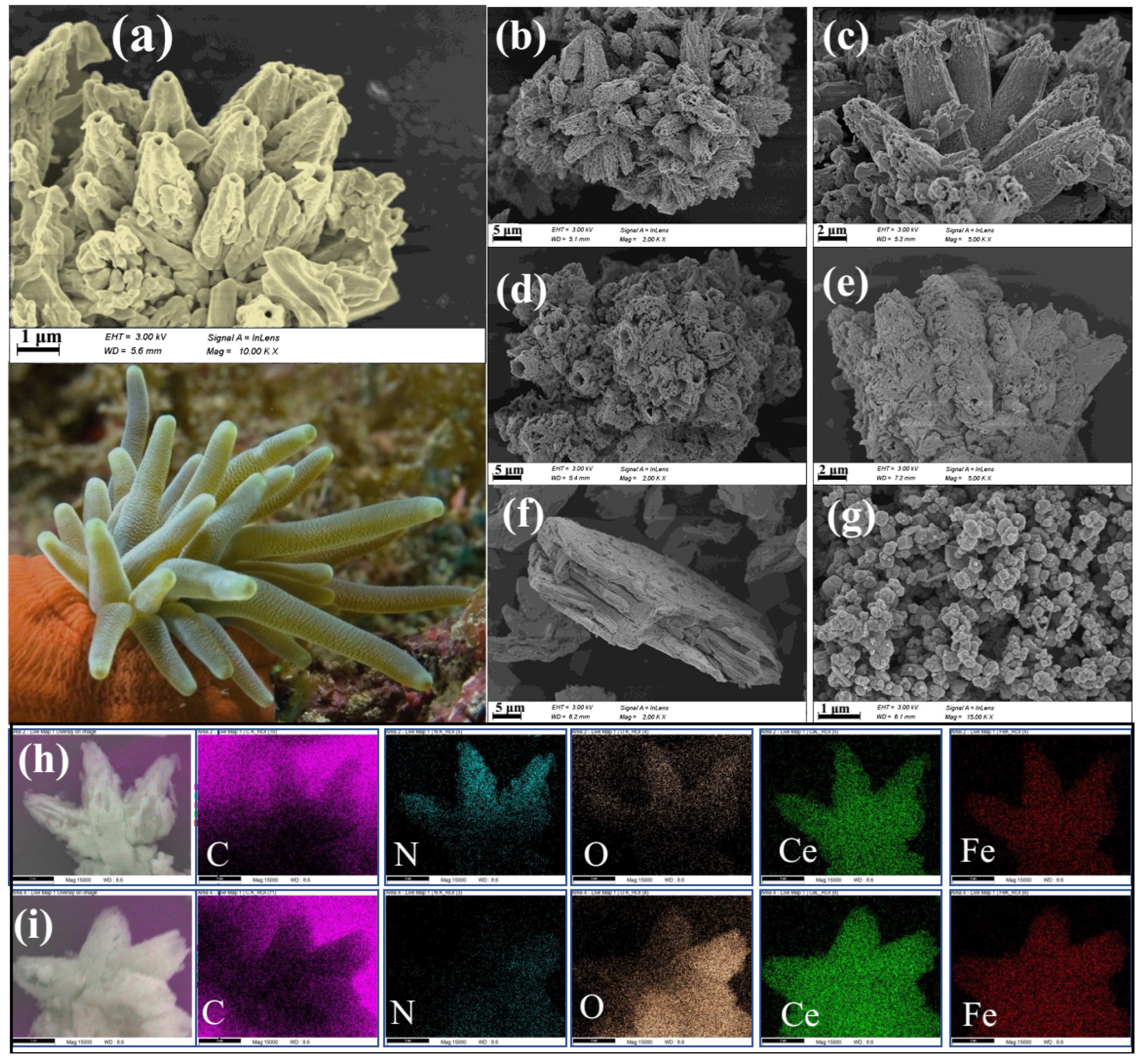
CMs are open three-dimensional (3D) frameworks that have great structural diversity as well as valuable physicochemical properties [26]. The morphologies of the fabricated CeFe-CM precursor and its corresponding pyrolysis product of CeFe-CM-T were characterized by SEM, as shown in Figure 1. An interesting “sea anemone”-shaped 3D microstructure with dozens of conical tentacles was observed in the SEM images of CeFe-CM (Figure 1a–c), and every tentacle had an opening at its end. The CeFe-CM provided a precursor to synthesize the CeFe-based oxides, i.e., CeFe-CM-T, whose morphology in Figure 1d–e well-maintained the 3D “sea anemone” structure but was rich in a porous and rough structure on the “sea anemone” body. This observation verifies the excellent self-templating role of CeFe-CM in fabricating porous CeFe-based oxides via a high-temperature pyrolysis process. It can be found that the special microstructure of the CeFe-based oxides in this study is significantly different from that of the commercial Ce-based and Fe-based oxides (Figure 1f,g). The commercial CeO2, in Figure 1f, shows a typical lamellar structure with tightly stacked nano-layers of large sizes, and the commercial Fe3O4 presented random particles with 100~300 nm sizes (Figure 1g). According to the element mapping in Figure 1h,i, the two metals, Ce and Fe, are uniformly distributed on the “sea anemone” structure of CeFe-CM, and the trace amount of O elements was derived from the structural water in the frameworks of CeFe-CM. After high-temperature oxidation, the amount of O element largely increased in the CeFe-CM-T framework, and most of the N element disappeared due to the decomposition of the CN ligand. The element signals of Ce and Fe were elevated to a certain extent on CeFe-CM-T, compared to its precursor.
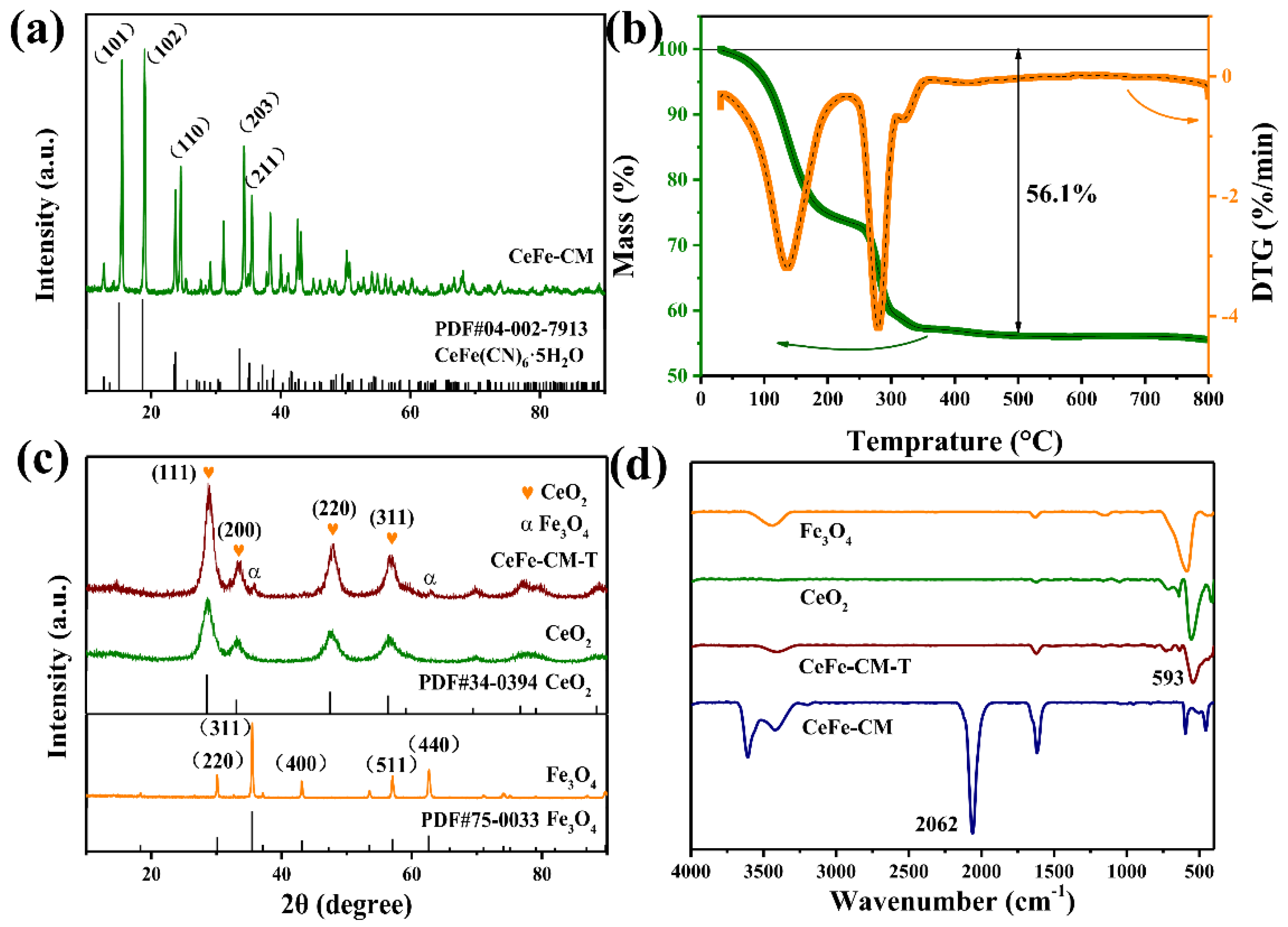
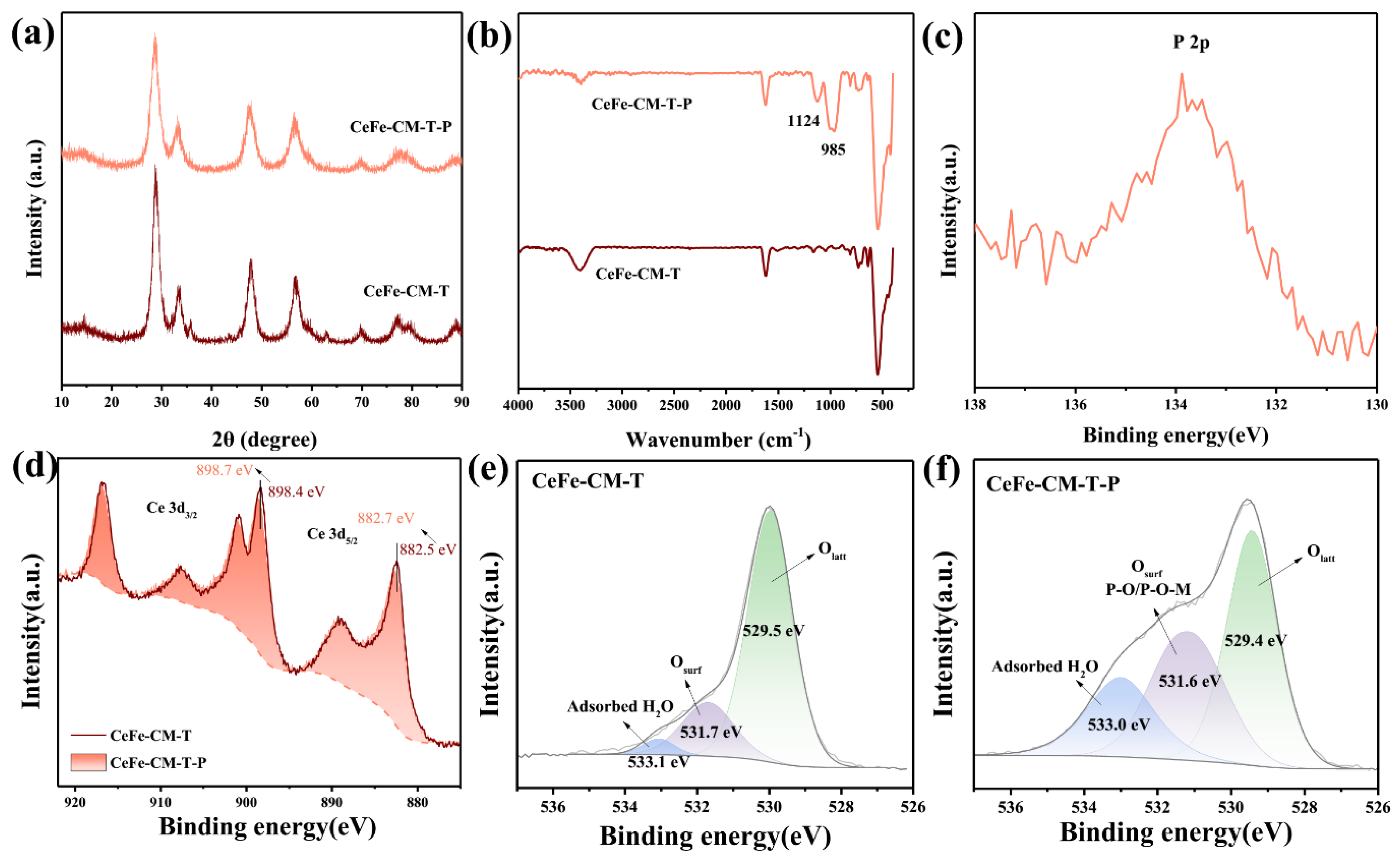
Figure 2a displays the XRD pattern of CeFe-CM with manifold diffraction peaks, indicating a complex crystal structure. Most of the diffraction peaks are well-coincided with the CeFe (CN)6·5H2O (PDF #04-002-7913), a Ce-Fe-based cyanometallate, including the obvious planes of (101), (102), (110), (203), (211) with sharp peaks, which illustrates a high crystallinity of the synthesized CeFe-CM. Before the high-temperature pyrolysis was conducted on the precursor, the thermal decomposition behavior of CeFe-CM was examined under an air atmosphere. As presented in Figure 2b, the TG curve shows two mass loss steps. The first step, before 250 °C, is ascribed to the loss of structural water in its framework, and the second loss step, between 250 and 350 °C, is due to the thermal decomposition of the C≡N ligands [20]. Then, it reached a weight equilibrium after 400 °C and formed stable metal oxides with a yield of about 56%. Thus, a pyrolysis temperature of 500 °C was performed on CeFe-CM to fabricate the stable CeFe-based oxides in this study. The gasification of structural water and the decomposition of the cyanide group during the two pyrolysis steps contributed to the formation of the porous structure of the resulting CeFe-CM-T. The XRD pattern of CeFe-CM-T, in Figure 2c, shows similar diffraction peaks to commercial CeO2 at 2θ of 28.6°, 35.5°, 33.1°, 47.5°, and 56.3°, which corresponded to (111), (200), (220), and (311), respectively, consisting well with a standard card of CeO2 (PDF #34-0394). In contrast, the diffraction peaks derived from the Fe-based oxide are very weak on CeFe-CM-T, and only the weak signals indexed to the (311) and (440) planes of Fe3O4 can be observed on this bimetallic oxide. The XRD results demonstrated that the Ce-based oxide (i.e., CeO2) dominated the phase state in the oxidation product (CeFe-CM-T) after high-temperature pyrolysis of the bimetallic CeFe-CM template under an air atmosphere.
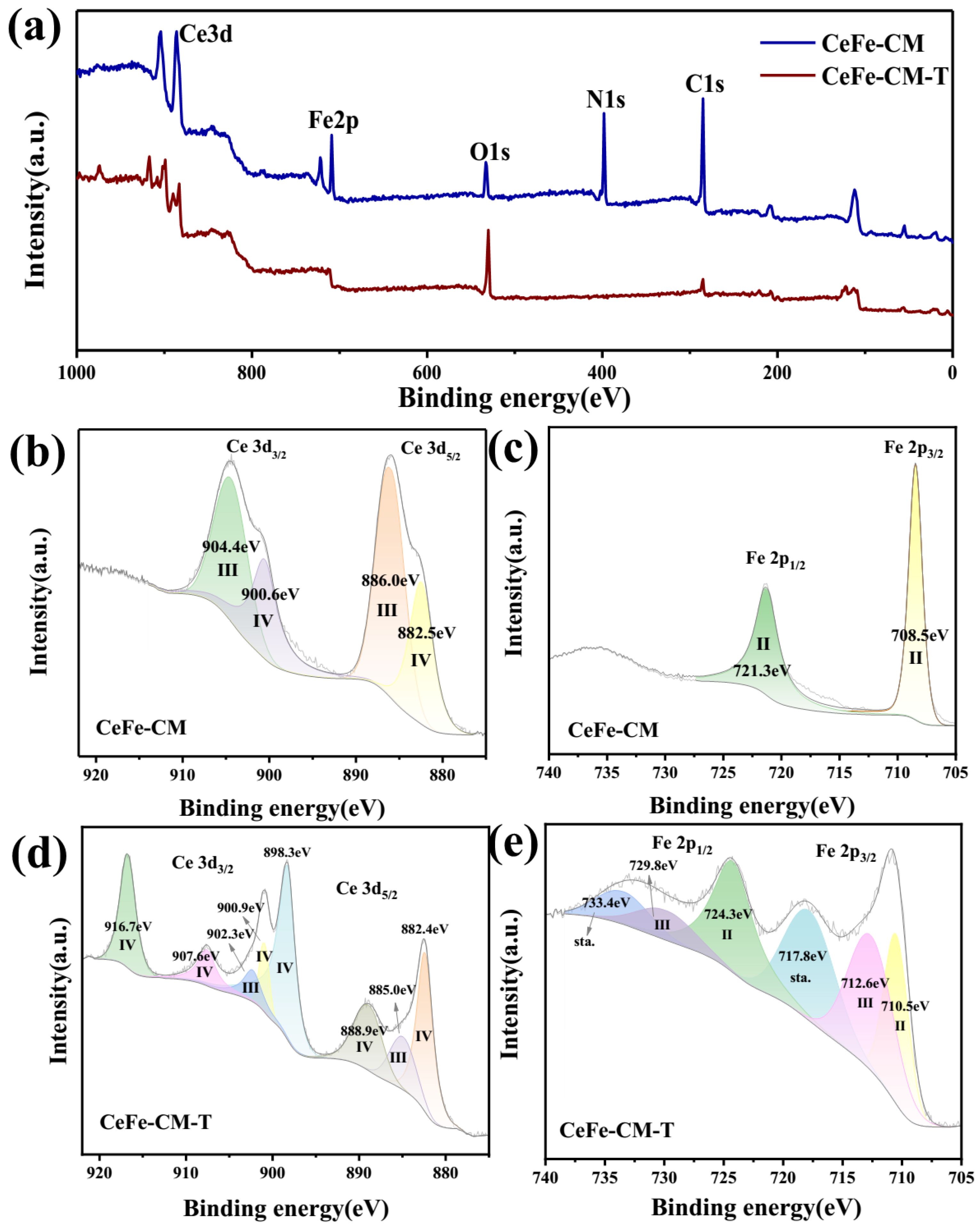
The FTIR spectra of the precursor and final product were recorded in the range of 4000–400 cm−1, as presented in Figure 2d. Significantly, the characteristic peak of the C≡N group at 2062 cm−1 [30], which can be observed on CeFe-CM, had completely disappeared on CeFe-CM-T, indicating a complete decomposition of the CN ligand under high-temperature pyrolysis. Meanwhile, the metal–oxygen band appeared on CeFe-CM-T at 400–600 cm−1 [11], and especially, the vibrational peak at 593 cm−1 is highly consistent with that of the CeO2 sample, which further verified the successful conversion of the CeFe-based oxides from the CM template. The chemical composition and the state of the metal elements in the adsorbents were analyzed by XPS. As shown in Figure 3a, the Ce, Fe, O, N, and C elements were detected on the full XPS spectrum of the CeFe-CM precursor, while the N signal disappeared and the O signal increased on the spectra of CeFe-CM-T, which was consistent with the EDS result in Figure 1h,i. High-resolution XPS spectra were conducted on the two metal elements of Ce and Fe, in Figure 3b–e, to give an insight into the change in the chemical state before and after high-temperature pyrolysis.
The high-resolution XPS spectra of Ce 3d are shown in Figure 3b,d. A typical double-peak pattern can be found on the CeFe-CM sample, which can be assigned to the Ce 3d5/2 and Ce 3d3/2 orbitals [31]. After peak-differentiating, four peaks, located at 882.5 eV, 886.0 eV, 900.6 eV and 904.4 eV, were fitted for the Ce 3d spectra, as shown in Figure 3b, where 882.5 eV/900.6 eV were assigned to Ce4+ and 886.0 eV/904.4 eV were assigned to Ce3+ [31]. This illustrates that Ce3+ was the main species in the precursor of CeFe-CM, while a small portion of Ce3+ was oxidized to Ce4+ during its coordination process. After high-temperature pyrolysis (Figure 3d), Ce 3d was deconvoluted into four pairs of the spin-orbital doublet-peak [16]: 882.4 eV/900.9 eV, 885.0 eV/902.3 eV, 888.9 eV/907.6 eV, and 898.3 eV/916.7 eV, where the pair of 885.0 eV/902.3 eV has corresponded to Ce3+, and the remaining three pairs are attributed to Ce4+. The strong signal of the Ce4+ peaks illustrates that a large portion of Ce3+ in CeFe-CM was converted to Ce4+ [32], and Ce4+ became the dominant form in CeFe-CM-T. Even so, part of Ce3+ remained in the CeFe-CM-T, unlike other Ce-based oxides, which are mainly in Ce4+. For the XPS spectra of the Fe element, characteristic peaks at 708.5 eV and 721.3 eV were detected on the precursor (Figure 3c), which are ascribed to the Fe 2p3/2 and Fe 2p1/2 orbitals of Fe2+. Similarly, most of the Fe2+ was oxidized to Fe3+ in CeFe-CM-T (Figure 3e), where the characteristic double-peak pattern (712.6 eV/729.8 eV) of Fe3+ was fitted [33], and the remaining pair of peaks, located at 717.8 eV and 733.4 eV, were satellite peaks.
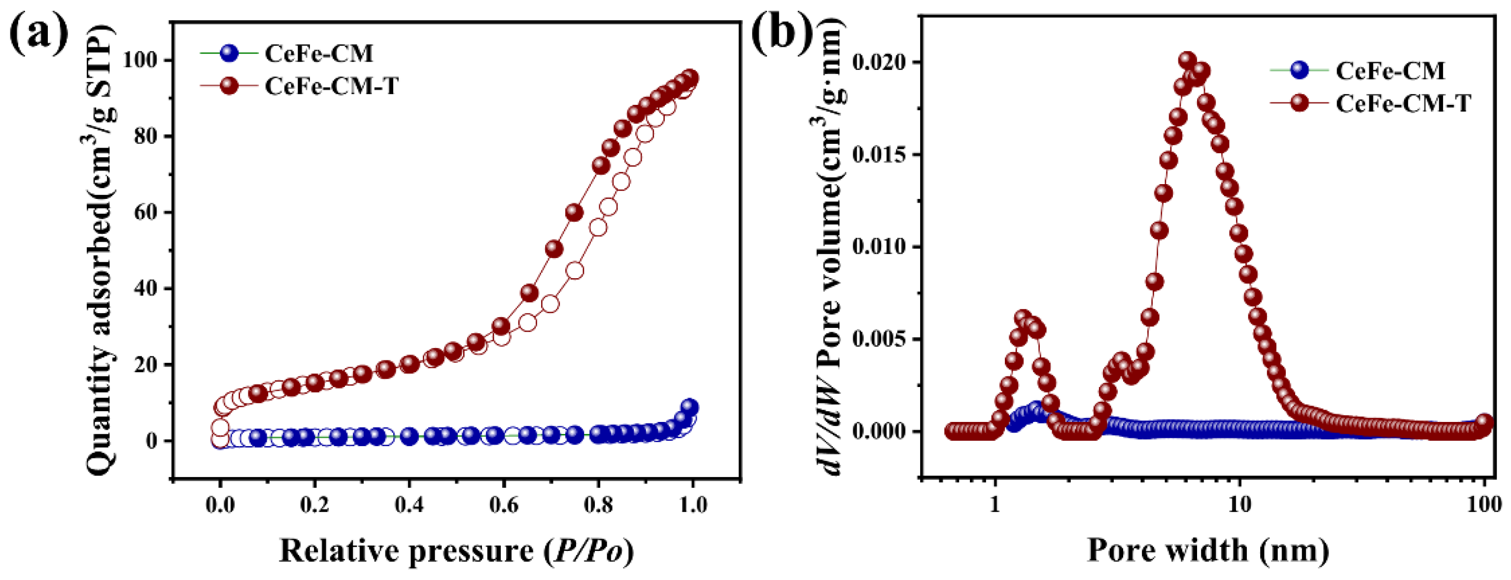
The specific surface area and pore size distribution of the samples were analyzed by the N2 adsorption–desorption method. As can be seen in Figure 4a, the precursor of CeFe-CM shows a very low surface area (SA) value of only 3.1 m2/g, and high-temperature pyrolysis rendered a remarkable increase in the SA (54.7 m2/g) on CeFe-CM-T, due to the pore-opening process caused by an outward gas flow, as mentioned in the TG analysis. There is a small number of micropores (1~2 nm) on CeFe-CM that originated from the framework cavities that were unoccupied by structural water during the coordination process, while it created abundant micropores and mesopores on the pyrolytic product of CeFe-CM-T. What was particularly obvious in the mesoporous region was that the pore volume largely increased in the range of 3–20 nm, which provided a structural advantage for the binding-site acquisition and ion diffusion in the following adsorption process [34].
3.2. Phosphate Adsorption Performance of CeFe-Based Adsorbents
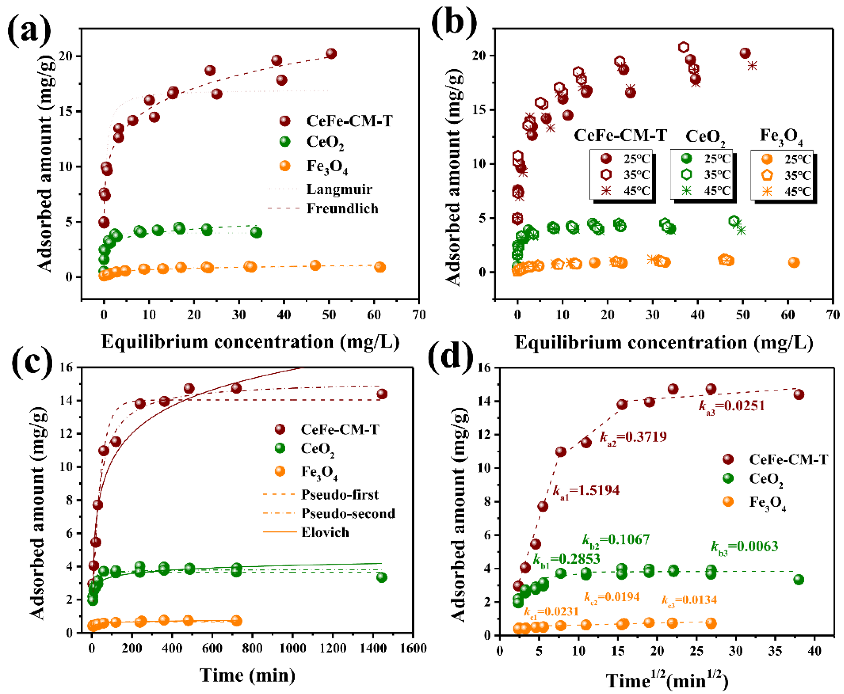
A severe dissolution of metal ions, as presented in Figure S1, prevented the precursor of CeFe-CM from being used as an adsorbent for phosphate removal. Thus, the stable product of CeFe-CM-T was examined for its adsorption performance in comparison with two other commercial oxides. Isothermal adsorption experiments were performed, and the results are displayed in Figure 5a. The typical Langmuir and Freundlich models were fitted to the corresponding data, and the regression parameters are listed in Table S1. Clearly, CeFe-CM-T exhibits a significantly superior phosphate adsorption capacity with a saturated adsorption amount (Q0) of 17.0 mg/g, one order of magnitude higher than that of commercial CeO2 (4.0 mg/g) and Fe3O4 (0.99 mg/g), which demonstrates the successful application of the self-templating strategy to fabricate the Ce-based oxides as phosphate adsorbents in this work. Furthermore, a thermodynamic test was performed by isothermal adsorptions at different temperatures (25, 35, and 45 °C). The results in Figure 5b show that temperature had no significant effect on phosphate adsorption, including the adsorption onto CeFe-CM-T, CeO2, and Fe3O4, which reveals a broad temperature adaptation of these metal oxides working for phosphate removal. A similar result was also reported for other materials [35,36], and the temperature-independent adsorption behavior is considered a combined reflection of physical and chemisorption mechanisms.
To further evaluate the adsorption behavior of phosphorus on CeFe-CM-T, kinetic experiments were conducted. Three typical kinetic models were used to fit the result, as presented in Figure 5c, including the pseudo-first-order, pseudo-second-order, and Elovich models, and the regression parameters are listed in Table S2. The pseudo-second-order model showed the best fitting with an R2 of 0.99, indicating that the phosphate adsorption process on CeFe-CM-T was dominated by chemisorption [37]. Although CeFe-CM-T possessed an obviously higher phosphate adsorption capacity than the other two commercial adsorbents (CeO2 and Fe3O4), its fitted kinetic rate parameters of k2 are lower, meaning CeFe-CM-T took a longer time to achieve an adsorption equilibrium. It can be ascribed to the hierarchical porous structure of CeFe-CM-T, as described in Figure 4, which renders phosphate ions diffused into the abundant micropores and mesopores and finally reaches the exposed adsorption sites. The commercial CeO2 possessed a tightly stacked structure, and only the outermost adsorption sites could be quickly obtained for phosphate adsorption. The Weber-Morris model was further used to examine the diffusion process (Figure 5d), where the adsorption process is divided into three steps: outer particle diffusion, inner particle adsorption, and adsorption equilibrium [25,38]. According to the regression parameters in Table S3, the first diffusion stage displayed a remarkably high ki1, one and two orders of magnitude higher than the second (ki2) and the third (ki3) stage, respectively. This result verified that both the external and intraparticle were the two main diffusion pathways during phosphate adsorption onto the porous CeFe-CM-T. Additionally, the closed ki2 and ki3 values were fitted on CeO2 and Fe3O4, indicating there was only an external surface adsorption process occurring on the two commercial adsorbents and the internal sites cannot be accessed by phosphate ions.
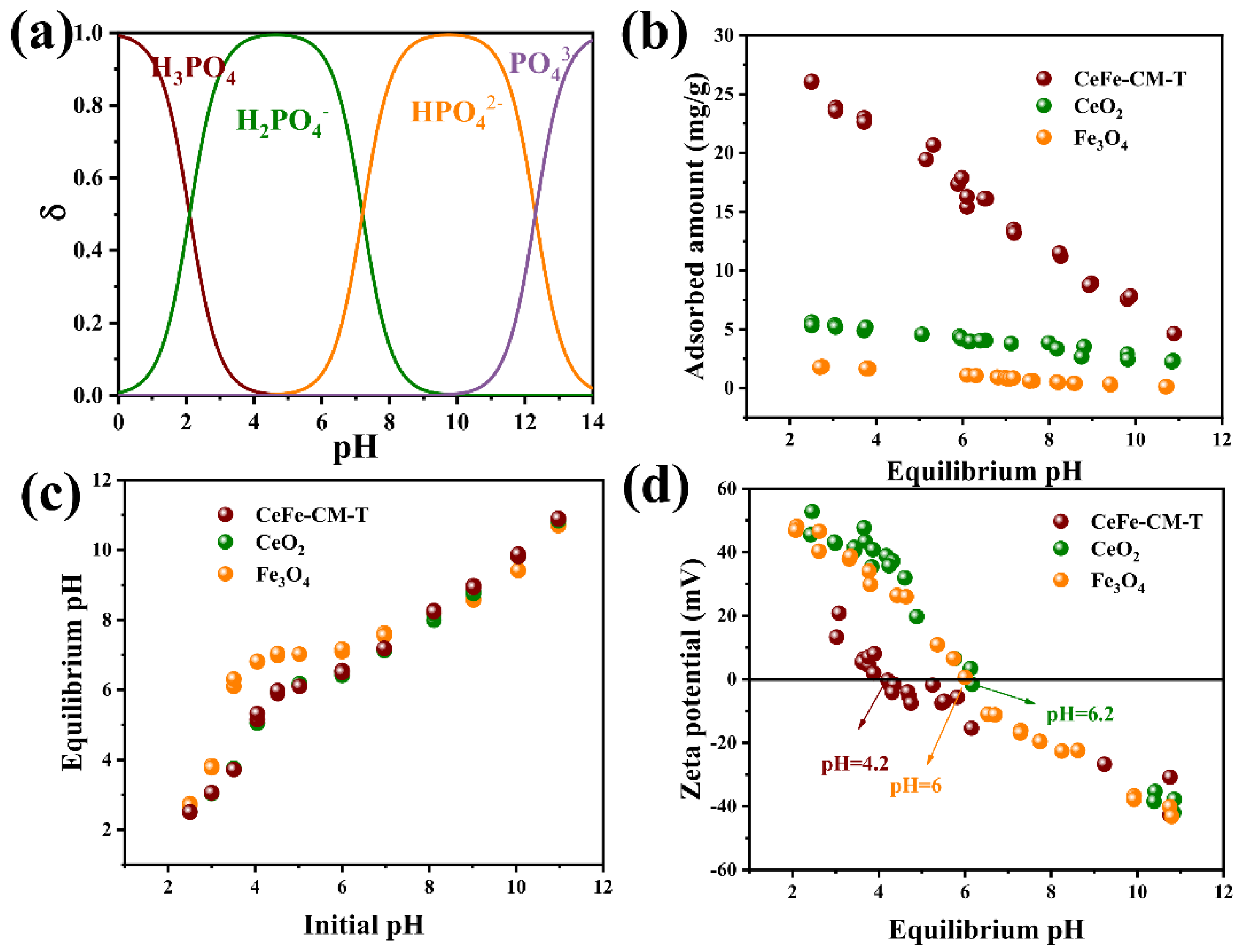
Both phosphate species and adsorbent surface properties in an aqueous solution are significantly influenced by pH [1], which determines the adsorption behavior of phosphate onto metal oxides. Figure 6a depicts the distribution of the phosphate species with respect to pH, including H3PO4, H2PO4−, HPO42−, and PO43−. The results in Figure 6b show that the phosphate adsorption of the prepared CeFe oxides was sensitive to the solution pH, and the adsorbed amount of phosphate dramatically decreased from 26.0 mg/g at pH 2.5 to 4.6 mg/g at pH 10.9, reflecting a fitness of this type of adsorbent for treating acidic wastewater. A pH isoelectric point (pHPZC) at 4.2 on the zeta-potential curve of CeFe-CM-T, as depicted in Figure 6d, was found. Below the pHPZC, the CeFe-CM-T surface was electropositive, which was favorable for the adsorption of electronegative phosphate (especially for the species of H2PO4−). Above the pHPZC, the adsorbent surface was electronegative, and electrostatic repulsion between the deprotonated surface and anions would largely prevent phosphate adsorption [37]. Indeed, the adsorbed amount of phosphate was continuously decreased with an increase in the pH. However, there remained a relatively high adsorption amount of phosphate when the solution pH was above pHPZC, indicating that other adsorption mechanisms, rather than electrostatic attraction, contributed to phosphate adsorption. Usually, ligand exchange is considered the main adsorption process of phosphate occurring on metal oxide surfaces, where the surface hydroxyl groups bonded with metal elements were exchanged by phosphate ions [8,39]. Additionally, the increased solution pH after adsorption, as shown in Figure 6c, probably originating from the exchanged hydroxyls, supported the ligand-exchange mechanism. On the other hand, the phenomenon of weak adsorption under alkali conditions can be properly used for adsorbent regeneration.
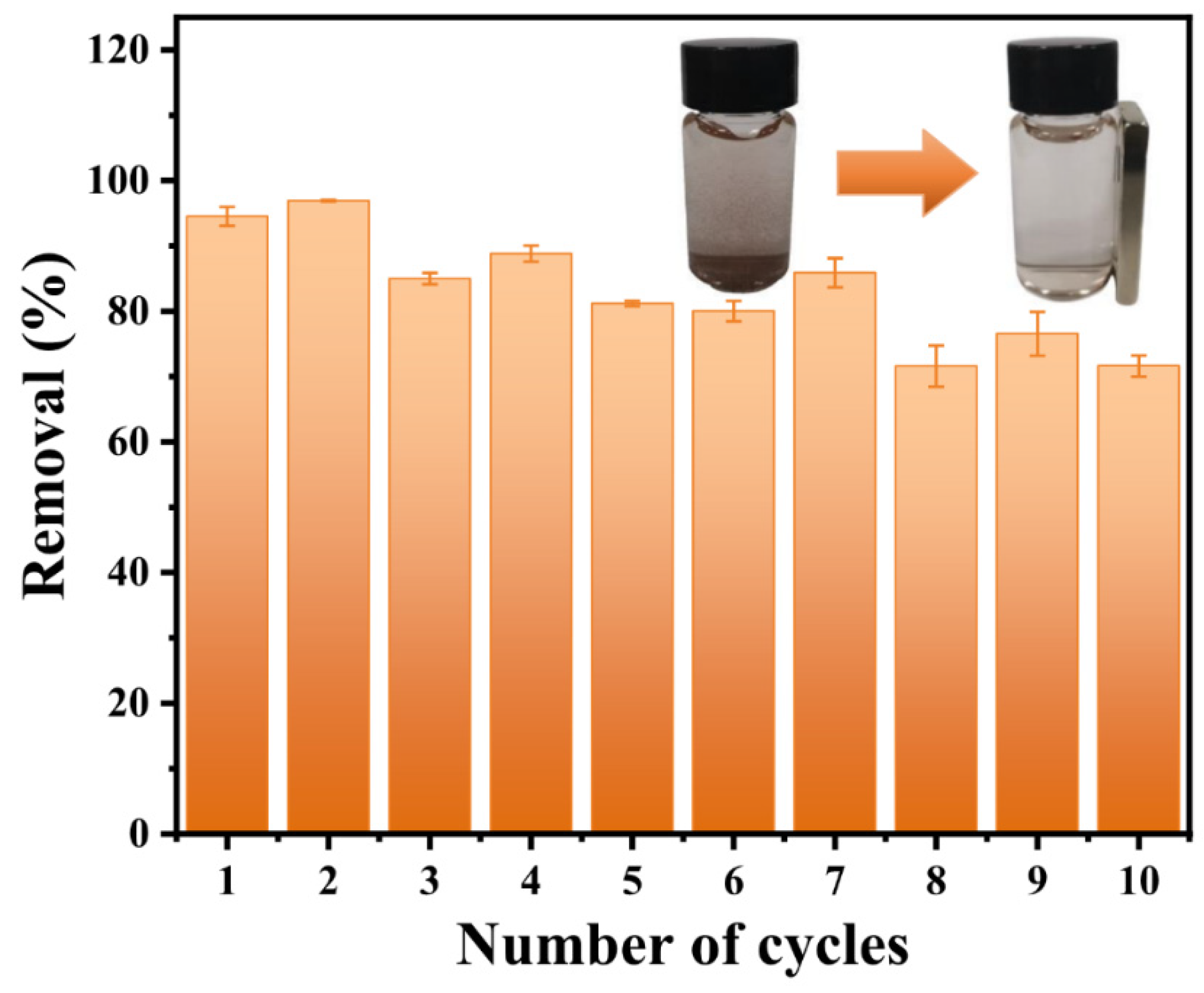
Therefore, a recycle test was performed on CeFe-CM-T, using NaOH as the regenerative reagent. As displayed in Figure 7, CeFe-CM-T can maintain phosphate removal above 90% during the first and second cycles, and even after ten cycles, the removal rate of phosphate still reaches 72%. The result reveals the excellent cycle performance of the fabricated CeFe-based oxides, where the exhausted adsorbent can be well-recovered of its phosphate capacity by alkali solution regeneration. What is more, it can be seen in the insert image of Figure 7 that CeFe-CM-T is magnetic because it contains Fe-based oxides, and this property can be fully used for adsorbent material recovery and is convenient for recycling applications via magnetic separation.
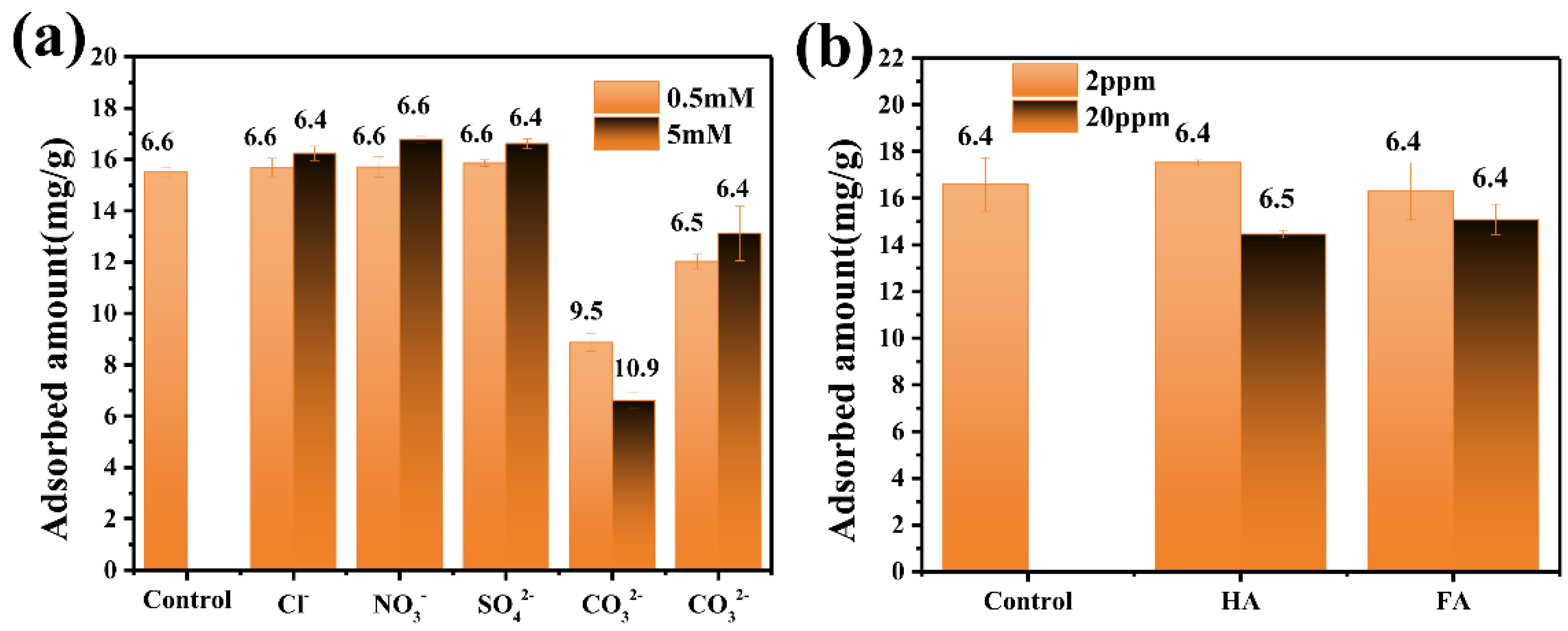
The effect of co-existing anions on phosphate adsorption was examined, as they might compete for the adsorption sites on CeFe-CM-T. Monovalent ions (Cl− and NO3−) and bivalent ions (CO32− and SO42−) were selected as the representatives [40]. The result in Figure 8a shows that the two monovalent ions (Cl− and NO3−) and bivalent SO42− had little effect on phosphate adsorption, regardless of the low concentration (0.5 M) or high concentration (5 M), which illustrated the high selectivity of CeFe-CM-T for phosphate adsorption during the practical application. There was no difference between the effect from monovalent and bivalent anions, which excluded the main role of electrostatic attraction for phosphate capture, and there was no difference between the oxoanion (NO3−) and non-oxoanion (Cl−), which further excluded the adsorption mechanism of the hydrogen bond, which usually occurred on the oxoanion (serving as an H-bond receptor). However, bivalent CO32− exhibited an obvious inhibition of phosphate adsorption on CeFe-CM-T, and the pH detection showed a remarkable alkaline buffer effect of CO32−, which rendered a solution pH of 9.5 and 10.9 at low and high concentrations of CO32−, respectively. According to the discussion on the pH effect in Figure 6b, we deduced it was not due to CO32− competition but to the influence of the solution pH. In fact, a pH adjustment to neutral thus offsets the negative effect on phosphate with the co-existence of CO32− to a certain degree, as displayed in Figure 8a.
Natural organic matters (NOMs) are widely widespread in surface water, whose effect on phosphate removal is also an issue of great concern. Herein, two typical NOMs, humic acid and fulvic acid, were evaluated for their influence on phosphate adsorption. As shown in Figure 8b, there was no significant effect of NOMs on phosphate adsorption at a low concentration (2 mg/L), while both humic acid and fulvic acid showed a weak inhibition at a high concentration (20 mg/L). This should be ascribed to the shielding effect of NOMs, where a high concentration of NOMs coated the outside of the adsorbent particle and shielded the surface adsorption sites. However, CeFe-CM-T displayed high selectivity and applicability for phosphate removal in a co-existing aqueous environment.
3.3. Phosphate Adsorption Mechanism on CeFe-Based Adsorbent
The above adsorption test indicated that chemisorption had a dominant role in the phosphate adsorption process, and to further reveal the adsorption mechanism, XRD, FTIR, and XPS were used to examine the CeFe-CM-T sample before and after the phosphate adsorption. No significant changes were obtained from the XRD patterns before and after adsorption, as shown in Figure 9a, indicating that CeFe-CM-T maintained its crystal structure well, and phosphate adsorption cannot be reflected in the change in XRD patterns. In contrast, two characteristic peaks, located at 1124 and 985 cm−1, appeared on the FTIR spectrum of CeFe-CM-T-P after the phosphate adsorption, which are assigned to the asymmetric and symmetric stretching vibrations of O-P-O, respectively. This provides proof of the adsorbed phosphate. Moreover, Figure 9c gives more direct proof that a characteristic P2p peak, located at 133.8 eV, was detected on the high-resolution XPS spectra of the CeFe-CM-T-P sample.
As described in Figure 5a, Ce4+ in commercial CeO2 rendered its poor adsorption capacity of phosphate. Fe-based oxides of Fe3O4 also possessed an even lower adsorption capacity, while CeFe-CM-T exhibited an obviously superior performance of phosphate removal due to its reserved Ce3+, which is considered the major active site for phosphate binding via the ligand-exchange mechanism to form a stable CePO4 compound. The high-resolution XPS spectra of Ce 3d, seen in Figure 9d, clearly show that no change occurred in the overall peak pattern and strength when comparing the CeFe-CM-T and CeFe-CM-T-P. This observation confirms that the ratio of Ce3+ and Ce4+ was not changed by the phosphate adsorption and that Ce3+ can be well-maintained in CeFe-CM-T to serve as active adsorption sites for phosphate. However, taking a close look at the spectra, a shift to higher energy can be found on the two main peaks of Ce 3d3/2 and Ce 3d5/2, from 898.4 to 898.7 eV and 882.5 to 882.7 eV, respectively. This demonstrates the formation of the CePO4 species, where the charge transfer occurred between Ce and phosphate via a charge reconciliation process [41]. Therefore, the Ce3+ in CeFe-CM-T is stable and plays a leading role in phosphate removal to form stable Ce-PO4 compounds.
The XPS O1s spectrum of CeFe-CM-T (Figure 9e) was deconvoluted into three peaks at 529.5 eV, 531.8 eV, and 533.1 eV, which are attributed to lattice oxygen (Olatt), surface oxygen (Osurf) and adsorbed structural H2O, respectively [15], where the Olatt is indexed to the oxygen in the form of metal-O in the lattice structure, and the Osurf is indexed to the active oxygen or hydroxyls on the adsorbent surface. Given the comparison to the peak-differentiating results in Figure 9e,f, we found a sharp increase in the peak strength at 531.6 eV and 533.0 eV on CeFe-CM-T-P. According to Table S4, it can be calculated that the rate of peak at 531.6 eV increased from 19.13% to 34.58%, and correspondingly, the rate of peak at 529.4 eV decreased from 77.40% to 44.66% after the phosphate adsorption. This is due to the partial conversion of metal-O to metal-O-P via the ligand exchange of phosphate with the surface oxygen-containing groups [42]. Moreover, the peak located at 533.0 eV (the adsorbed H2O) was also largely enhanced, and the binding of water along with the adsorbed phosphate might have attributed to it.
4. Conclusions
In summary, a “sea anemone”-like CeFe cyanometallate (CM) with a 3D microstructure was fabricated, which provided a precursor to synthesize CeFe-based oxides (CeFe-CM-T) via a self-templating strategy by high-temperature pyrolysis under an air atmosphere. The as-prepared CeFe-CM-T maintained the “sea anemone” morphology well and had abundant micropores and mesopores. The XRD results demonstrated that the Ce-based oxide (i.e., CeO2) dominated the phase state in CeFe-CM-T, and both Ce3+ and Ce4+ were reserved within the oxides. The CeFe-CM-T exhibited an obviously superior phosphate adsorption capacity to the commercial CeO2 and Fe3O4 materials, with 1~2 orders of magnitude higher than the saturated adsorption amount. The sensitivity of the phosphate adsorption on CeFe-CM-T to the solution pH provided excellent recycling performance using the alkali solution as a regenerative reagent, and its magnetic property makes it convenient for the recycle application via magnetic separation. Moreover, the fabricated CeFe-CM-T showed high selectivity for phosphate removal when it co-existed with other anions and NOMs. Finally, the reserved Ce3+ in CeFe-CM-T was demonstrated to be the main active site for phosphate capture, which formed stable Ce-PO4 compounds via a ligand-exchange mechanism with the surface oxygen-containing groups.
Supplementary Materials
The following supporting information can be downloaded at: https://www.mdpi.com/article/10.3390/w14152445/s1, Figure S1: Ions releasing of the as-prepared CeFe based materials (CeFe-CM and CeFe-CM-T) at the same solid/aqueous rate with adsorption of 10 mg/60 mL; Table S1: Langmuir and Freundlich model parameters; Table S2: Regression parameters of the three kinetic models; Table S3: Regression Parameters of the Weber-Morris model; Table S4: Atomic rates of different states deconvoluted from O1s XPS spectrum of CeFe-CM-T before and after phosphate adsorption
Author Contributions
Conceptualization, Q.F. and H.M.; methodology, P.D. and X.W.; software, X.T.; validation, X.T., P.D., and J.L.; investigation, X.T. and P.D.; resources, W.H. and Q.F.; data curation, X.T. and Q.F.; writing—original draft preparation, X.T. and P.D.; writing—review and editing, W.H., Q.F., J.L. and Q.L.; visualization, Q.F.; supervision, Q.F.; project administration, Q.F.; funding acquisition, Q.F. All authors have read and agreed to the published version of the manuscript.
Funding
This research was funded by the Zhuhai Basic and Applied Basic Research Foundation, grant number ZH22017003210025PWC, and the startup fund to Qile Fang from the Advanced Institute of Natural Sciences, Beijing Normal University, Zhuhai.
Institutional Review Board Statement
Not applicable.
Informed Consent Statement
Not applicable.
Data Availability Statement
Not applicable.
Conflicts of Interest
The authors declare no conflict of interest.
References
- He, J.; Pei, C.; Yang, Y.; Lai, B.; Sun, Y.; Yang, L. The structural design and valence state control of cerium-based metal-organic frameworks for their highly efficient phosphate removal. J. Clean. Prod. 2021, 321, 128778. [Google Scholar] [CrossRef]
- Downing, J.A.; Polasky, S.; Olmstead, S.M.; Newbold, S.C. Protecting local water quality has global benefits. Nat. Commun. 2021, 12, 2709. [Google Scholar] [CrossRef]
- Wu, B.; Wan, J.; Zhang, Y.; Pan, B.; Lo, I.M.C. Selective phosphate removal from water and wastewater using sorption: Process fundamentals and removal mechanisms. Environ. Sci. Technol. 2020, 54, 50–66. [Google Scholar] [CrossRef] [PubMed]
- Yang, Y.; Shi, X.; Ballent, W.; Mayer, B.K. Biological phosphorus recovery: Review of current progress and future needs. Water Environ. Res. 2017, 89, 2122–2135. [Google Scholar] [CrossRef] [PubMed]
- Ye, Y.; Ngo, H.H.; Guo, W.; Liu, Y.; Li, J.; Liu, Y.; Zhang, X.; Jia, H. Insight into chemical phosphate recovery from municipal wastewater. Sci. Total Environ. 2017, 576, 159–171. [Google Scholar] [CrossRef]
- Zhang, S.; Ding, J.; Tian, D. Incorporation of MIL-101 (Fe or Al) into chitosan hydrogel adsorbent for phosphate removal: Performance and mechanism. J. Solid State Chem. 2022, 306, 122709. [Google Scholar] [CrossRef]
- Jiao, G.-J.; Ma, J.; Li, Y.; Jin, D.; Ali, Z.; Zhou, J.; Sun, R. Recent advances and challenges on removal and recycling of phosphate from wastewater using biomass-derived adsorbents. Chemosphere 2021, 278, 130377. [Google Scholar] [CrossRef] [PubMed]
- Liu, R.; Chi, L.; Wang, X.; Sui, Y.; Wang, Y.; Arandiyan, H. Review of metal (hydr)oxide and other adsorptive materials for phosphate removal from water. J. Environ. Chem. Eng. 2018, 6, 5269–5286. [Google Scholar] [CrossRef]
- Liu, R.; Chi, L.; Wang, X.; Wang, Y.; Sui, Y.; Xie, T.; Arandiyan, H. Effective and selective adsorption of phosphate from aqueous solution via trivalent-metals-based amino-MIL-101 MOFs. Chem. Eng. J. 2019, 357, 159–168. [Google Scholar] [CrossRef]
- Wang, L.; Wang, J.; He, C.; Lyu, W.; Zhang, W.; Yan, W.; Yang, L. Development of rare earth element doped magnetic biochars with enhanced phosphate adsorption performance. Colloid. Surface. Asp. 2019, 561, 236–243. [Google Scholar] [CrossRef]
- Deng, H.; Yu, X. Adsorption of fluoride, arsenate and phosphate in aqueous solution by cerium impregnated fibrous protein. Chem. Eng. J. 2012, 184, 205–212. [Google Scholar] [CrossRef]
- Bacelo, H.; Pintor, A.M.A.; Santos, S.C.R.; Boaventura, R.A.R.; Botelho, C.M.S. Performance and prospects of different adsorbents for phosphorus uptake and recovery from water. Chem. Eng. J. 2020, 381, 122566. [Google Scholar] [CrossRef]
- Zhang, K.; Hu, L.; Wang, C.; Zhang, K. Middle-low-temperature oxidation and adsorption of arsenic from flue gas by Fe-Ce-based composite catalyst. Chemosphere 2022, 288, 132425. [Google Scholar] [CrossRef] [PubMed]
- Montini, T.; Melchionna, M.; Monai, M.; Fornasireo, P. Fundamentals and Catalytic Applications of CeO2-Based Materials. Chem. Rev. 2016, 116, 5987–6041. [Google Scholar] [CrossRef] [PubMed]
- Wang, Y.; Xie, X.; Chen, X.; Huang, C.; Yang, S. Biochar-loaded Ce3+-enriched ultra-fine ceria nanoparticles for phosphate adsorption. J. Hazard. Mater. 2020, 396, 122626. [Google Scholar] [CrossRef]
- He, J.; Xu, Y.; Wang, W.; Hu, B.; Wang, Z.; Yang, X.; Wang, Y.; Yang, L. Ce(III) nanocomposites by partial thermal decomposition of Ce-MOF for effective phosphate adsorption in a wide pH range. Chem. Eng. J. 2020, 379, 122431. [Google Scholar] [CrossRef]
- Wu, B.; Lo, I.M.C. Surface functional group engineering of CeO2 particles for enhanced phosphate adsorption. Environ. Sci. Technol. 2020, 54, 4601–4608. [Google Scholar] [CrossRef]
- Feng, Y.; Lu, H.; Liu, Y.; Xue, L.; Dionysiou, D.D.; Yang, L.; Xing, B. Nano-cerium oxide functionalized biochar for phosphate retention: Preparation, optimization and rice paddy application. Chemosphere 2017, 185, 816–825. [Google Scholar] [CrossRef]
- Li, C.; Zhang, Y.; Wang, X.; Yin, X.; Luo, N.; Khayambashi, A.; Wei, Y. The synthesis and characterization of hydrous cerium oxide nanoparticles loaded on porous silica micro-sphere as novel and efficient adsorbents to remove phosphate radicals from water. Micropor. Mesopor. Mater. 2019, 279, 73–81. [Google Scholar] [CrossRef]
- Zhang, L.; Wu, H.B.; Madhavi, S.; Hng, H.H.; Lou, X.W.D. Formation of Fe2O3 microboxes with hierarchical shell structures from metal-organic frameworks and their lithium storage properties. J. Am. Chem. Soc. 2012, 134, 17388–17391. [Google Scholar] [CrossRef]
- Wang, Y.; Zhao, W.; Qi, Z.; Zhang, L.; Zhang, Y.; Huang, H.; Peng, Y. Designing ZIF-8/hydroxylated MWCNT nanocomposites for phosphate adsorption from water: Capability and mechanism. Chem. Eng. J. 2020, 394, 124992. [Google Scholar] [CrossRef]
- Li, S.; Lei, T.; Jiang, F.; Liu, M.; Wang, Y.; Wang, S.; Yang, X. Tuning the morphology and adsorption capacity of Al-MIL-101 analogues with Fe3+ for phosphorus removal from water. J. Colloid Interf. Sci. 2020, 560, 321–329. [Google Scholar] [CrossRef] [PubMed]
- Li, M.; Liu, Y.; Li, F.; Shen, C.; Kaneti, Y.V.; Yamauchi, Y.; Yuliarto, B.; Chen, B.; Wang, C.-C. Defect-rich hierarchical porous UiO-66 (Zr) for tunable phosphate removal. Environ. Sci. Technol. 2021, 55, 13209–13218. [Google Scholar] [CrossRef]
- Zhang, Y.; Kang, X.; Guo, P.; Tan, H.; Zhang, S.-H. Studies on the removal of phosphate in water through adsorption using a novel Zn-MOF and its derived materials. Arab. J. Chem. 2022, 15, 103955. [Google Scholar] [CrossRef]
- Dong, P.; Jing, X.; Li, Y.; Shen, Y.; Li, Q.; Fang, Q. “Twin lotus flower” adsorbents derived from LaFe cyanometallate for high-performance phosphorus removal. Sep. Purif. Technol. 2022, 291, 120924. [Google Scholar] [CrossRef]
- Alexandrov, E.V.; Virovets, A.V.; Blatov, V.A.; Peresypkina, E. Topological Motifs in Cyanometallates: From Building Units to Three-Periodic Frameworks. Chem. Rev. 2015, 115, 12286–12319. [Google Scholar] [CrossRef] [Green Version]
- Zakrzewski, J.J.; Liberka, M.; Zychowicz, M.; Chorazy, S. Diverse physical functionalities of rare-earth hexacyanidometallate frameworks and their molecular analogues. Inorg. Chem. Front. 2021, 8, 452–483. [Google Scholar] [CrossRef]
- Nai, J.; Lou, X.W. Hollow structures based on Prussian blue and its analogs for electrochemical energy storage and conversion. Adv. Mater. 2019, 31, 1706825. [Google Scholar] [CrossRef]
- Litvinova, Y.M.; Gayfulin, Y.M.; Leusen, J.; Samsonenko, D.G.; Lazarenko, V.A.; Zubavichus, Y.V.; Kögerler, P.; Mironov, Y.V. Metal-organic frameworks based on polynuclear lanthanide complexes and octahedral rhenium clusters. Inorg. Chem. Front. 2019, 6, 1518–1526. [Google Scholar] [CrossRef]
- Li, Y.; Jing, X.; Li, Q.; Shen, Y.; Fang, Q. Well-defined bimetal oxides derived from Prussian blue analogues with regulable active sites for phosphate removal. J. Colloid Interf. Sci. 2022, 622, 390–401. [Google Scholar] [CrossRef]
- Peng, B.; Cui, J.; Wang, Y.; Liu, J.; Zheng, H.; Jin, L.; Zhang, X.; Zhang, Y.; Wu, Y. CeO2-x/C/rGO nanocomposites derived from Ce-MOF and graphene oxide as robust platform for highly sensitive uric acid detection. Nanoscale 2018, 10, 1939–1945. [Google Scholar] [CrossRef]
- Schelter, E.J. Cerium under the lens. Nat. Chem. 2013, 5, 348. [Google Scholar] [CrossRef]
- Qu, F.; Cao, A.; Yang, Y.; Mahmud, S.; Su, P.; Yang, J.; He, Z.; Lai, Q.; Zhu, L.; Tu, Z.; et al. Hierarchically superhydrophilic poly(vinylidene fluoride) membrane with self-cleaning fabricated by surface mineralization for stable separation of oily wastewater. J. Membr. Sci. 2021, 640, 119864. [Google Scholar] [CrossRef]
- Jiao, G.; Ma, J.; Zhang, Y.; Jin, D.; Li, Y.; Hu, C.; Guo, Y.; Wang, Z.; Zhou, J.; Sun, R. Nitrogen-doped lignin-derived biochar with enriched loading of CeO2 nanoparticles for highly efficient and rapid phosphate capture. Int. J. Biol. Macromol. 2021, 182, 1484–1494. [Google Scholar] [CrossRef]
- Wang, N.; Feng, J.; Chen, J.; Wang, J.; Yan, W. Adsorption mechanism of phosphate by polyaniline/TiO2 composite from wastewater. Chem. Eng. J. 2017, 316, 33–40. [Google Scholar] [CrossRef]
- Huang, Y.; Lee, X.; Grattieri, M.; Yuan, M.; Cai, R.; Macazo, F.C.; Minteer, S.D. Modified biochar for phosphate adsorption in environmentally relevant conditons. Chem. Eng. J. 2020, 380, 122375. [Google Scholar] [CrossRef]
- He, J.; Xu, Y.; Shao, P.; Yang, L.; Sun, Y.; Yang, Y.; Cui, F.; Wang, W. Modulation of coordinative unsaturation degree and valence state for cerium-based adsorbent to boost phosphate adsorption. Chem. Eng. J. 2020, 394, 124912. [Google Scholar] [CrossRef]
- Abdellaoui, Y.; Oualid, H.A.; Hsini, A.; Ibrahimi, B.E.; Laabd, M.; Ouardi, M.E.; Giacoman-Vallejos, G.; Gamero-Melo, P. Synthesis of zirconium-modified merlinoite from fly ash for enhanced removal of phosphate in aqueous medium: Experimental studies supported by Monte Carlo/SA simulaitons. Chem. Eng. J. 2021, 404, 126600. [Google Scholar] [CrossRef]
- Zhang, Y.; Pan, B.; Shan, C.; Gao, X. Enhanced phosphate removal by nanosized hydrated La(III) oxide confined in cross-linked polystyrene networks. Environ. Sci. Technol. 2016, 50, 1447–1454. [Google Scholar] [CrossRef]
- Liu, M.; Li, S.; Tang, N.; Wang, Y.; Yang, X.; Wang, S. Highly efficient capture of phosphate from water via cerium-doped metal-organic frameworks. J. Clean. Prod. 2020, 265, 121782. [Google Scholar] [CrossRef]
- Xiang, C.; Ji, Q.; Zhang, G.; Wang, H.; Qu, J. In situ creation of oxygen vacancies in porous bimetallic La/Zr sorbent for aqueous phosphate: Hierarchical pores control mass transport and vacancy sites determine interaction . Environ. Sci. Technol. 2020, 54, 437–445. [Google Scholar] [CrossRef]
- Xiong, W.; Tong, J.; Yang, Z.; Zeng, G.; Zhou, Y.; Wang, D.; Song, P.; Xu, R.; Zhang, C.; Cehng, M. Adsorption of phosphate from aqueous solution using iron-zirconium modified activated carbon nanofiber: Performance and mechanism. J. Colloid. Interf. Sci. 2017, 493, 17–23. [Google Scholar] [CrossRef]
Figure 1.
SEM images of (a–c) CeFe-CM, (d,e) CeFe-CM-T, (f) CeO2, and (g) Fe3O4. Element mapping of (h) CeFe-CM and (i) CeFe-CM-T.
Figure 1.
SEM images of (a–c) CeFe-CM, (d,e) CeFe-CM-T, (f) CeO2, and (g) Fe3O4. Element mapping of (h) CeFe-CM and (i) CeFe-CM-T.

Figure 2.
XRD patterns of (a) CeFe-CM and (c) CeFe-CM-T, CeO2, Fe3O4; (b) TG and DTG curves of CeFe-CM; (d) FTIR spectra of CeFe-CM, CeFe-CM-T, CeO2, and Fe3O4.
Figure 2.
XRD patterns of (a) CeFe-CM and (c) CeFe-CM-T, CeO2, Fe3O4; (b) TG and DTG curves of CeFe-CM; (d) FTIR spectra of CeFe-CM, CeFe-CM-T, CeO2, and Fe3O4.

Figure 3.
(a) XPS spectra of CeFe-CM and CeFe-CM-T; high-resolution XPS spectra of Ce 3d and Fe 2p of CeFe-CM (b,c) and CeFe-CM-T (d,e).
Figure 3.
(a) XPS spectra of CeFe-CM and CeFe-CM-T; high-resolution XPS spectra of Ce 3d and Fe 2p of CeFe-CM (b,c) and CeFe-CM-T (d,e).

Figure 4.
(a) N2 adsorption–desorption curves and (b) pore distribution curves of CeFe-CM and CeFe-CM-T.
Figure 4.
(a) N2 adsorption–desorption curves and (b) pore distribution curves of CeFe-CM and CeFe-CM-T.

Figure 5.
Adsorption isotherm curves of phosphate onto CeFe-CM-T, CeO2, and Fe3O4 at (a) 25 °C and (b) different temperatures (25 °C, 35 °C and 45 °C); (c,d) adsorption kinetics of phosphate onto CeFe-CM-T, CeO2, and Fe3O4.
Figure 5.
Adsorption isotherm curves of phosphate onto CeFe-CM-T, CeO2, and Fe3O4 at (a) 25 °C and (b) different temperatures (25 °C, 35 °C and 45 °C); (c,d) adsorption kinetics of phosphate onto CeFe-CM-T, CeO2, and Fe3O4.

Figure 6.
(a) Distribution of phosphate species at different solution pH; (b) Effect of solution pH on phosphate adsorption onto CeFe-CM-T, CeO2, and Fe3O4; (c) change in solution pH before and after phosphate adsorption; (d) zeta potentials of CeFe-CM-T, CeO2, and Fe3O4 at different equilibrium pHs.

Figure 7.
The recycle performance of CeFe-CM-T for phosphate adsorption, and the inset is the display of magnetic separation.
Figure 7.
The recycle performance of CeFe-CM-T for phosphate adsorption, and the inset is the display of magnetic separation.

Figure 8.
Effect of (a) co-existing anions and (b) natural organic matters on the phosphate adsorption onto CeFe-CM-T.
Figure 8.
Effect of (a) co-existing anions and (b) natural organic matters on the phosphate adsorption onto CeFe-CM-T.

Figure 9.
(a) XRD patterns and (b) FTIR spectra of CeFe-CM-T before and after phosphate adsorption; high-resolution XPS spectra of (c) P 2p of CeFe-CM-T-P, (d) Ce 3d, and (e,f) O1s before and after phosphate adsorption.
Figure 9.
(a) XRD patterns and (b) FTIR spectra of CeFe-CM-T before and after phosphate adsorption; high-resolution XPS spectra of (c) P 2p of CeFe-CM-T-P, (d) Ce 3d, and (e,f) O1s before and after phosphate adsorption.

Publisher’s Note: MDPI stays neutral with regard to jurisdictional claims in published maps and institutional affiliations. |
© 2022 by the authors. Licensee MDPI, Basel, Switzerland. This article is an open access article distributed under the terms and conditions of the Creative Commons Attribution (CC BY) license (https://creativecommons.org/licenses/by/4.0/).
Share and Cite
MDPI and ACS Style
Tan, X.; Dong, P.; Min, H.; Luo, J.; Huang, W.; Wang, X.; Li, Q.; Fang, Q. “Sea Anemone”-like CeFe Oxides for High-Efficient Phosphate Removal. Water 2022, 14, 2445. https://doi.org/10.3390/w14152445
AMA Style
Tan X, Dong P, Min H, Luo J, Huang W, Wang X, Li Q, Fang Q. “Sea Anemone”-like CeFe Oxides for High-Efficient Phosphate Removal. Water. 2022; 14(15):2445. https://doi.org/10.3390/w14152445
Chicago/Turabian StyleTan, Xiaoying, Pingping Dong, Hongping Min, Jinxue Luo, Wenhai Huang, Xiaodong Wang, Qingqing Li, and Qile Fang. 2022. "“Sea Anemone”-like CeFe Oxides for High-Efficient Phosphate Removal" Water 14, no. 15: 2445. https://doi.org/10.3390/w14152445
Note that from the first issue of 2016, this journal uses article numbers instead of page numbers. See further details here.