
Biosorption and Bioaccumulation Capacity of Arthospiraplatensis toward Europium Ions
by
 , and
, and
Nikita Yushin
1,2, 
Inga Zinicovscaia
1,3,4,*,
Liliana Cepoi
5,
Tatiana Chiriac
5,
Ludmila Rudi
5 and
and
Dmitrii Grozdov
1 1
Joint Institute for Nuclear Research, 141980 Dubna, Russia
2
Doctoral School Biological, Geonomic, Chemical and Technological Science, State University of Moldova, MD-2006 Chisinau, Moldova
3
Horia Hulubei National Institute for R&D in Physics and Nuclear Engineering, 077125 Magurele, Romania
4
Institute of Chemistry, MD-2028 Chisinau, Moldova
5
Institute of Microbiology and Biotechnology, MD-2028 Chisinau, Moldova
*
Author to whom correspondence should be addressed.
Water 2022, 14(13), 2128; https://doi.org/10.3390/w14132128
Submission received: 5 June 2022
/
Revised: 26 June 2022
/
Accepted: 1 July 2022
/
Published: 4 July 2022
(This article belongs to the Special Issue Plant- and Microbial-Based Novel Biosorbents)
Abstract
:Europium recovery from wastewater is determined by its high significance for industry and toxicity for living organisms. The capacity of cyanobacteria Arthospira platensis (Spirulina) to remove Eu(III) through biosorption and bioaccumulation was evaluated. In biosorption experiments, the effects of four variables pH, metal concentration, time, and temperature on metal removal were studied. In bioaccumulation experiments, the effect of Eu(III) concentrations on biomass bioaccumulation capacity and biochemical composition was assessed. The efficiency of Eu(III) uptake in both experiments was determined using ICP-AES techniques. Maximum biosorption of Eu(III) was achieved at pH 3.0. Equilibrium data fitted well with the Langmuir and Freundlich models, with maximum adsorption capacity of 89.5 mg/g. The pseudo-first-, pseudo-second-order, and Elovich models were found to correlate well with the experimental data. According to thermodynamic studies the sorption was feasible, spontaneous, and endothermic in nature. At addition of Eu(III) ions in the cultivation medium in concentrations of 10–30 mg/L, its accumulation in biomass was 9.8–29.8 mg/g (removal efficiency constituting 98–99%). Eu(III) did not affect productivity and content of carbohydrates and pigments in biomass but led to the decrease of the content of protein and an increase in the amount of MDA. The high Eu(III) biosorption and bioaccumulation efficiency of Arthrospira platensis may constitute an effective and eco-friendly strategy to recover it from contaminated environment.
1. Introduction
Eu(III), with a content of circa 0.000106% in earth, is one of the most critical rare earth elements [1]. Recently Eu(III) has been widely requested in the production of LED and LCD flat screen displays, batteries, fiber optics, portable wireless installations, superconductors, magnets, light emitting diodes and fluorescent lighting, and catalysts [2,3,4]. Beside wide application in industry, Eu(III) complexes are of interest for sensing and targeting specific DNA structures, bioimaging, melamine detection, and cellular imaging [3]. Eu(III) is also used in studies investigating trivalent metal ion surface interactions as a non-radioactive analog for Am(III) and Cm(III) due to complexities in dealing with transuranic elements [5].
In view of the high commercial value of Eu(III), it is important to apply innovative technologies to recover it from secondary resources, for example, electronic wastes or wastewater [4,6]. Equally important is the need to reduce the impact of Eu(III) ions on water, soil, agricultural crops, and consequently human health. It is known that Eu(III) can enter into reaction with biologically active compounds by replacing calcium, zinc, and magnesium irons [2,7]. High concentrations of europium resulted in the decrease of rat body, hyperkeratosis of the forestomach, and eosinocyte infiltration of the stomach submucosa [8].
Several methods are traditionally used for Eu(III) recovery, in particular solvent extraction, ion exchange, co-precipitation, electrochemical reduction, and leaching in sulfuric acid [4,9,10]. However, these techniques have several serious limitations, such as the requirement of large volumes of solvents and other chemicals, high energy consumption, and generation of large amount of hazardous waste products. They are usually time-consuming and labor-intensive [9,10].
As an alternative to the abovementioned techniques can be considered the application of biological objects (living or dead biomass). Use of biological objects for metal recovery has many advantages over the conventional methods, such as high sorption/accumulation capacity, ability of microorganisms to grow in polluted media for several life cycles and adaptation to high metals concentrations, applicability for remediation of dilute solutions, low price, low quantity of sewage sludge disposed, simple design of experiments, possibility of integration in existing schemes of wastewater treatment, and possibility of multiple use of biosorbents [10,11].
The use of biological object for Eu(III) sorption and accumulation has attracted interests in recent years. However, in comparison with heavy metals, the potential of biological sorbents for rare earth elements removal is not so well-scrutinized.
Brown marine alga, Turbinaria conoides, was applied for lanthanum, cerium, europium, and ytterbium sorption [12]. Sargassum polycystum Ca-loaded biomass used for lanthanum, europium, and ytterbium sorption from single-component and multi-component batch systems showed the highest affinity for europium ions [13]. Bacteria Bacillus thuringiensis demonstrated excellent biosorption capacity (160 mg/g) toward Eu(III) (Pan et al. 2017). Equally good results were obtained when Deinococcus radiodurans was used for Eu(III) recovery from aquatic solutions [4]. Studying the bioaccumulation of Eu(III) by a thermophilic bacterium, Thermus scotoductus SA-01, authors revealed that strain can survive at Eu levels up to 1 mM [3].
The scope of the present study was to explore the biosorption and bioaccumulation potential of cyanobacteria Arthospira (Spirulina) platensis (A. platensis). To achieve the aim of the study the following objectives were addressed: (i) the effect of pH, initial Eu(III) concentration, time, and temperature on A. platensis sorption capacity was evaluated; (ii) the equilibrium, kinetics, and thermodynamics of the process were described; (iii) the bioaccumulation capacity of A. platensis grown in Eu-loaded medium was assessed; and (iv) the effect of Eu(III) on biochemical composition of biomass was examined.
2. Materials and Methods
2.1. Arthospira platensis
Cyanobacteria Arthospira platensis (spirulina) CNMN-CB-02 used in the present study was obtained from collection of non-pathogenic microorganisms (Institute Microbiology and Biotechnology, Chisinau, Moldova). For biomass cultivation the medium with the following composition, major components (in g/L), namely NaNO3—2.5; NaHCO3—8.0; NaCl—1.0; K2SO4—1.0; Na2HPO4—0.2; MgSO4·7H2O—0.2; and in 1 mL/L of solution of microelements (mg/L), namely H3BO3—2.86; MnCl2·4H2O—1.81; CuSO4·5H2O—0.08; MoO3 −0.000015; FeEDTA—1 mL/L, were prepared. For biosorption experiments, biomass was grown at temperature of 25–28 °C, illumination ~37 µM photons/m2/s, and pH 8–9 during first two days of growth and temperature of 30–32 °C, pH −9–10, illumination ~55 µM photons/m2/s during next four days of cultivation. Then, it was separated from the culture medium by filtration, dried at 105 °C, homogenized, and used as biosorbent. Cultivation of biomass in bioaccumulation experiments is presented in details in Section 2.3.
2.2. Biosorption Experiment
The solutions with desired Eu(III) concentrations were prepared by dissolving a suit-able amount of Eu2(SO4)3·8H2O (Sigma Aldrich, Darmstadt, Germany). Biosorption experiments were performed in conic flask of 50 mL volume using 20 mL of solution and 100 mg of biosorbent. To obtain biosorption isotherms, Eu(III) initial concentration varied from 10 to 100 mg/L at fixing pH (3.0), contact time (30 min), and temperature (22 °C). To evaluate the kinetics of biosorption, samples were withdrawn at predetermined time intervals (3, 7, 15, 30, 45, 60, and 120 min), maintaining pH (3.0), Eu(III) concentration (10 mg/L), and temperature (22 °C) constant. The thermodynamic of the process was determined by varying the temperature of the solution from 20 to 50°, keeping other parameters constant. The effect of pH on Eu(III) removal was assessed in the pH range 2.0–6.0. Concentrated HNO3 and NaOH solutions were used to obtain required values of pH. The concentration of Eu(III) in initial and experimental solution was determined by ICP-OES PlasmaQuant PQ 9000 Elite spectrometer (Analytik Jena, Jena, Germany).
The sorption capacity q (mg/g) of A. platensis and efficiency of metal removal E (%) were calculated using Equations (1)–(2):
where V is the volume of the solution, ml; Ci and Cf are the initial and final metal concentrations in mg/L; and m is the mass of sorbent in g.
2.3. Bioaccumulation Experiment
In bioaccumulation experiment, on the first day of biomass growth in the cultivation medium described in the Section 2.1, Eu was added (standard for AAS, Merck, Germany) in concentrations 10–30 mg/L by metal. Spirulina was grown in europium-loaded medium during six days maintaining the following parameters: temperature of 25–28 °C, illumination ~37 µM photons/m2/s, and pH 8–9 during first two days of growth and temperature of 30–32 °C, pH −9–10, illumination ~55 µM photons/m2/s during next four days of cultivation. Biomass cultivation was carried out in 250 mL Erlenmeyer flasks with a working volume of 100 mL. Biomass used as reference was cultivated in the same cultivation medium, but without addition of Eu(III). At the end of the cultivation cycle, the spirulina from the experimental and control variants was filtered, and the resulting biomass was standardized at a concentration of 10 mg/mL and subjected to freezing-thawing for three times in order to prepare biomass for biochemical analyzes. The concentration of Eu(III) in control and experimental solution was determined by ICP-OES.
2.4. Amount of Biomass
The amount of biomass, expressed in g/L, was determined according to procedure described in [11].
2.5. Biochemical Tests
The protein content was measured according to the Lowry method, while phycobiliprotein content was determined by the method described by Siegelman and Kycia. The carbohydrates and lipids were determined spectrophotometrically based on the interaction with anthrone reagent and phospho-vanillin reagent, respectively. The content of chlorophyll a and β-carotene was determined spectrophotometrically at 665 nm for chlorophyll a and at 450 nm for β-carotene. Malondialdehyde (MDA) content was determined spectrophotometrically based on the reactive products of thiobarbituric acid. More details about biochemical test can be found in our previous study [14].
2.6. Antioxidant Activity
Ethanolic and water extracts were prepared for antioxidant tests. The antioxidative activity was determined by application of non-biological tests with radicals ABTS (2.2′-azino-bis(3-ethylbenzothiazoline-6-sulphonic acid) [15].
2.7. Statistical Analysis
The experiments were performed in triplicate, and the data are presented as average of three experiments ± standard deviation. One-way analysis of variance (ANOVA) was performed using Student’s t-test.
3. Results and Discussion
3.1. Biosorption
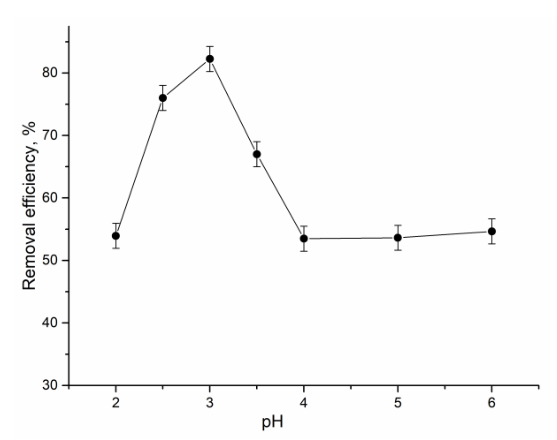
The pH is one the key parameters in the adsorption experiments since it affects the charge of the binding sites of the biosorbent and chemical speciation of metals [4]. Figure 1 shows the pH dependence of Eu(III) adsorption on A. platensis at pH range 2.0–6.0. The experiments were not performed at higher pH values since, at pH > 6.5, Eu(III) is present in solution in the form of Eu(OH)2 or Eu(OH)3 [4]. At pH 2.0, the relatively low efficiency of Eu(III) removal (53%) can be explained by competition of metal ions and H+ for binding sites. Main europium species at pH < 6.5 are Eu3+ and Eu(OH)2+ [16].
The highest rate of Eu(III) biosorption was attained at pH 3.0 (82%), and at the pH range of 4.0–6.0, it was reduced to 53%. High Eu(III) biosorption at pH 3 can be associated with the protonation of the functional groups and decrease of competing H+ ions [2]. Decrease of Eu(III) biosorption between pH 4.0 and 6.0 can be explained by the reduced effect of protonation of the metal binding sites, associated with the increase of the number of hydroxyl groups due to the addition of sodium hydroxide to increase the pH of experimental solutions [2]. Reduction of Eu(III) removal with the increase of pH is possible in the case when ionic binding on cell surface is the main mechanism of metal adsorption [17].
The maximum Eu3+ biosorption by Bacillus thuringiensis [1] was at pH 8.0. About 99% of Eu(III) was adsorbed on TiO2 at pH > 6 [16]. Experiments on Eu(III) biosorption by Deinococcus radiodurans were conducted at pH 6.0 [14]. Thus, in the present study, the kinetics, isotherm, and thermodynamic studies were performed at pH 3.0.
The evaluation of the sorption kinetics is important since it provides valuable information about the mechanism of a sorption reaction [12]. Results presented in Figure 2a showed that sorption of Eu(III) on A. platensis was very quick process, 75% of ions being removed from solution in the first 3 min of sorbent–sorbate interaction. This initial quick phase of sorption associated with the presence of a large number of exchangeable binding sites was followed by the slow attainment of equilibrium due to saturation of the binding sites.
Three classical kinetic models were applied to describe Eu(III) biosorption (Figure 2a). The Lagergren-first-order model (PFO) describes adsorption in solid–liquid systems, representing a reversible sorption process [2,18]. It is assumed that one europium ion is sorbed on one sorption site on the A. platensis surface:
The pseudo-second-order (PFO) model is applied to analyze chemisorption kinetics from liquid solutions [18].
The Elovich equation is used to describe chemical adsorption mechanism in nature [19].
where qe and qt are the content of metal (mg/g) adsorbed at equilibrium and at t time, k2 (g/mg·min) and k1 (1/min) are rates constant of pseudo-first order and pseudo-second order, and α (g/mg∙min) and β (g/mg) are the Elovich equation constants.
The parameters of applied models are listed in Table 1. Extremely high values of k2 in PSO model and α in Elovich model indicate that these models are not applicable for description of experimentally obtained data.
High correlation coefficient conveys that the adsorption of Eu(III) onto A. platensis follows the PFO model. Good agreement between q values, both calculated and experimental supports this fact. Applicability of PFO model suggest that the adsorption proceed by diffusion of Eu(III) through the boundary layer on the adsorbent surface, and this may be the rate-determining step of the overall process [12].
Eu(III) removal by other sorbents, e.g., bacteria Bacillus thuringiensis [1] and cellulose–yeast surface [6], was better described by the PSO model.
On the next step, equilibrium experiments were performed to evaluate the adsorption capacity of A. platensis. The effect of initial Eu(III) concentration on adsorption capacity was studied by varying its concentration in solution from 10 to 100 mg/L, maintaining other parameters as constant. According to obtained data, the biosorption capacity of A. platensis increased with the increase of Eu(III) concentration in solution from 0.8 to 7.25 mg/g (Figure 2b), which can be explained by the provision of more Eu(III) for the available adsorption sites [2].
The adsorption of Eu(III) onto the A. platensis was described using Langmuir and Freundlich models. The Langmuir model is applicable for monolayer adsorption on the sorbent containing definite number of identical binding sites [16] and is presented as:
where Ce is Eu(III) concentration at equilibrium in mg/L, qe and qm are is amount of metal adsorbed at equilibrium and maximum adsorption capacity in mg/g, and b is Langmuir constant in L/mg.
The Freundlich expression is an empirical equation describing adsorption onto heterogeneous surface. It presupposes firstly that stronger binding sites are occupied and that the binding strength is reduced due to an increase in the degree of site occupation [6,16]. The equation is presented as:
where KF and n are Freundlich constants.
The correlation coefficient values of both applied models were 0.99, indicating their applicability for the description of experimentally obtained data (Table 1). Maximum sorption capacity calculated from Langmuir model was 89.5 mg/g, and b indicates high affinity between the sorbate and sorbent. The high R2 values of the Langmuir model indicate that that chemisorption may be the main mechanism of Eu(III) biosorption. The values of n from Freundlich model higher than 1 support this fact [2]. The applicability of Langmuir and Freundlich models for the description of Eu(III) sorption onto bare TiO2 was shown in [16].
Maximum sorption capacity of A. platensis was compared with the data presented in the literature (Table 2). According to present values, the sorption capacity of spirulina was lower compared to Bacillus thuringiensis [1] and Acutodesmus acuminatus [17] but higher than values obtained for other sorbents listed in the Table 2. The difference in the maximum Eu(III) sorption capacity of different sorbents can be explained by different conditions, temperature, pH, the sorbate and sorbent concentrations, and way of biomass pretreatment by which experiments were performed.
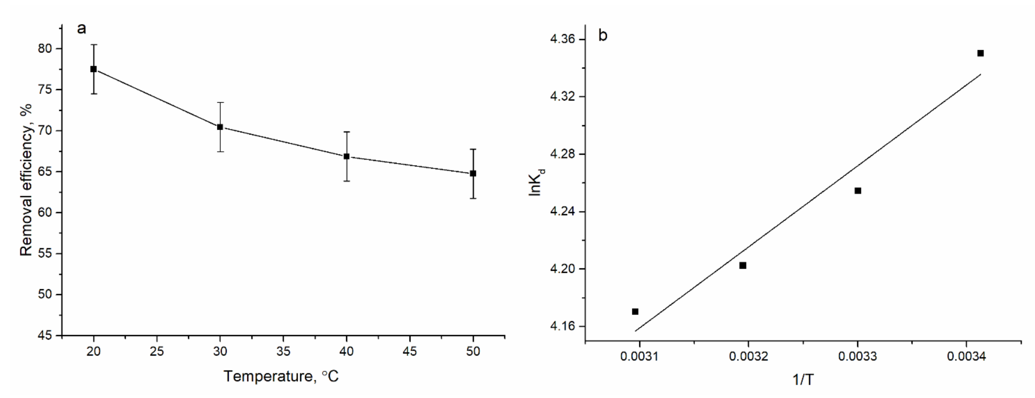
Temperature is another important factor influencing the sorption process. The effect of temperature on the Eu(III) biosorption on A. platensis was studied at range of 20–50 °C. An increase in the temperature (Figure 3a) resulted in the decrease of Eu(III) adsorption, indicating that the process is exothermic. Biosorption of Eu(III) onto the cellulose–yeast surface was spontaneous and exothermic [6].
The free energy, enthalpy, and entropy changes were obtained from the plot of lnKd against 1/T (Figure 3b) and are presented in Table 3.
where Kd is the distribution coefficient, and it is calculated according to Equation (15):
Negative values of ∆G◦ indicate spontaneous adsorption and the degree of spontaneity of the reaction increases with increasing temperature. The negative charge ∆H◦ indicate the exothermic character of the sorption, while the positive ∆S◦ reflects the affinity of the biosorbent toward Eu(III).
3.2. Bioaccummulation
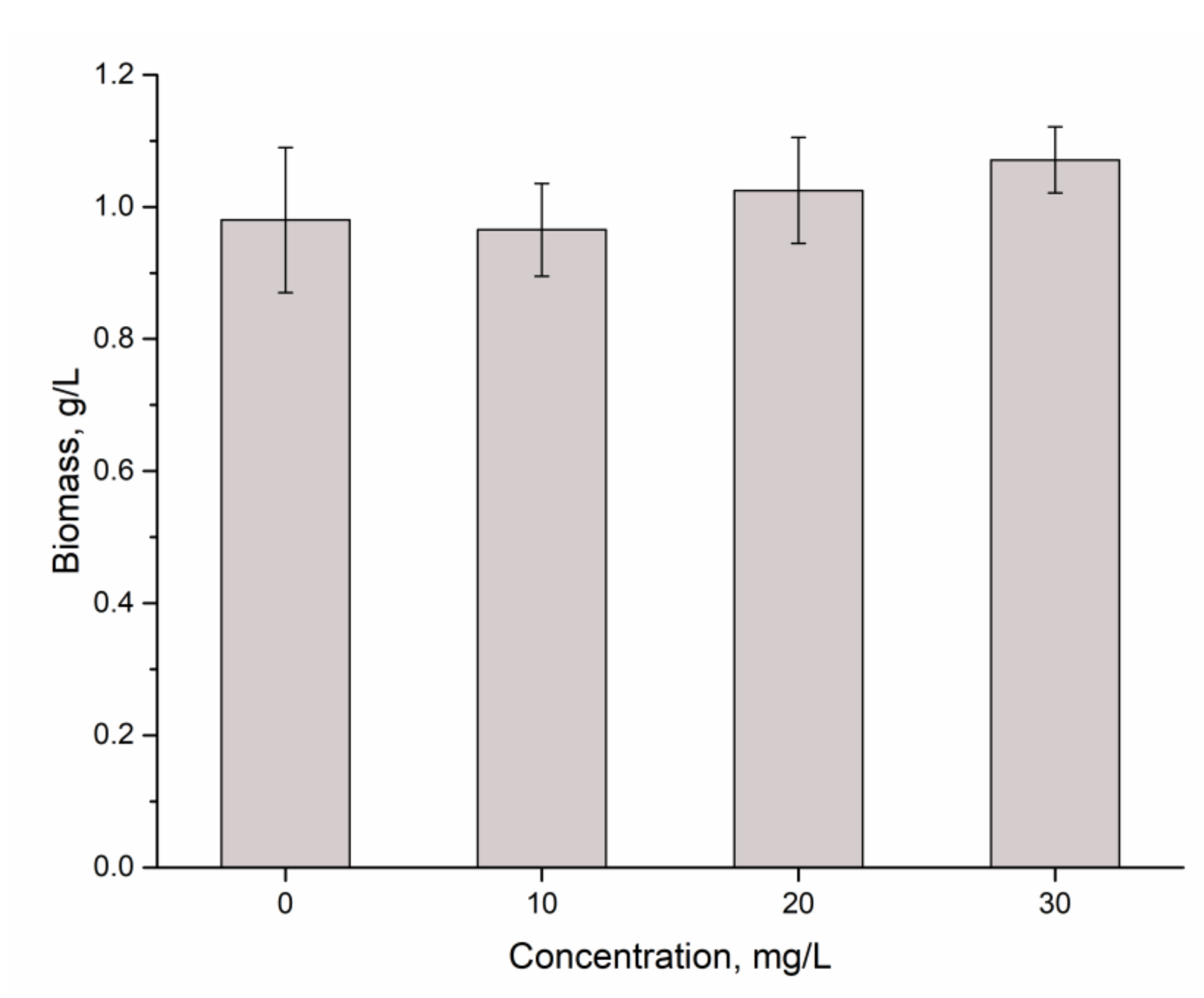
In bioaccumulation experiments, the effect of different concentrations of Eu(III) on A. platensis was assessed. According to ICP-AES data, the content of Eu in biomass increased directly proportionally to its concentration in medium. At Eu(III) concentration of 10 mg/L, its content in biomass constituted 9.8 mg/g and increased up to 29.8 mg/g at Eu(III) concentration in solution of 30 mg/L. The removal efficiency of A. platensis was at an extremely high level, constituting 98–99% at all studied Eu(III) concentrations. According to obtained results (Figure 4), the amount of biomass accumulated in the control and experimental samples was very close but without statistically significant differences and constituted 0.96–1.07 g/L dry biomass.
It is known that the effect of the rare earth elements on microorganisms depends on element and its concentration [20,21,22,23]. In our previous research, it has been shown that spirulina has a high degree of tolerance to rare earths elements in the concentration range of 10−30 mg/L. However, depending on the element added to the nutrient medium, different response patterns have been observed. Thus, Sm, Tb, and Nd at concentrations 10–30 mg/L did not influence amount of biomass, and Dy and La induced an increase of biomass up to 19.3%, while Yb, the on contrary, led to its decrease by 26.8% compared to control [14].
Although there were no pronounced effect of Eu(III) on biomass, it induced significant changes in the biochemical composition of biomass, especially at a concentration of 30 mg/L. The tendency of the decrease of protein content was observed at all applied concentrations, and at concentrations of 20 and 30 mg/L, the reduction of protein content was statistically significant (Figure 5). Thus, at Eu(III) concentration 20 mg/L, the content of protein in the biomass was 55.36%, which was 10.2% lower compared to control (p = 0.0213), and at the concentration of 30 mg/L, it comprised 50.7% of the biomass, which was 17.7% less than in control (p = 0.0097). Proteins are the basic components of spirulina biomass, constituting, depending on the conditions, 60–70% of biomass. Decreasing of their amount below 50% is associated with significant degradation of biomass. Usually, the content of protein below 45% is a result of a toxic effect, to which the culture is not able to adapt or adapts very poorly [24,25]. In the case of Eu(III), the toxic effect was directly proportional to increase of its concentration in the medium and at 30 mg/L reached the values close to the critical level for spirulina.
The content of carbohydrates (Figure 5) in spirulina biomass at Eu(III) concentrations of 10 and 20 mg/L did not differ significantly from that characteristic for control, while at a concentration of 30 mg/L, it was reduced significantly, by 27.4% compared to control (p = 0.0048). Comparing results obtained in the present study with data obtained for other rare earth elements the different response of the culture was observed. Thus, in the case of Sm and Tb, which did influence the amount of biomass, the content of carbohydrates increased [14], which is associated with elimination of toxic element from the cell. The effect of Eu(III) was similar to those of Nd, resulting in a decrease of the content of carbohydrates at all studied concentrations.
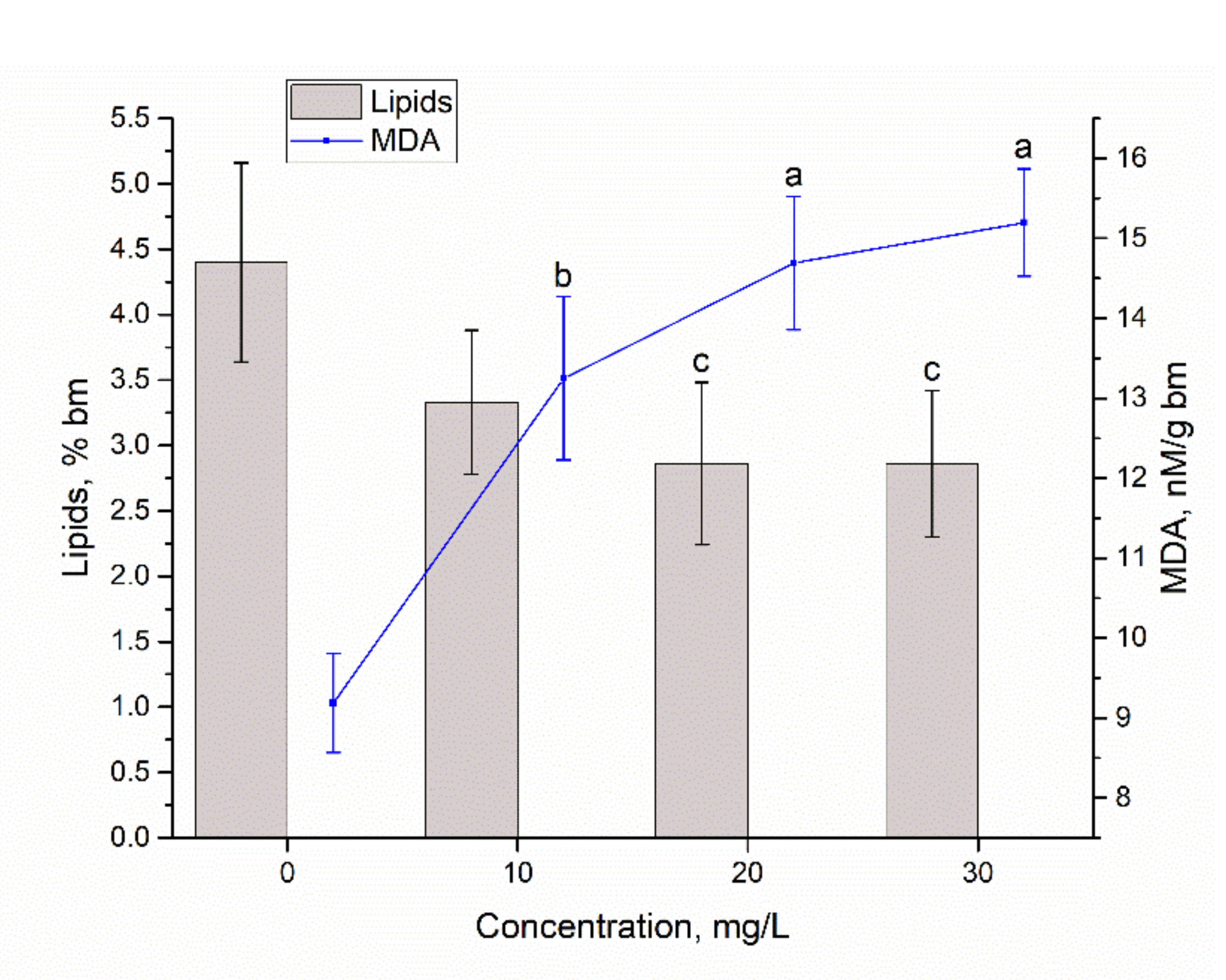
Spirulina is an organism with a low content of lipids, which are mainly located in the membranes and ensure the functioning of the cell as a whole. The control biomass contained 4.4% lipids, while in the experimental variants, their levels was reduced (Figure 6). At concentrations of 20 and 30 mg/L, the reduction was statistically significant (p = 0.0269 and p = 0.0242, respectively), and it was about 35% lower compared to the control. The content of the product of oxidative degradation of lipids (malonic dialdehyde) in the experimental samples increased by 41–73% compared to the control sample. Since MDA is considered a proper marker of oxidative stress, it can be suggested that presence of Eu(III) in the cultivation medium led to pronounced oxidative stress, which is associated with a decrease in the amount of total lipids. Under these conditions, the physiological processes in the spirulina cells can be compromised.
Other rare earth elements can change the content of lipids in spirulina biomass in a very different way. Thus, Sm and Nd at concentrations of 10–30 mg/L did not produce quantitative changes in the total lipids content, and La caused their slight increase, while Tb, Dy, and Yb resulted in the decrease of the content of lipids [14].
Eu(III) provoked effect very similar to Yb ions. Regarding the amount of MDA, the Eu(III) effect on biomass can also be compared with the effect of Yb and Nd ions, which led to a considerable increase in the amount of MDA at all applied concentrations.
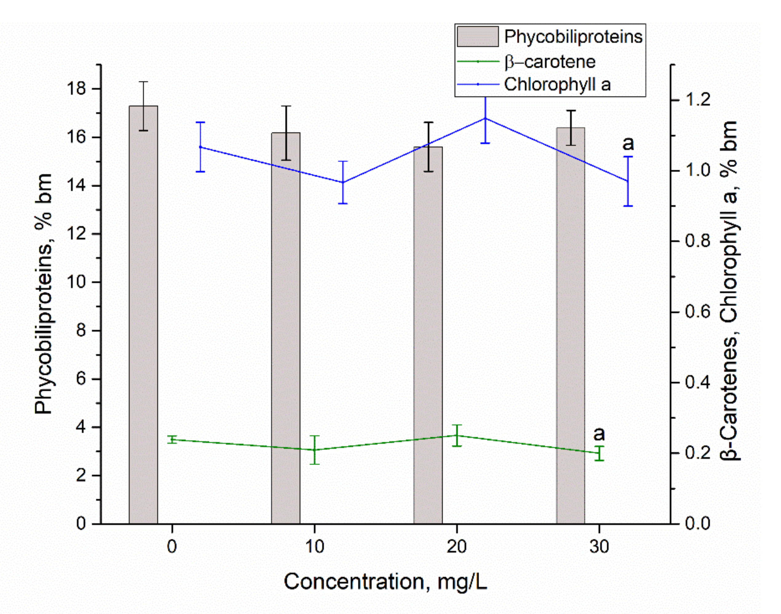
Maintaining of the productivity of Eu-loaded biomass on the level of control was possible, due to retention of the adequate level of photosynthetic pigments (Figure 7). Thus, the amount of α-chlorophyll and β-carotene at the first two concentrations of Eu(III) did not differ from the control. Their decrease was observed only at Eu(III) concentration of 30 mg/L. The total amount of phycobiliproteins also was not influenced significantly by addition of Eu(III) in the cultivation media. In this sense, Eu(III) behaved differently from other rare earth elements, which led to a significant reduction (by 10–50% compared to the control) of total phycobiliproteins [14].
The antioxidant activity of biomass was also maintained at a stable level (Figure 8). The activity of ethanolic extract obtained from spirulina biomass grown on medium loaded with Eu(III) was on the level of the control for all Eu(III) concentrations. The activity of the water extract at Eu(III) concentrations of 10 and 20 mg/L was higher compared to the control, while at concentration of 30 mg/L it was on the level of control. Data reported previously for La, Dy, Sm, and Tb showed a marked increase in the antioxidant activity of water extract from spirulina biomass, while Nd and Yb in concentrations of 10–30 mg/L caused its considerable decrease [14].
Biosorption and bioaccumulation showed to be suitable approaches for Eu(III) removal from wastewater. Both processes occur permanently in natural environment in uncontrolled conditions and are also applied in the industrial practice under controlled conditions [26]. In the present study, in bioaccumulation experiments, higher efficiency of Eu(III) uptake was achieved that can be explained by presence of the functional groups on the surface and inside the cell and increase of biomass concentration, which make possible the binding of more Eu(III). However, bioaccumulation require resources for biomass cultivation, and high metal concentrations can inhibit its growth; therefore, biosorption seems to be more suitable process for industrial application. The main limitation of biological sorbents is their short life in comparison with traditional sorbents [26].
Often, the parameters (pH, metal concentration) of real wastewater are different from batch systems; in this case, the efficiency of metal ions removal can be increased in several ways: by effluent dilution or application of a two-or-more-step scheme of metal removal with the addition of a new dosage of biosorbent, as it was shown in our previous studies [27,28].
4. Conclusions
Cyanobacteria Arthrospira platensis was tested for its ability to remove Eu(III) by means of biosorption and bioaccumulation experiments. Maximum Eu(III) removal in biosorption experiments can be achieved at pH 3.0, contact time 3 min, and temperature 20 °C. Equilibrium of the sorption can be described by both Langmiur and Freundlich models, while kinetics fits better to the pseudo-first-order model. From the thermodynamic point of view, it is an exothermic, spontaneous process.
In bioaccumulation experiments, Arthrospira platensis showed a high rate of Eu(III) uptake, up to 98–99% at its concentration in the medium of 10–30 mg/L. Although Eu-loaded spirulina maintained a high level of productivity and a normal content of carbohydrates and pigments, Eu(III)’s toxic effect was manifested by the decrease of the content of protein and an increase in the amount of MDA. These two factors denote a state of pronounced oxidative stress, which can significantly impair the quality of spirulina biomass in the long term or in repeated action of metal ions.
Author Contributions
Conceptualization, N.Y., L.C. and I.Z.; methodology, L.C. and I.Z.; software, D.G.; validation, L.C. and I.Z.; investigation, N.Y., T.C. and L.R.; writing—original draft preparation, L.C. and I.Z.; funding acquisition N.Y.; writing—review and editing, all authors; visualization, I.Z and N.Y. All authors have read and agreed to the published version of the manuscript.
Funding
The study was carried out with financial support of Association of Young Scientists and Specialist of the JINR Grant No 22-402-11.
Institutional Review Board Statement
Not applicable.
Informed Consent Statement
Not applicable.
Data Availability Statement
Not applicable.
Conflicts of Interest
The authors declare no conflict of interest.
References
- Pan, X.; Wu, W.; Lü, J.; Chen, Z.; Li, L.; Rao, W.; Guan, X. Biosorption and extraction of europium by Bacillus thuringiensis strain. Inorg. Chem. Commun. 2017, 75, 21–24. [Google Scholar] [CrossRef]
- Cadogan, E.I.; Lee, C.-H.; Popuri, S.R. Facile synthesis of chitosan derivatives and Arthrobacter sp. biomass for the removal of europium(III) ions from aqueous solution through biosorption. Int. Biodeterior. Biodegrad. 2015, 102, 286–297. [Google Scholar] [CrossRef]
- Maleke, M.; Valverde, A.; Vermeulen, J.-G.; Cason, E.D.; Gómez-Arias, A.; Moloantoa, K.; Coetsee-Hugo, L.; Swart, H.; Van Heerden, E.; Castillo, J. Biomineralization and Bioaccumulation of Europium by a Thermophilic Metal Resistant Bacterium. Front. Microbiol. 2019, 10, 81. [Google Scholar] [CrossRef] [PubMed]
- Jena, A.; Pradhan, S.; Mishra, S.; Sahoo, N.K. Evaluation of Europium Biosorption Using Deinococcus radiodurans. Environ. Process. 2021, 8, 251–265. [Google Scholar] [CrossRef]
- Baumer, T.; Hixon, A.E. Kinetics of europium sorption to four different aluminum (hydr)oxides: Corundum, γ-alumina, bayerite, and gibbsite. J. Environ. Radioact. 2018, 195, 20–25. [Google Scholar] [CrossRef]
- Arunraj, B.; Sathvika, T.; Rajesh, V.; Rajesh, N. Cellulose and Saccharomyces cerevisiae Embark to Recover Europium from Phosphor Powder. ACS Omega 2019, 4, 940–952. [Google Scholar] [CrossRef] [Green Version]
- Lazaris, D.; Liasko, R.; Leonardos, I.; Evangelou, A.; Kalfakakou, V. Toxic effects of europium chloride on developing zebrafish (Danio rerio) embryos. J. Biol. Res. 2012, 18, 291–296. [Google Scholar]
- Nörenberg, D.; Schmidt, F.; Schinke, K.; Frenzel, T.; Pietsch, H.; Giese, A.; Ertl-Wagner, B.; Levin, J. Investigation of potential adverse central nervous system effects after long term oral administration of gadolinium in mice. PLoS ONE 2020, 15, e0231495. [Google Scholar] [CrossRef] [Green Version]
- Das, N.; Das, D. Recovery of rare earth metals through biosorption: An overview. J. Rare Earths 2013, 31, 933–943. [Google Scholar] [CrossRef]
- Rabah, M.A. Recyclables recovery of europium and yttrium metals and some salts from spent fluorescent lamps. Waste Manag. 2008, 28, 318–325. [Google Scholar] [CrossRef]
- Cepoi, L.; Zinicovscaia, I.; Rudi, L.; Chiriac, T.; Djur, S.; Yushin, N.; Grozdov, D. Assessment of Metal Accumulation by Arthrospira platensis and Its Adaptation to Iterative Action of Nickel Mono- and Polymetallic Synthetic Effluents. Microorganisms 2022, 10, 1041. [Google Scholar] [CrossRef] [PubMed]
- Vijayaraghavan, K.; Sathishkumar, M.; Balasubramanian, R. Biosorption of Lanthanum, Cerium, Europium, and Ytterbium by a Brown Marine Alga, Turbinaria Conoides. Ind. Eng. Chem. Res. 2010, 49, 4405–4411. [Google Scholar] [CrossRef]
- Diniz, V.; Volesky, B. Biosorption of La, Eu and Yb using Sargassum biomass. Water Res. 2005, 39, 239–247. [Google Scholar] [CrossRef] [PubMed]
- Zinicovscaia, I.; Cepoi, L.; Rudi, L.; Chiriac, T.; Grozdov, D.; Pavlov, S.; Djur, S. Accumulation of dysprosium, samarium, terbium, lanthanum, neodymium and ytterbium by Arthrospira platensis and their effects on biomass biochemical composition. J. Rare Earths 2020, 39, 1133–1143. [Google Scholar] [CrossRef]
- Cepoi, L.; Rudi, L.; Miscu, V.; Cojocari, A.; Chiriac, T.; Sadovnic, D. Antioxidative activity of ethanol extracts from Spirulina platensis and Nostoc linckia measured by various methods. Analele Univ. Oradea Fasc. Biol. 2009, 16, 43–48. [Google Scholar]
- Tan, X.; Fang, M.; Li, J.; Lu, Y.; Wang, X. Adsorption of Eu(III) onto TiO2: Effect of pH, concentration, ionic strength and soil fulvic acid. J. Hazard. Mater. 2009, 168, 458–465. [Google Scholar] [CrossRef]
- Furuhashi, Y.; Honda, R.; Noguchi, M.; Hara-Yamamura, H.; Kobayashi, S.; Higashimine, K.; Hasegawa, H. Optimum conditions of pH, temperature and preculture for biosorption of europium by microalgae Acutodesmus acuminatus. Biochem. Eng. J. 2019, 143, 58–64. [Google Scholar] [CrossRef]
- Boparai, H.K.; Joseph, M.; O’Carroll, D.M. Kinetics and thermodynamics of cadmium ion removal by adsorption onto nano zerovalent iron particles. J. Hazard. Mater. 2011, 186, 458–465. [Google Scholar] [CrossRef]
- Wu, F.-C.; Tseng, R.-L.; Juang, R.-S. Characteristics of Elovich equation used for the analysis of adsorption kinetics in dye-chitosan systems. Chem. Eng. J. 2009, 150, 366–373. [Google Scholar] [CrossRef]
- Wenhua, L.; Ruming, Z.; Zhixiong, X.; Xiangdong, C.; Ping, S. Effects of La3+ on Growth, Transformation, and Gene Expression of Escherichia coli. Biol. Trace Element Res. 2003, 94, 167–177. [Google Scholar] [CrossRef]
- Jin, X.; Chu, Z.; Yan, F.; Zeng, Q. Effects of lanthanum(III) and EDTA on the growth and competition of Microcystis aeruginosa and Scenedesmus quadricauda. Limnologica 2009, 39, 86–93. [Google Scholar] [CrossRef] [Green Version]
- Wang, Y.; Li, Y.; Luo, X.; Ren, Y.; Gao, E.; Gao, H. Effects of yttrium and phosphorus on growth and physiological characteristics of Microcystis aeruginosa. J. Rare Earths 2018, 36, 781–788. [Google Scholar] [CrossRef]
- Wang, Y.; Li, J.; Lü, Y.; Jin, H.; Deng, S.; Zeng, Y. Effects of cerium on growth and physiological characteristics of Anabaena flosaquae. J. Rare Earths 2012, 30, 1287–1292. [Google Scholar] [CrossRef]
- Cepoi, L.; Zinicovscaia, I.; Rudi, L.; Chiriac, T.; Miscu, V.; Djur, S.; Strelkova, L.; Vergel, K.; Nekhoroshkov, P. Growth and heavy metals accumulation by Spirulina platensis biomass from multicomponent copper containing synthetic effluents during repeated cultivation cycles. Ecol. Eng. 2020, 142, 105637. [Google Scholar] [CrossRef]
- Zinicovscaia, I.; Cepoi, L.; Rudi, L.; Chiriac, T.; Grozdov, D.; Vergel, K. Effect of zinc-containing systems on Spirulina platensis bioaccumulation capacity and biochemical composition. Environ. Sci. Pollut. Res. 2021, 28, 52216–52224. [Google Scholar] [CrossRef]
- Chojnacka, K. Biosorption and bioaccumulation—The prospects for practical applications. Environ. Int. 2010, 36, 299–307. [Google Scholar] [CrossRef]
- Zinicovscaia, I.; Yushin, N.; Grozdov, D.; Abdusamadzoda, D.; Safonov, A.; Rodlovskaya, E. Zinc-containing effluent treatment using Shewanella xiamenensis biofilm formed on zeolite. Materials 2021, 14, 1760. [Google Scholar] [CrossRef]
- Zinicovscaia, I.; Yushin, N.; Grozdov, D.; Vergel, K.; Popova, N.; Artemiev, G.; Safonov, A. Metal removal from nickel-containing effluents using mineral–organic hybrid adsorbent. Materials 2020, 13, 4462. [Google Scholar] [CrossRef]
Figure 1.
Effect of pH on Eu(III) biosorption by S. platensis (Ci 10 mg/L, time 60 min, temperature 22 °C).
Figure 1.
Effect of pH on Eu(III) biosorption by S. platensis (Ci 10 mg/L, time 60 min, temperature 22 °C).

Figure 2.
(a) Kinetics and (b) isotherms of Eu(III) biosorption on S. platensis biomass.

Figure 3.
(a) Effect of temperature on Eu(III) removal and (b) lnKd versus 1/T.

Figure 4.
Influence of Eu(III) applied at a concentration of 10–30 mg/L on the A. platensis biomass.
Figure 4.
Influence of Eu(III) applied at a concentration of 10–30 mg/L on the A. platensis biomass.

Figure 5.
The content of protein and carbohydrates in A. platensis biomass exposed to Eu(III) in concentrations 10–30 mg/L (a—p < 0.01, b—p < 0.05, c—p < 0.005 for the difference between sample and control).
Figure 5.
The content of protein and carbohydrates in A. platensis biomass exposed to Eu(III) in concentrations 10–30 mg/L (a—p < 0.01, b—p < 0.05, c—p < 0.005 for the difference between sample and control).

Figure 6.
The content of lipids and MDA in A. platensis biomass exposed to Eu(III) in concentrations 10–30 mg/L (a—p < 0.01, b—p < 0.05, c—p < 0.005 for the difference between sample and control).
Figure 6.
The content of lipids and MDA in A. platensis biomass exposed to Eu(III) in concentrations 10–30 mg/L (a—p < 0.01, b—p < 0.05, c—p < 0.005 for the difference between sample and control).

Figure 7.
The content of pigments in A. platensis biomass exposed to Eu(III) in concentrations 10–30 mg/L (a—p <0.01 for the difference between sample and control).
Figure 7.
The content of pigments in A. platensis biomass exposed to Eu(III) in concentrations 10–30 mg/L (a—p <0.01 for the difference between sample and control).

Figure 8.
The antioxidant activity of extracts obtained from A. platensis biomass exposed to Eu(III) in concentrations 10–30 mg/L (a—p < 0.01, b—p < 0.05for the difference between sample and control).
Figure 8.
The antioxidant activity of extracts obtained from A. platensis biomass exposed to Eu(III) in concentrations 10–30 mg/L (a—p < 0.01, b—p < 0.05for the difference between sample and control).


Table 1.
Parameters of the kinetics and isotherm of Eu(III) sorption on A. platensis.
| Kinetics | |||||||||
| Model | PFO | PSO | Elovich | ||||||
| Parameters | qe | k1 | R2 | qe | k2 | R2 | α | β | R2 |
| Eu(III) | 0.99 | 1.79 | 0.99 | 0.99 | 2.83·1044 | 0.99 | 1.07 ·1044 | 108 | 0.99 |
| Isotherms | |||||||||
| Model | Langmuir | Freundlich | |||||||
| Parameters | qm | b | R2 | KF | n | R2 | |||
| Eu(III) | 89.5 | 0.0009 | 0.99 | 0.09 | 1.06 | 0.98 | |||
Table 2.
Comparison of A. platensis sorption capacity with the literature data.
| Sorbent | q, mg/g | Reference |
|---|---|---|
| Spirulina platensis | 89.5 | Present study |
| Bacillus thuringiensis | 160 | [1] |
| Native cellulose | 18.5 | [6] |
| Saccharomyces cerevisiae | 14.2 | [6] |
| Yeast-immobilized cellulose | 25.9 | [6] |
| Arthrobacter sp. | 9.53 | [2] |
| Chitosan powder | 48.3 | [2] |
| Chitosan beads | 18.4 | [2] |
| Acutodesmus acuminatus | 174.2 | [17] |
Table 3.
Thermodynamics parameters for Eu(III) biosorption on A. platensis.
| Temperature, K | ∆G◦, kJ/mol | ∆H◦, kJ/mol | ∆S◦, J/mol·K |
|---|---|---|---|
| 293 | −10.5 | −4.7 | 19.9 |
| 303 | −10.7 | ||
| 313 | −10.9 | ||
| 323 | −11.1 |
Publisher’s Note: MDPI stays neutral with regard to jurisdictional claims in published maps and institutional affiliations. |
© 2022 by the authors. Licensee MDPI, Basel, Switzerland. This article is an open access article distributed under the terms and conditions of the Creative Commons Attribution (CC BY) license (https://creativecommons.org/licenses/by/4.0/).
Share and Cite
MDPI and ACS Style
Yushin, N.; Zinicovscaia, I.; Cepoi, L.; Chiriac, T.; Rudi, L.; Grozdov, D. Biosorption and Bioaccumulation Capacity of Arthospiraplatensis toward Europium Ions. Water 2022, 14, 2128. https://doi.org/10.3390/w14132128
AMA Style
Yushin N, Zinicovscaia I, Cepoi L, Chiriac T, Rudi L, Grozdov D. Biosorption and Bioaccumulation Capacity of Arthospiraplatensis toward Europium Ions. Water. 2022; 14(13):2128. https://doi.org/10.3390/w14132128
Chicago/Turabian StyleYushin, Nikita, Inga Zinicovscaia, Liliana Cepoi, Tatiana Chiriac, Ludmila Rudi, and Dmitrii Grozdov. 2022. "Biosorption and Bioaccumulation Capacity of Arthospiraplatensis toward Europium Ions" Water 14, no. 13: 2128. https://doi.org/10.3390/w14132128
Note that from the first issue of 2016, this journal uses article numbers instead of page numbers. See further details here.