The “first cut” in a gross anatomy class, when a student uses a scalpel to slice into the skin of a cadaver, is a nerve-wracking experience for anyone embarking on a medical career. But it’s almost sacred, too. As students begin their hands-on anatomy education, they also have to ruminate on life and death, and their relationship with their future patients.
In today’s American medical education, all but a handful of medical schools make cadaver dissection or prosection (dissection done by an expert as demonstration for students) part of their first- and second-year anatomy courses. But now, with COVID-19 upending the higher education experience around the world, those teaching and studying medicine are wondering how to proceed with the training. Harvard Medical School has already announced it will not be having first-year medical students come to campus this fall. Baylor College of Medicine in Texas will have medical students do classes online but come to campus in small groups to learn anatomy in person. Some schools, though, are already prepared for these constraints because they have created a way to do most of their anatomy education without ever touching a human body: They have found a way to move dissection labs to a virtual reality world.
The use of anatomical dissection has been a cornerstone of medical education for hundreds of years. In centuries past, bodies were procured through illegal back channels, but since the middle of the 20th century, people in the U.S. have been able to donate their bodies to medical schools for educational purposes. Medical schools around the country get hundreds of donations each year, and body donation services in the U.S. are seeing a rise in the number of donations.
The physical act of dissecting a human body may seem impossible to replace, and in some senses, it is. Cadaver dissection is a rite of passage in medical schools—the deceased person is a medical student’s introduction to the human aspect of medicine outside the pages of a textbook, and it is often referred to as their first patient. This provides a unique emotional experience, just as valuable as the anatomy lessons it accompanies, that is unlikely to happen while wearing a VR headset. However, there are benefits to be gained in that trade-off. Medical educators have been working for years to make virtual anatomy learning interactive so students can experience “living anatomy” instead of struggling to see anatomical structures for the first time in an embalmed cadaver.
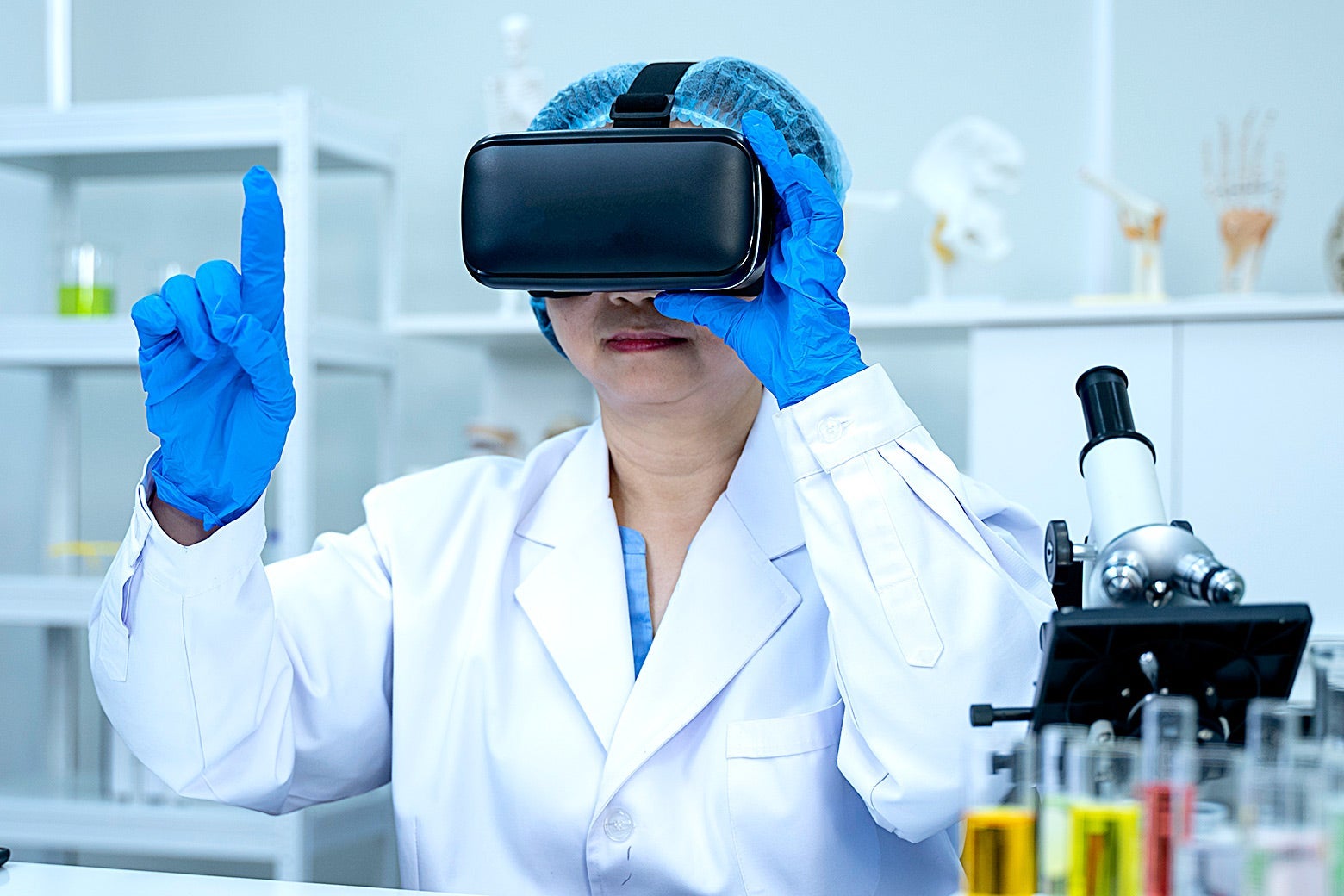
At Case Western Reserve University School of Medicine, instructors have been teaching anatomy for years with software called HoloAnatomy. It runs on Microsoft’s mixed reality HoloLens headset—a glasseslike setup that overlays the VR components on the user’s surroundings. Mark Griswold, a professor of radiology at the school and faculty leader of the HoloAnatomy program, says that students usually don their headsets and learn anatomy together in the same room on campus, but when classes went online, they were able to ship out 185 HoloLenses to first-year students so their education could continue uninterrupted.
Ellen Kendall is a first-year medical student at Case Western Reserve who took her anatomy class last year primarily via HoloLens headset. She says she didn’t miss a beat when classes went remote because of COVID-19: “I had Zoom open on my computer and I was connected to my professor who was lecturing, and I had a HoloLens with a ‘holo body’ in my living room.
“If you look at it from the outside, it’s super funny because we’re all just staring at each other,” Kendall says. “But really, we’re all looking at different holograms of anatomy.” While students are immersed in the program, they can zoom in on and virtually dissect parts of the “holo body” to see intricate details of structures like lymph nodes and nerves that are near-impossible to spot in a cadaver.
Case Western Reserve built its new medical campus without a cadaver lab, so technology like this is a crucial part of the educational experience (though students still take a two-week-long gross anatomy boot camp on the older part of campus, which still has an anatomy lab). Other schools are moving in the same direction. The Kaiser Permanente Medical School in Pasadena, California, which opens for its first class of students in July, also decided not to build a cadaver lab. Instead, it too will teach basic anatomy using touch screens and VR headsets. Reasons for this choice include cost of the laboratory—a cadaver lab costs millions of dollars to build—and the cost of the cadavers themselves. The bodies and care of the labs cost schools tens of thousands a year and expose students and staff to hazardous embalming chemicals like formaldehyde.
But the decision to move from cadavers to VR or mixed reality is not a simple cost-benefit analysis. The buy-in for HoloAnatomy was not immediate, Griswold says, because it is difficult to diverge from a centuries-old tradition. Old-school anatomists were skeptical that students could learn as much from a virtual tool as they could from a real human body. Now, though, he says they have been converted because of how effective a teaching tool mixed reality is. In one recent study at Case Western Reserve, medical students were divided into two groups: One group learned the anatomy of the upper and lower limb through cadaver dissection and mixed reality education, and the other group learned entirely on a cadaver. Both groups took an exam at the end and got statistically indistinguishable scores.
David Axelrod and Scott Ceresnak, both pediatric cardiologists at Stanford University School of Medicine, know that virtual reality anatomy education works for their students and pediatric cardiologists in training. They worked with a company called Lighthaus, where Axelrod is also the lead medical consultant, to design virtual reality software called the Stanford Virtual Heart that runs on the Oculus Rift headset. The software is free and open to anyone who wants to use it, and so far, Axelrod says, 35 schools and medical centers around the U.S. and beyond are using it for education. Once a user puts on the Oculus Rift, they can use hand-held controls to rotate and zoom in on the virtual heart, allowing cardiologists to better understand abnormalities associated with various heart conditions that are difficult to see up close in a living patient. Ceresnak says that the first time he saw the virtual heart he was blown away: “I mean, you can stand in the ventricle and appreciate the anatomy in a completely different, new, novel way.”
Doctors often have to assess their patients’ three-dimensional bodies through two-dimensional images like X-rays and scans. Virtual anatomy programs can help them learn how to bridge that gap. Kendall says that with HoloAnatomy, an X-ray can be virtually juxtaposed with the “living” hologram body, which helps students better understand what they are actually looking at during the notoriously difficult process of interpreting an X-ray.
Incorporating new methods of education for foundational subjects like anatomy can save time as the amount of knowledge the students need to learn grows, says Erin Henninger, the executive director of Interactive Commons at Case Western Reserve: “If we can learn [anatomy] faster, maybe our students can spend some more time learning the rapidly changing medical concepts they need to know.”
Despite all of these technological benefits, anatomy educators say there is no need to leave the practice of cadaver dissection completely in the past, even considering the wrench COVID-19 throws into their plans. Most instructors call the virtual or mixed reality experiences a supplement to in-person education. Kendall says the tactile and emotional experience of a cadaver lab is important because the students learn to process and collect their thoughts about confronting the death of a patient, which are later shared with the families of the donors. Many medical schools have ceremonies and practices like this to honor the donors and their families.
Yet opponents of virtual-only dissection are not simply traditionalists stuck in the past. “Cadaveric dissection is a very active and exploratory learning experience,” says Stefanie Attardi, an anatomical sciences professor at Oakland University William Beaumont School of Medicine in Michigan, adding that she and other educators do not want to replace that with an online or virtual-only experience. She says that in-person anatomy labs lead to important lessons of teamwork and start teaching some of the hallmarks of medicine, like problem-solving and careful work: “Years down the road, many students will still remember their dissection teammates. It is such a special learning experience to share with others.”
Attardi suggests that schools without a robust online or virtual anatomy program may be able to figure out ways around campus restrictions during COVID-19 and still show students how to dissect a cadaver: “Should teachers be allowed on campus, some instructors are exploring the possibility of livestreaming or recording from the anatomy lab.” This would be a way to ensure new students get some experience with cadaver dissection, but if it’s not possible this year, instructors would be able to make a place for an anatomy lab experience later on in their lessons in another class.
The pandemic will eventually end and students will be back on campus one day, but Axelrod thinks this unprecedented teaching moment could show instructors the benefits of virtual anatomy: “I think they will end up demanding these resources and the power of this kind of instruction. So I’m optimistic, I think it’s going to take off.”
Future Tense is a partnership of Slate, New America, and Arizona State University that examines emerging technologies, public policy, and society.